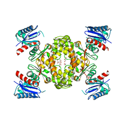
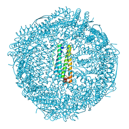
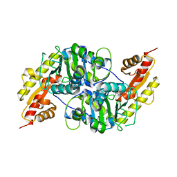
4AAM
 
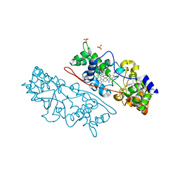 | | MacA wild-type mixed-valence | | 分子名称: | CALCIUM ION, CYTOCHROME C551 PEROXIDASE, HEME C, ... | | 著者 | Seidel, J. | | 登録日 | 2011-12-05 | | 公開日 | 2012-10-17 | | 最終更新日 | 2024-10-23 | | 実験手法 | X-RAY DIFFRACTION (2.17 Å) | | 主引用文献 | Maca is a Second Cytochrome C Peroxidase of Geobacter Sulfurreducens.
Biochemistry, 51, 2012
|
|
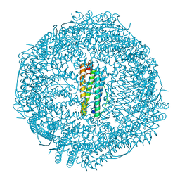
4AAL
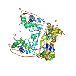 | | MacA wild-type oxidized | | 分子名称: | ACETATE ION, CALCIUM ION, CYTOCHROME C551 PEROXIDASE, ... | | 著者 | Seidel, J. | | 登録日 | 2011-12-05 | | 公開日 | 2012-10-17 | | 最終更新日 | 2024-10-23 | | 実験手法 | X-RAY DIFFRACTION (1.84 Å) | | 主引用文献 | Maca is a Second Cytochrome C Peroxidase of Geobacter Sulfurreducens.
Biochemistry, 51, 2012
|
|
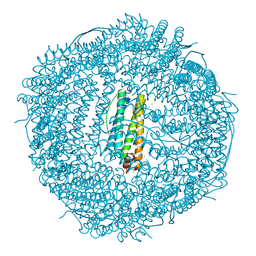
4V26
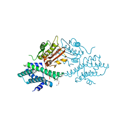 | | VER-246608, a novel pan-isoform ATP competitive inhibitor of pyruvate dehydrogenase kinase, disrupts Warburg metabolism and induces context- dependent cytostasis in cancer cells | | 分子名称: | MAGNESIUM ION, N-(2-AMINOETHYL)-2-{3-CHLORO-4-[(4-ISOPROPYLBENZYL)OXY]PHENYL} ACETAMIDE, N-[4-(2-CHLORO-5-METHYLPYRIMIDIN-4-YL)PHENYL]-2,4-DIHYDROXY-N-(4-{[(TRIFLUOROACETYL)AMINO]METHYL}BENZYL)BENZAMIDE, ... | | 著者 | Moore, J.D, Staniszewska, A, Shaw, T, D'Alessandro, J, Davis, B, Surgenor, A, Baker, L, Matassova, N, Murray, J, Macias, A, Brough, P, Wood, M, Mahon, P.C. | | 登録日 | 2014-10-06 | | 公開日 | 2014-12-03 | | 最終更新日 | 2024-05-01 | | 実験手法 | X-RAY DIFFRACTION (2.49 Å) | | 主引用文献 | VER-246608, a novel pan-isoform ATP competitive inhibitor of pyruvate dehydrogenase kinase, disrupts Warburg metabolism and induces context-dependent cytostasis in cancer cells.
Oncotarget, 5, 2014
|
|
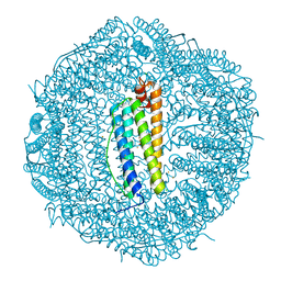
4V25
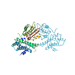 | | VER-246608, a novel pan-isoform ATP competitive inhibitor of pyruvate dehydrogenase kinase, disrupts Warburg metabolism and induces context- dependent cytostasis in cancer cells | | 分子名称: | MAGNESIUM ION, N-(2-AMINOETHYL)-2-{3-CHLORO-4-[(4-ISOPROPYLBENZYL)OXY]PHENYL} ACETAMIDE, N-[4-(2-chloro-5-methylpyrimidin-4-yl)phenyl]-N-(4-{[(difluoroacetyl)amino]methyl}benzyl)-2,4-dihydroxybenzamide, ... | | 著者 | Moore, J.D, Staniszewska, A, Shaw, T, D'Alessandro, J, Davis, B, Surgenor, A, Baker, L, Matassova, N, Murray, J, Macias, A, Brough, P, Wood, M, Mahon, P.C. | | 登録日 | 2014-10-06 | | 公開日 | 2014-12-03 | | 最終更新日 | 2024-05-01 | | 実験手法 | X-RAY DIFFRACTION (2.6 Å) | | 主引用文献 | VER-246608, a novel pan-isoform ATP competitive inhibitor of pyruvate dehydrogenase kinase, disrupts Warburg metabolism and induces context-dependent cytostasis in cancer cells.
Oncotarget, 5, 2014
|
|
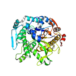
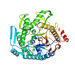
3VOV
 | | Crystal Structure of ROK Hexokinase from Thermus thermophilus | | 分子名称: | GLYCEROL, Glucokinase, ZINC ION | | 著者 | Nakamura, T, Kashima, Y, Mine, S, Oku, T, Uegaki, K. | | 登録日 | 2012-02-21 | | 公開日 | 2012-06-27 | | 最終更新日 | 2024-03-20 | | 実験手法 | X-RAY DIFFRACTION (2.02 Å) | | 主引用文献 | Characterization and crystal structure of the thermophilic ROK hexokinase from Thermus thermophilus
J.Biosci.Bioeng., 2012
|
|
6UVD
 | | Crystal structure of BCL-XL bound to compound 2: (2R)-3-(Benzylsulfanyl)-2-({[(4-methylphenyl)methyl] [(4 phenylphenyl)carbonyl] carbamoyl}amino) propanoic acid | | 分子名称: | (2R)-3-(Benzylsulfanyl)-2-({[(4-methylphenyl)methyl] [(4 phenylphenyl)carbonyl] carbamoyl}amino) propanoic acid, 1,2-ETHANEDIOL, Bcl-2-like protein 1, ... | | 著者 | Roy, M.J, Birkinshaw, R, Lessene, G, Czabotar, P.E. | | 登録日 | 2019-11-02 | | 公開日 | 2021-05-05 | | 最終更新日 | 2023-10-11 | | 実験手法 | X-RAY DIFFRACTION (2.15 Å) | | 主引用文献 | Structure-Guided Development of Potent Benzoylurea Inhibitors of BCL-X L and BCL-2.
J.Med.Chem., 64, 2021
|
|
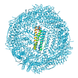
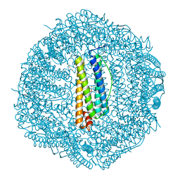
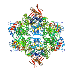
1SX3
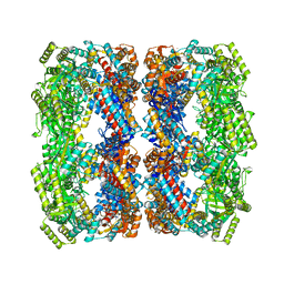 | | GroEL14-(ATPgammaS)14 | | 分子名称: | MAGNESIUM ION, PHOSPHOTHIOPHOSPHORIC ACID-ADENYLATE ESTER, POTASSIUM ION, ... | | 著者 | Chaudhry, C, Horwich, A.L, Brunger, A.T, Adams, P.D. | | 登録日 | 2004-03-30 | | 公開日 | 2005-03-01 | | 最終更新日 | 2024-02-14 | | 実験手法 | X-RAY DIFFRACTION (2 Å) | | 主引用文献 | Exploring the structural dynamics of the E.coli chaperonin GroEL using translation-libration-screw crystallographic refinement of intermediate states.
J.Mol.Biol., 342, 2004
|
|
8U7F
 | | Crystal structure of CIB_12 beta-galactosidase from Cuniculiplasma divulgatum | | 分子名称: | CIB_12 Beta-galactosidase, GLYCEROL | | 著者 | Stogios, P.J, Skarina, T, Di Leo, R, Yakunin, A, Golyshin, P, Savchenko, A. | | 登録日 | 2023-09-15 | | 公開日 | 2024-07-24 | | 最終更新日 | 2025-02-05 | | 実験手法 | X-RAY DIFFRACTION (2.55 Å) | | 主引用文献 | Moderately thermostable GH1 beta-glucosidases from hyperacidophilic archaeon Cuniculiplasma divulgatum S5.
Fems Microbiol.Ecol., 100, 2024
|
|
7O66
 | | Crystal structure of human mitochondrial ferritin (hMTF) Fe(II)-loaded for 60 minutes showing either a dioxygen or a superoxide anion coordinated to iron ions in the ferroxidase site | | 分子名称: | CHLORIDE ION, FE (II) ION, Ferritin, ... | | 著者 | Pozzi, C, Ciambellotti, S, Tassone, G, Turano, P, Mangani, S. | | 登録日 | 2021-04-09 | | 公開日 | 2021-10-13 | | 最終更新日 | 2024-01-31 | | 実験手法 | X-RAY DIFFRACTION (1.6 Å) | | 主引用文献 | Iron Binding in the Ferroxidase Site of Human Mitochondrial Ferritin.
Chemistry, 27, 2021
|
|
8U7G
 | |
1S31
 | | Crystal Structure Analysis of the human Tub protein (isoform a) spanning residues 289 through 561 | | 分子名称: | TRIETHYLENE GLYCOL, tubby isoform a | | 著者 | Boutboul, S, Carroll, K.J, Basdevant, A, Gomez, C, Nandrot, E, Clement, K, Shapiro, L, Abitbol, M. | | 登録日 | 2004-01-12 | | 公開日 | 2005-01-25 | | 最終更新日 | 2023-08-23 | | 実験手法 | X-RAY DIFFRACTION (2.704 Å) | | 主引用文献 | A novel human obesity and sensory deficit syndrome resulting from a mutation in the TUB gene
To be Published
|
|
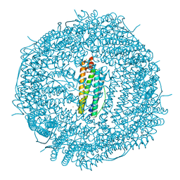
1SX4
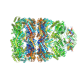 | | GroEL-GroES-ADP7 | | 分子名称: | ADENOSINE-5'-DIPHOSPHATE, MAGNESIUM ION, groEL protein, ... | | 著者 | Chaudhry, C, Horwich, A.L, Brunger, A.T, Adams, P.D. | | 登録日 | 2004-03-30 | | 公開日 | 2005-03-01 | | 最終更新日 | 2024-02-14 | | 実験手法 | X-RAY DIFFRACTION (3 Å) | | 主引用文献 | Exploring the structural dynamics of the E.coli chaperonin GroEL using translation-libration-screw crystallographic refinement of intermediate states.
J.Mol.Biol., 342, 2004
|
|
4UHV
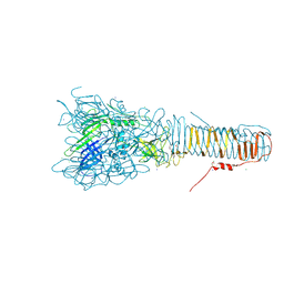 | | The structure of VgrG1, the needle tip of the bacterial Type VI Secretion System | | 分子名称: | CHLORIDE ION, SODIUM ION, VGRG1, ... | | 著者 | Spinola-Amilibia, M, Davo-Siguero, I, Ruiz, F.M, Santillana, E, Medrano, F.J, Romero, A. | | 登録日 | 2015-03-25 | | 公開日 | 2016-01-27 | | 最終更新日 | 2024-11-13 | | 実験手法 | X-RAY DIFFRACTION (2 Å) | | 主引用文献 | The Structure of Vgrg1 from Pseudomonas Aeruginosa, the Needle Tip of the Bacterial Type Vi Secretion System
Acta Crystallogr.,Sect.D, 72, 2016
|
|
7O65
 | | Crystal structure of human mitochondrial ferritin (hMTF) Fe(II)-loaded for 90 minutes showing either a dioxygen or a superoxide anion coordinated to iron ions in the ferroxidase site | | 分子名称: | CHLORIDE ION, FE (II) ION, Ferritin, ... | | 著者 | Pozzi, C, Ciambellotti, S, Tassone, G, Turano, P, Mangani, S. | | 登録日 | 2021-04-09 | | 公開日 | 2021-10-13 | | 最終更新日 | 2024-01-31 | | 実験手法 | X-RAY DIFFRACTION (1.7 Å) | | 主引用文献 | Iron Binding in the Ferroxidase Site of Human Mitochondrial Ferritin.
Chemistry, 27, 2021
|
|
7O64
 | | Crystal structure of human mitochondrial ferritin (hMTF) Fe(II)-loaded for 1 minute. | | 分子名称: | CHLORIDE ION, FE (II) ION, Ferritin, ... | | 著者 | Pozzi, C, Ciambellotti, S, Tassone, G, Turano, P, Mangani, S. | | 登録日 | 2021-04-09 | | 公開日 | 2021-10-13 | | 最終更新日 | 2024-01-31 | | 実験手法 | X-RAY DIFFRACTION (1.96 Å) | | 主引用文献 | Iron Binding in the Ferroxidase Site of Human Mitochondrial Ferritin.
Chemistry, 27, 2021
|
|
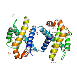
7O6C
 | | Crystal structure of human mitochondrial ferritin (hMTF) Fe(II)-loaded for 15 minutes under anaerobic environment | | 分子名称: | CHLORIDE ION, FE (II) ION, Ferritin, ... | | 著者 | Pozzi, C, Ciambellotti, S, Tassone, G, Turano, P, Mangani, S. | | 登録日 | 2021-04-09 | | 公開日 | 2021-10-13 | | 最終更新日 | 2024-01-31 | | 実験手法 | X-RAY DIFFRACTION (1.2 Å) | | 主引用文献 | Iron Binding in the Ferroxidase Site of Human Mitochondrial Ferritin.
Chemistry, 27, 2021
|
|
7O68
 | | Crystal structure of human mitochondrial ferritin (hMTF) Fe(II)-loaded for 120 minutes showing either a dioxygen or a superoxide anion coordinated to iron ions in the ferroxidase site. | | 分子名称: | CHLORIDE ION, FE (II) ION, Ferritin, ... | | 著者 | Pozzi, C, Ciambellotti, S, Tassone, G, Turano, P, Mangani, S. | | 登録日 | 2021-04-09 | | 公開日 | 2021-10-13 | | 最終更新日 | 2024-01-31 | | 実験手法 | X-RAY DIFFRACTION (1.68 Å) | | 主引用文献 | Iron Binding in the Ferroxidase Site of Human Mitochondrial Ferritin.
Chemistry, 27, 2021
|
|
7O63
 | | High resolution crystal structure of human mitochondrial ferritin (hMTF) | | 分子名称: | CHLORIDE ION, Ferritin, mitochondrial, ... | | 著者 | Pozzi, C, Ciambellotti, S, Tassone, G, Turano, P, Mangani, S. | | 登録日 | 2021-04-09 | | 公開日 | 2021-10-13 | | 最終更新日 | 2024-01-31 | | 実験手法 | X-RAY DIFFRACTION (1.16 Å) | | 主引用文献 | Iron Binding in the Ferroxidase Site of Human Mitochondrial Ferritin.
Chemistry, 27, 2021
|
|
7O6A
 | | Crystal structure of human mitochondrial ferritin (hMTF) Fe(II)-loaded for 5 minutes under anaerobic environment | | 分子名称: | CHLORIDE ION, FE (II) ION, Ferritin, ... | | 著者 | Pozzi, C, Ciambellotti, S, Tassone, G, Turano, P, Mangani, S. | | 登録日 | 2021-04-09 | | 公開日 | 2021-10-13 | | 最終更新日 | 2024-01-31 | | 実験手法 | X-RAY DIFFRACTION (1.4 Å) | | 主引用文献 | Iron Binding in the Ferroxidase Site of Human Mitochondrial Ferritin.
Chemistry, 27, 2021
|
|
7O6D
 | | Crystal structure of human mitochondrial ferritin (hMTF) Fe(II)-loaded for 3 minutes under anaerobic environment | | 分子名称: | CHLORIDE ION, FE (II) ION, Ferritin, ... | | 著者 | Pozzi, C, Ciambellotti, S, Tassone, G, Turano, P, Mangani, S. | | 登録日 | 2021-04-09 | | 公開日 | 2021-10-13 | | 最終更新日 | 2024-01-31 | | 実験手法 | X-RAY DIFFRACTION (1.47 Å) | | 主引用文献 | Iron Binding in the Ferroxidase Site of Human Mitochondrial Ferritin.
Chemistry, 27, 2021
|
|
7O69
 | | Crystal structure of human mitochondrial ferritin (hMTF) Fe(II)-loaded for 5 minutes showing a peroxide anion as bridging species of iron ions in the ferroxidase site | | 分子名称: | CHLORIDE ION, FE (II) ION, Ferritin, ... | | 著者 | Pozzi, C, Ciambellotti, S, Tassone, G, Turano, P, Mangani, S. | | 登録日 | 2021-04-09 | | 公開日 | 2021-10-13 | | 最終更新日 | 2024-01-31 | | 実験手法 | X-RAY DIFFRACTION (1.35 Å) | | 主引用文献 | Iron Binding in the Ferroxidase Site of Human Mitochondrial Ferritin.
Chemistry, 27, 2021
|
|
7O67
 | | Crystal structure of human mitochondrial ferritin (hMTF) Fe(II)-loaded for 15 minutes showing either a dioxygen or a superoxide anion coordinated to iron ions in the ferroxidase site | | 分子名称: | CHLORIDE ION, FE (II) ION, Ferritin, ... | | 著者 | Pozzi, C, Ciambellotti, S, Tassone, G, Turano, P, Mangani, S. | | 登録日 | 2021-04-09 | | 公開日 | 2021-10-13 | | 最終更新日 | 2024-01-31 | | 実験手法 | X-RAY DIFFRACTION (1.86 Å) | | 主引用文献 | Iron Binding in the Ferroxidase Site of Human Mitochondrial Ferritin.
Chemistry, 27, 2021
|
|
7NS7
 | |
8OY0
 | | ATP phosphoribosyltransferase (HisZG ATPPRT) from Acinetobacter baumanii | | 分子名称: | ATP phosphoribosyltransferase, ATP phosphoribosyltransferase regulatory subunit, GLYCEROL, ... | | 著者 | Alphey, M.S, Read, B, da Silva, R.G. | | 登録日 | 2023-05-03 | | 公開日 | 2023-11-29 | | 最終更新日 | 2024-03-13 | | 実験手法 | X-RAY DIFFRACTION (2.4 Å) | | 主引用文献 | Crystal Structure, Steady-State, and Pre-Steady-State Kinetics of Acinetobacter baumannii ATP Phosphoribosyltransferase.
Biochemistry, 63, 2024
|
|
5EXU
 | |


