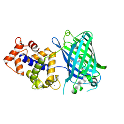
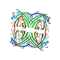
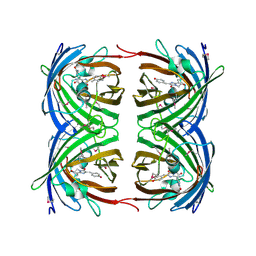
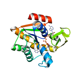
2TOD
 
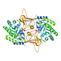 | | ORNITHINE DECARBOXYLASE FROM TRYPANOSOMA BRUCEI K69A MUTANT IN COMPLEX WITH ALPHA-DIFLUOROMETHYLORNITHINE | | 分子名称: | ALPHA-DIFLUOROMETHYLORNITHINE, PROTEIN (ORNITHINE DECARBOXYLASE), PYRIDOXAL-5'-PHOSPHATE | | 著者 | Grishin, N.V, Osterman, A.L, Brooks, H.B, Phillips, M.A, Goldsmith, E.J. | | 登録日 | 1999-05-18 | | 公開日 | 1999-11-17 | | 最終更新日 | 2023-08-30 | | 実験手法 | X-RAY DIFFRACTION (2 Å) | | 主引用文献 | X-ray structure of ornithine decarboxylase from Trypanosoma brucei: the native structure and the structure in complex with alpha-difluoromethylornithine.
Biochemistry, 38, 1999
|
|
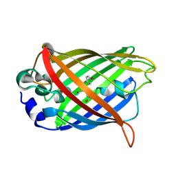
7YV3
 | |
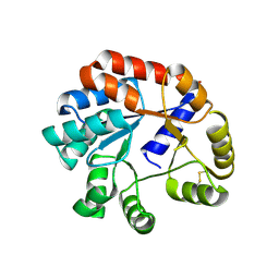
2EKC
 | |
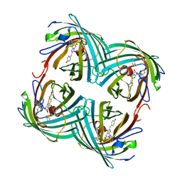
2BTJ
 | |
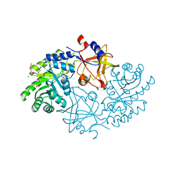
3SG2
 | | Crystal Structure of GCaMP2-T116V,D381Y | | 分子名称: | CALCIUM ION, Myosin light chain kinase, Green fluorescent protein, ... | | 著者 | Schreiter, E.R, Akerboom, J, Looger, L.L. | | 登録日 | 2011-06-14 | | 公開日 | 2012-06-20 | | 最終更新日 | 2023-12-06 | | 実験手法 | X-RAY DIFFRACTION (2 Å) | | 主引用文献 | Optimization of a GCaMP calcium indicator for neural activity imaging.
J.Neurosci., 32, 2012
|
|
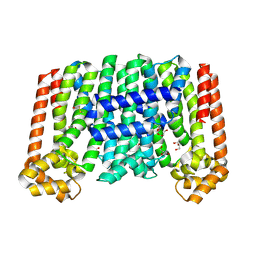
2IB6
 | | Structural characterization of a blue chromoprotein and its yellow mutant from the sea anemone cnidopus japonicus | | 分子名称: | PHOSPHATE ION, Yellow mutant chromo protein | | 著者 | Chan, M.C.Y, Bosanac, I, Ho, D, Prive, G, Ikura, M. | | 登録日 | 2006-09-10 | | 公開日 | 2006-10-10 | | 最終更新日 | 2023-11-15 | | 実験手法 | X-RAY DIFFRACTION (2 Å) | | 主引用文献 | Structural Characterization of a Blue Chromoprotein and Its Yellow Mutant from the Sea Anemone Cnidopus Japonicus
J.Biol.Chem., 281, 2006
|
|
8F8F
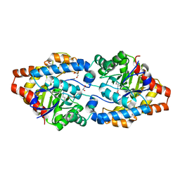
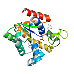
 | | The structure of Rv2173 from M. tuberculosis (APO form) | | 分子名称: | (2E,6E)-farnesyl diphosphate synthase, GLYCEROL, MAGNESIUM ION | | 著者 | Johnston, J.M, Allison, T.M, Titterington, J. | | 登録日 | 2022-11-22 | | 公開日 | 2023-11-29 | | 実験手法 | X-RAY DIFFRACTION (2 Å) | | 主引用文献 | The structure of Rv2173 from M. tuberculosis in APO-, IPP-, and DMAP-bound forms.
To be Published
|
|
5GJN
 | |
2VVJ
 | | IrisFP fluorescent protein in its red form, cis conformation | | 分子名称: | Green to red photoconvertible GFP-like protein EosFP, SULFATE ION, SULFITE ION | | 著者 | Adam, V, Lelimousin, M, Boehme, S, Desfonds, G, Nienhaus, K, Field, M.J, Wiedenmann, J, McSweeney, S, Nienhaus, G.U, Bourgeois, D. | | 登録日 | 2008-06-09 | | 公開日 | 2008-08-12 | | 最終更新日 | 2023-12-13 | | 実験手法 | X-RAY DIFFRACTION (2 Å) | | 主引用文献 | Structural Characterization of Irisfp, an Optical Highlighter Undergoing Multiple Photo-Induced Transformations.
Proc.Natl.Acad.Sci.USA, 105, 2008
|
|
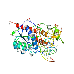
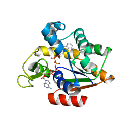
3UR2
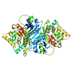
 | | Crystal Structure of PTE mutant H254G/H257W/L303T/K185R/I274N/A80V | | 分子名称: | 1,2-ETHANEDIOL, COBALT (II) ION, IMIDAZOLE, ... | | 著者 | Tsai, P, Fox, N.G, Li, Y, Barondeau, D.P, Raushel, F.M. | | 登録日 | 2011-11-21 | | 公開日 | 2012-08-01 | | 最終更新日 | 2023-12-06 | | 実験手法 | X-RAY DIFFRACTION (2 Å) | | 主引用文献 | Enzymes for the homeland defense: optimizing phosphotriesterase for the hydrolysis of organophosphate nerve agents.
Biochemistry, 51, 2012
|
|
4R6B
 | | Rational Design of Enhanced Photoresistance in a Photoswitchable Fluorescent Protein | | 分子名称: | Green to red photoconvertible GFP-like protein EosFP, SULFATE ION, SULFITE ION | | 著者 | Duan, C, Adam, V, Byrdin, M, Bourgeois, D. | | 登録日 | 2014-08-23 | | 公開日 | 2015-02-04 | | 最終更新日 | 2023-11-15 | | 実験手法 | X-RAY DIFFRACTION (2 Å) | | 主引用文献 | Rational design of enhanced photoresistance in a photoswitchable fluorescent protein.
Methods Appl Fluoresc, 3, 2015
|
|
3FB4
 | |
3OSR
 | | Maltose-bound maltose sensor engineered by insertion of circularly permuted green fluorescent protein into E. coli maltose binding protein at position 311 | | 分子名称: | Maltose-binding periplasmic protein,Green fluorescent protein, alpha-D-glucopyranose-(1-4)-alpha-D-glucopyranose | | 著者 | Echevarria, I.M, Marvin, J.S, Looger, L.L, Schreiter, E.R. | | 登録日 | 2010-09-09 | | 公開日 | 2011-10-26 | | 最終更新日 | 2024-07-10 | | 実験手法 | X-RAY DIFFRACTION (2 Å) | | 主引用文献 | A genetically encoded, high-signal-to-noise maltose sensor.
Proteins, 79, 2011
|
|
5AB5
 | |
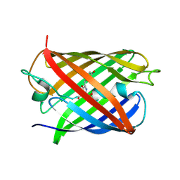
1UIS
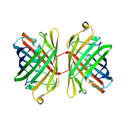 | | The 2.0 crystal structure of eqFP611, a far-red fluorescent protein from the sea anemone Entacmaea quadricolor | | 分子名称: | ACETIC ACID, CALCIUM ION, red fluorescent protein FP611 | | 著者 | Petersen, J, Wilmann, P.G, Beddoe, T, Oakley, A.J, Devenish, R.J, Prescott, M, Rossjohn, J. | | 登録日 | 2003-07-21 | | 公開日 | 2003-10-21 | | 最終更新日 | 2023-12-27 | | 実験手法 | X-RAY DIFFRACTION (2 Å) | | 主引用文献 | The 2.0A crystal structure of eqFP611, a far-red fluorescent protein from the sea anemone Entacmaea quadricolor
J.Biol.Chem., 278, 2003
|
|
1G7K
 | | CRYSTAL STRUCTURE OF DSRED, A RED FLUORESCENT PROTEIN FROM DISCOSOMA SP. RED | | 分子名称: | FLUORESCENT PROTEIN FP583 | | 著者 | Yarbrough, D, Wachter, R.M, Kallio, K, Matz, M.V, Remington, S.J. | | 登録日 | 2000-11-10 | | 公開日 | 2000-12-06 | | 最終更新日 | 2023-11-15 | | 実験手法 | X-RAY DIFFRACTION (2 Å) | | 主引用文献 | Refined crystal structure of DsRed, a red fluorescent protein from coral, at 2.0-A resolution.
Proc.Natl.Acad.Sci.USA, 98, 2001
|
|
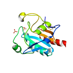
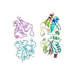
2I3Y
 | | Crystal structure of human glutathione peroxidase 5 | | 分子名称: | 1,2-ETHANEDIOL, Epididymal secretory glutathione peroxidase | | 著者 | Kavanagh, K.L, Johansson, C, Rojkova, A, Umeano, C, Bunkoczi, G, Gileadi, O, von Delft, F, Weigelt, J, Arrowsmith, C, Sundstrom, M, Edwards, A, Oppermann, U, Structural Genomics Consortium (SGC) | | 登録日 | 2006-08-21 | | 公開日 | 2006-09-12 | | 最終更新日 | 2023-08-30 | | 実験手法 | X-RAY DIFFRACTION (2 Å) | | 主引用文献 | Crystal structure of human glutathione peroxidase 5
To be published
|
|
8DJ8
 | |
7FC2
 | | Crystal Structure of GPX6 | | 分子名称: | Glutathione peroxidase 6, SULFATE ION | | 著者 | Sun, J. | | 登録日 | 2021-07-13 | | 公開日 | 2022-07-20 | | 最終更新日 | 2023-11-29 | | 実験手法 | X-RAY DIFFRACTION (2 Å) | | 主引用文献 | Crystal Structure of GPX6
To Be Published
|
|
1JC0
 | | CRYSTAL STRUCTURE ANALYSIS OF A REDOX-SENSITIVE GREEN FLUORESCENT PROTEIN VARIANT IN A REDUCED FORM | | 分子名称: | GREEN FLUORESCENT PROTEIN | | 著者 | Hanson, G.T, Aggeler, R, Oglesbee, D, Cannon, M, Capaldi, R.A, Tsien, R.Y, Remington, S.J. | | 登録日 | 2001-06-07 | | 公開日 | 2003-09-09 | | 最終更新日 | 2023-11-15 | | 実験手法 | X-RAY DIFFRACTION (2 Å) | | 主引用文献 | Investigating mitochondrial redox potential with redox-sensitive green fluorescent protein indicators.
J.Biol.Chem., 279, 2004
|
|
2UYC
 | |
1AKE
 | |
3HPQ
 | |
1GP1
 | |
2H8Q
 | |


