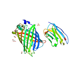
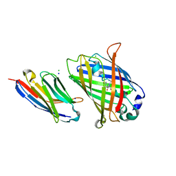
8J2J
 
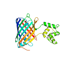
 | | Crystal structure of a bright green fluorescent protein (StayGold) with single mutation (Y187F) in jellyfish Cytaeis uchidae from Biortus | | 分子名称: | 1,2-ETHANEDIOL, SULFATE ION, StayGold(Y187F) | | 著者 | Wu, J, Wang, F, Gui, W, Cheng, W, Yang, Y. | | 登録日 | 2023-04-14 | | 公開日 | 2023-12-13 | | 実験手法 | X-RAY DIFFRACTION (1.9 Å) | | 主引用文献 | Crystal structure of a bright green fluorescent protein (StayGold) in jellyfish Cytaeis uchidae from Biortus
To Be Published
|
|
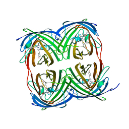
8J1E
 | | AtSLAC1 in open state | | 分子名称: | CHLORIDE ION, CHOLESTEROL HEMISUCCINATE, Guard cell S-type anion channel SLAC1,Green fluorescent protein | | 著者 | Lee, Y, Lee, S. | | 登録日 | 2023-04-12 | | 公開日 | 2023-11-22 | | 最終更新日 | 2023-11-29 | | 実験手法 | ELECTRON MICROSCOPY (3.84 Å) | | 主引用文献 | Cryo-EM structures of the plant anion channel SLAC1 from Arabidopsis thaliana suggest a combined activation model.
Nat Commun, 14, 2023
|
|
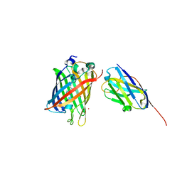
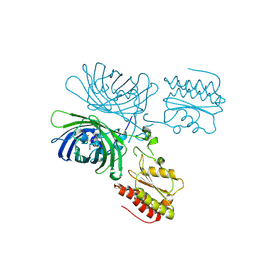
8SFX
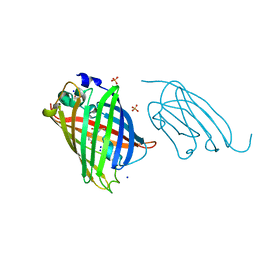
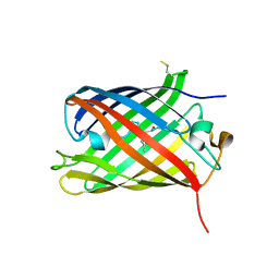
 | | High Affinity nanobodies against GFP | | 分子名称: | D-MALATE, GLYCEROL, Green fluorescent protein, ... | | 著者 | Ketaren, N.E, Rout, M.P, Bonanno, J.B, Almo, S.C. | | 登録日 | 2023-04-11 | | 公開日 | 2024-05-22 | | 実験手法 | X-RAY DIFFRACTION (1.95 Å) | | 主引用文献 | High Affinity nanobodies against GFP
To Be Published
|
|
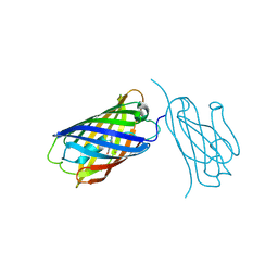
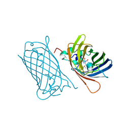
8SFV
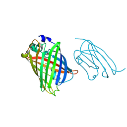
 | | High affinity nanobodies to GFP | | 分子名称: | GLYCEROL, Green fluorescent protein, LaG19, ... | | 著者 | Ketaren, N.E, Rout, M.P, Bonanno, J.B, Almo, S.C. | | 登録日 | 2023-04-11 | | 公開日 | 2024-05-22 | | 実験手法 | X-RAY DIFFRACTION (1.83 Å) | | 主引用文献 | High affinity nanobodies to GFP
To Be Published
|
|
8SG3
 | |
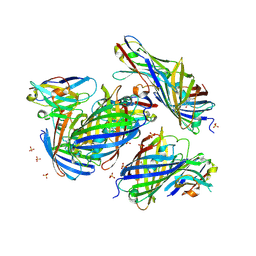
8SFS
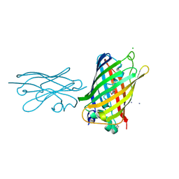 | | High Affinity nanobodies against GFP | | 分子名称: | AMMONIUM ION, CHLORIDE ION, GLYCEROL, ... | | 著者 | Ketaren, N.E, Rout, M.P, Bonnano, J.B, Almo, S.C. | | 登録日 | 2023-04-11 | | 公開日 | 2024-05-22 | | 実験手法 | X-RAY DIFFRACTION (2.37 Å) | | 主引用文献 | High Affinity nanobodies against GFP
To Be Published
|
|
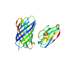
8SFZ
 | | High Affinity nanobodies against GFP | | 分子名称: | Green fluorescent protein, LaG35, POTASSIUM ION, ... | | 著者 | Ketaren, N.E, Rout, M.P, Bonanno, J.B, Almo, S.C. | | 登録日 | 2023-04-11 | | 公開日 | 2024-05-22 | | 実験手法 | X-RAY DIFFRACTION (1.9 Å) | | 主引用文献 | High Affinity nanobodies against GFP
To Be Published
|
|
8J0J
 | | AtSLAC1 8D mutant in closed state | | 分子名称: | CHLORIDE ION, CHOLESTEROL HEMISUCCINATE, Guard cell S-type anion channel SLAC1,Green fluorescent protein | | 著者 | Lee, Y, Lee, S. | | 登録日 | 2023-04-11 | | 公開日 | 2023-11-22 | | 最終更新日 | 2023-11-29 | | 実験手法 | ELECTRON MICROSCOPY (2.7 Å) | | 主引用文献 | Cryo-EM structures of the plant anion channel SLAC1 from Arabidopsis thaliana suggest a combined activation model.
Nat Commun, 14, 2023
|
|
8IMX
 | | Cryo-EM structure of GPI-T with a chimeric GPI-anchored protein | | 分子名称: | 1-palmitoyl-2-oleoyl-sn-glycero-3-phosphocholine, 2-acetamido-2-deoxy-beta-D-glucopyranose, 2-acetamido-2-deoxy-beta-D-glucopyranose-(1-4)-2-acetamido-2-deoxy-beta-D-glucopyranose, ... | | 著者 | Xu, Y, Li, T, Qu, Q, Li, D. | | 登録日 | 2023-03-07 | | 公開日 | 2023-08-16 | | 最終更新日 | 2023-11-01 | | 実験手法 | ELECTRON MICROSCOPY (2.85 Å) | | 主引用文献 | Structures of liganded glycosylphosphatidylinositol transamidase illuminate GPI-AP biogenesis.
Nat Commun, 14, 2023
|
|
8IM0
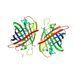
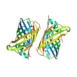
 | | mCherry-LaM8 complex | | 分子名称: | LaM8, MCherry fluorescent protein | | 著者 | Liang, H, Liu, R, Ding, Y. | | 登録日 | 2023-03-05 | | 公開日 | 2023-06-21 | | 実験手法 | X-RAY DIFFRACTION (1.31 Å) | | 主引用文献 | Structural Insights into the Binding of Red Fluorescent Protein mCherry-Specific Nanobodies.
Int J Mol Sci, 24, 2023
|
|
8ILX
 | | mCherry-LaM3 complex | | 分子名称: | LAM3, MCherry fluorescent protein | | 著者 | Liang, H, Liu, R, Ding, Y. | | 登録日 | 2023-03-05 | | 公開日 | 2023-06-21 | | 実験手法 | X-RAY DIFFRACTION (3.29 Å) | | 主引用文献 | Structural Insights into the Binding of Red Fluorescent Protein mCherry-Specific Nanobodies.
Int J Mol Sci, 24, 2023
|
|
8IM1
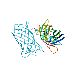
 | | mCherry-LaM1 complex | | 分子名称: | LaM1, MCherry fluorescent protein, SULFATE ION | | 著者 | Liang, H, Liu, R, Ding, Y. | | 登録日 | 2023-03-05 | | 公開日 | 2023-06-21 | | 実験手法 | X-RAY DIFFRACTION (2.05 Å) | | 主引用文献 | Structural Insights into the Binding of Red Fluorescent Protein mCherry-Specific Nanobodies.
Int J Mol Sci, 24, 2023
|
|
8G4E
 | |
8G0I
 | |
8I4J
 | |
8C7I
 | |
8C1X
 | |
8C0T
 | | NRS 1.2: Fluorescent Sensors for Imaging Interstitial Calcium | | 分子名称: | CALCIUM ION, SULFATE ION, mNeonGreen,Optimized Ratiometric Calcium Sensor | | 著者 | Basquin, J, Griesbeck, O, Valiente-Gabioud, A. | | 登録日 | 2022-12-19 | | 公開日 | 2023-11-15 | | 実験手法 | X-RAY DIFFRACTION (1.28 Å) | | 主引用文献 | Fluorescent sensors for imaging of interstitial calcium.
Nat Commun, 14, 2023
|
|
8C0N
 | | Crystal structure of the red form of the mTagFT fluorescent timer | | 分子名称: | Blue-to-red TagFT fluorescent timer | | 著者 | Boyko, K.M, Nikolaeva, A.Y, Vlaskina, A.V, Agapova, Y.K, Subach, O.M, Popov, V.O, Subach, F.V. | | 登録日 | 2022-12-19 | | 公開日 | 2023-03-08 | | 最終更新日 | 2023-11-15 | | 実験手法 | X-RAY DIFFRACTION (2.9 Å) | | 主引用文献 | Blue-to-Red TagFT, mTagFT, mTsFT, and Green-to-FarRed mNeptusFT2 Proteins, Genetically Encoded True and Tandem Fluorescent Timers.
Int J Mol Sci, 24, 2023
|
|
8BXT
 | |
8BXP
 | |
8FED
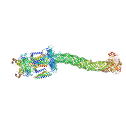 | | Structure of Mce1-LucB complex from Mycobacterium smegmatis (Map1) | | 分子名称: | ABC transporter, ATP-binding protein,Green fluorescent protein chimera, ABC-transporter integral membrane protein, ... | | 著者 | Chen, J, Bhabha, G, Ekiert, D.C. | | 登録日 | 2022-12-06 | | 公開日 | 2023-02-22 | | 最終更新日 | 2023-08-23 | | 実験手法 | ELECTRON MICROSCOPY (2.76 Å) | | 主引用文献 | Structure of an endogenous mycobacterial MCE lipid transporter.
Nature, 620, 2023
|
|
8FEE
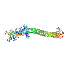 | | Structure of Mce1 transporter from Mycobacterium smegmatis in the absence of LucB (Map2) | | 分子名称: | ABC transporter, ATP-binding protein,Green fluorescent protein chimera, ABC-transporter integral membrane protein, ... | | 著者 | Chen, J, Bhabha, G, Ekiert, D.C. | | 登録日 | 2022-12-06 | | 公開日 | 2023-02-22 | | 最終更新日 | 2023-08-23 | | 実験手法 | ELECTRON MICROSCOPY (2.9 Å) | | 主引用文献 | Structure of an endogenous mycobacterial MCE lipid transporter.
Nature, 620, 2023
|
|
8FEF
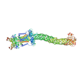 | | Structure of Mce1 transporter from Mycobacterium smegmatis (Map0) | | 分子名称: | ABC transporter, ATP-binding protein,Green fluorescent protein chimera, ABC-transporter integral membrane protein, ... | | 著者 | Chen, J, Bhabha, G, Ekiert, D.C. | | 登録日 | 2022-12-06 | | 公開日 | 2023-02-22 | | 最終更新日 | 2023-08-23 | | 実験手法 | ELECTRON MICROSCOPY (2.71 Å) | | 主引用文献 | Structure of an endogenous mycobacterial MCE lipid transporter.
Nature, 620, 2023
|
|
8BVG
 | |


