3BZA
 
 | |
3ZCH
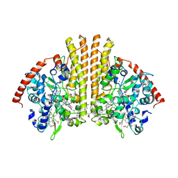
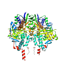
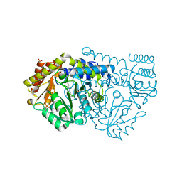
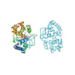 | | Ascorbate peroxidase W41A-H42M mutant | | 分子名称: | 4-(2-HYDROXYETHYL)-1-PIPERAZINE ETHANESULFONIC ACID, ASCORBATE PEROXIDASE, POTASSIUM ION, ... | | 著者 | Gumiero, A, Raven, E.L, Moody, P.C.E. | | 登録日 | 2012-11-20 | | 公開日 | 2012-12-19 | | 最終更新日 | 2023-12-20 | | 実験手法 | X-RAY DIFFRACTION (2 Å) | | 主引用文献 | Probing the Conformational Mobility of the Active Site of Ascorbate Peroxidase
Dalton Trans, 42, 2013
|
|
6ZQS
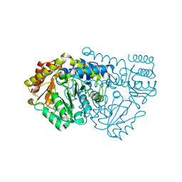
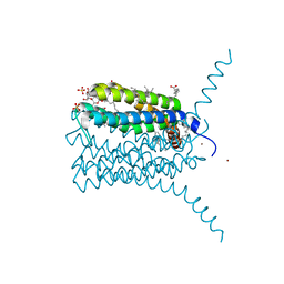
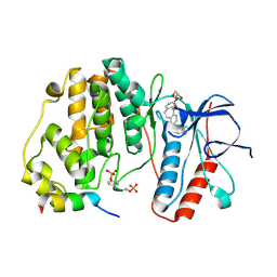 | | Crystal structure of double-phosphorylated p38alpha with ATF2(83-102) | | 分子名称: | 2-[(2,4-difluorophenyl)amino]-7-{[(2R)-2,3-dihydroxypropyl]oxy}-10,11-dihydro-5H-dibenzo[a,d][7]annulen-5-one, Cyclic AMP-dependent transcription factor ATF-2, Mitogen-activated protein kinase 14 | | 著者 | Kirsch, K, Sok, P, Poti, A.L, Remenyi, A. | | 登録日 | 2020-07-10 | | 公開日 | 2020-11-18 | | 最終更新日 | 2024-11-06 | | 実験手法 | X-RAY DIFFRACTION (1.95 Å) | | 主引用文献 | Co-regulation of the transcription controlling ATF2 phosphoswitch by JNK and p38.
Nat Commun, 11, 2020
|
|
2RDZ
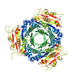
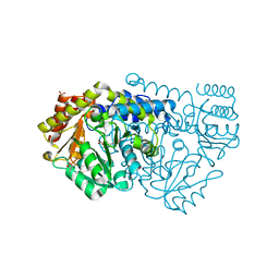
 | | High Resolution Crystal Structure of the Escherichia coli Cytochrome c Nitrite Reductase. | | 分子名称: | 1,2-ETHANEDIOL, CALCIUM ION, Cytochrome c-552, ... | | 著者 | Clarke, T.A, Hemmings, A.M, RIchardson, D.J. | | 登録日 | 2007-09-25 | | 公開日 | 2008-03-25 | | 最終更新日 | 2024-10-30 | | 実験手法 | X-RAY DIFFRACTION (1.74 Å) | | 主引用文献 | Role of a Conserved Glutamine Residue in Tuning the Catalytic Activity of Escherichia coli Cytochrome c Nitrite Reductase.
Biochemistry, 47, 2008
|
|
2RF7
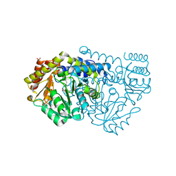
 | | Crystal structure of the escherichia coli nrfa mutant Q263E | | 分子名称: | 1,2-ETHANEDIOL, CALCIUM ION, Cytochrome c-552, ... | | 著者 | Clarke, T.A, Richardson, D.J, Hemmings, A.M. | | 登録日 | 2007-09-28 | | 公開日 | 2008-03-25 | | 最終更新日 | 2023-08-30 | | 実験手法 | X-RAY DIFFRACTION (2.04 Å) | | 主引用文献 | Role of a Conserved Glutamine Residue in Tuning the Catalytic Activity of Escherichia coli Cytochrome c Nitrite Reductase.
Biochemistry, 47, 2008
|
|
2VA1
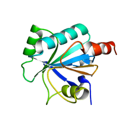
 | | Crystal structure of UMP kinase from Ureaplasma parvum | | 分子名称: | PHOSPHATE ION, URIDYLATE KINASE | | 著者 | Egeblad-Welin, L, Welin, M, Wang, L, Eriksson, S. | | 登録日 | 2007-08-28 | | 公開日 | 2007-09-18 | | 最終更新日 | 2023-12-13 | | 実験手法 | X-RAY DIFFRACTION (2.5 Å) | | 主引用文献 | Structural and Functional Investigations of Ureaplasma Parvum Ump Kinase - a Potential Antibacterial Drug Target
FEBS J., 274, 2007
|
|
2VUP
 | |
2W7G
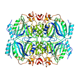
 | | Crystal structure of Y51FbsSHMT L-allo-Threonine extrnal aldimine | | 分子名称: | (4S)-2-METHYL-2,4-PENTANEDIOL, ALLO-THREONINE, PHOSPHATE ION, ... | | 著者 | Rajaram, V, Bhavani, B.S, Bisht, S, Kaul, P, Prakash, V, Appaji Rao, N, Savithri, H.S, Murthy, M.R.N. | | 登録日 | 2008-12-22 | | 公開日 | 2010-08-18 | | 最終更新日 | 2023-12-13 | | 実験手法 | X-RAY DIFFRACTION (1.92 Å) | | 主引用文献 | Importance of Tyrosine Residues of Bacillus Stearothermophilus Serine Hydroxymethyltransferase in Cofactor Binding and L-Allo-Thr Cleavage.
FEBS J., 275, 2008
|
|
2VZ2
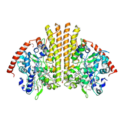
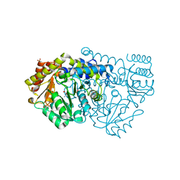
 | | Human MAO B in complex with mofegiline | | 分子名称: | (1Z)-4-(4-FLUOROPHENYL)-2-METHYLIDENEBUTAN-1-IMINE, AMINE OXIDASE [FLAVIN-CONTAINING] B, FLAVIN-ADENINE DINUCLEOTIDE, ... | | 著者 | Bonivento, D, Mattevi, A. | | 登録日 | 2008-07-29 | | 公開日 | 2008-12-16 | | 最終更新日 | 2024-11-06 | | 実験手法 | X-RAY DIFFRACTION (2.3 Å) | | 主引用文献 | Structural and mechanistic studies of mofegiline inhibition of recombinant human monoamine oxidase B.
J. Med. Chem., 51, 2008
|
|
2W7I
 | | Crystal structure of Y61AbsSHMT internal aldimine | | 分子名称: | PYRIDOXAL-5'-PHOSPHATE, SERINE HYDROXYMETHYLTRANSFERASE | | 著者 | Rajaram, V, Bhavani, B.S, Bisht, S, Kaul, P, Prakash, V, Appaji Rao, N, Savithri, H.S, Murthy, M.R.N. | | 登録日 | 2008-12-22 | | 公開日 | 2010-08-18 | | 最終更新日 | 2023-12-13 | | 実験手法 | X-RAY DIFFRACTION (2.72 Å) | | 主引用文献 | Importance of Tyrosine Residues of Bacillus Stearothermophilus Serine Hydroxymethyltransferase in Cofactor Binding and L-Allo-Thr Cleavage.
FEBS J., 275, 2008
|
|
2W7H
 | | Crystal structure of Y51FbsSHMT obtained in the presence of Gly and 5- Formyl Tetrahydrofolate | | 分子名称: | (4S)-2-METHYL-2,4-PENTANEDIOL, GLYCINE, PHOSPHATE ION, ... | | 著者 | Rajaram, V, Bhavani, B.S, Bisht, S, Kaul, P, Prakash, V, Appaji Rao, N, Savithri, H.S, Murthy, M.R.N. | | 登録日 | 2008-12-22 | | 公開日 | 2010-08-18 | | 最終更新日 | 2023-12-13 | | 実験手法 | X-RAY DIFFRACTION (1.67 Å) | | 主引用文献 | Importance of Tyrosine Residues of Bacillus Stearothermophilus Serine Hydroxymethyltransferase in Cofactor Binding and L-Allo-Thr Cleavage.
FEBS J., 275, 2008
|
|
2W7E
 | | Crystal structure of Y51FbsSHMT obtained in the presence of Glycine | | 分子名称: | (4S)-2-METHYL-2,4-PENTANEDIOL, GLYCINE, PHOSPHATE ION, ... | | 著者 | Rajaram, V, Bhavani, B.S, Bisht, S, Kaul, P, Prakash, V, Appaji Rao, N, Savithri, H.S, Murthy, M.R.N. | | 登録日 | 2008-12-22 | | 公開日 | 2010-08-18 | | 最終更新日 | 2023-12-13 | | 実験手法 | X-RAY DIFFRACTION (1.69 Å) | | 主引用文献 | Importance of Tyrosine Residues of Bacillus Stearothermophilus Serine Hydroxymethyltransferase in Cofactor Binding and L-Allo-Thr Cleavage.
FEBS J., 275, 2008
|
|
2WKL
 | | Velaglucerase alfa | | 分子名称: | 2-acetamido-2-deoxy-beta-D-glucopyranose, 2-acetamido-2-deoxy-beta-D-glucopyranose-(1-4)-2-acetamido-2-deoxy-beta-D-glucopyranose, GLUCOSYLCERAMIDASE, ... | | 著者 | Brumshtein, B, Salinas, P, Peterson, B, Chan, V, Silman, I, Sussman, J.L, Savickas, P.J, Robinson, G.S, Futerman, A.H. | | 登録日 | 2009-06-15 | | 公開日 | 2009-09-22 | | 最終更新日 | 2024-11-20 | | 実験手法 | X-RAY DIFFRACTION (2.7 Å) | | 主引用文献 | Characterization of Gene-Activated Human Acid-Beta-Glucosidase: Crystal Structure, Glycan Composition and Internalization Into Macrophages.
Glycobiology, 20, 2010
|
|
2W7K
 | | Crystal structure of Y61AbsSHMT L-Serine external aldimine | | 分子名称: | PYRIDOXAL-5'-PHOSPHATE, SERINE, SERINE HYDROXYMETHYLTRANSFERASE | | 著者 | Rajaram, V, Bhavani, B.S, Bisht, S, Kaul, P, Prakash, V, Appaji Rao, N, Savithri, H.S, Murthy, M.R.N. | | 登録日 | 2008-12-22 | | 公開日 | 2010-08-18 | | 最終更新日 | 2023-12-13 | | 実験手法 | X-RAY DIFFRACTION (2.42 Å) | | 主引用文献 | Importance of Tyrosine Residues of Bacillus Stearothermophilus Serine Hydroxymethyltransferase in Cofactor Binding and L-Allo-Thr Cleavage.
FEBS J., 275, 2008
|
|
2W7L
 | | Crystal structure of Y61AbsSHMT L-allo-Threonine external aldimine | | 分子名称: | ALLO-THREONINE, PYRIDOXAL-5'-PHOSPHATE, SERINE HYDROXYMETHYLTRANSFERASE | | 著者 | Rajaram, V, Bhavani, B.S, Bisht, S, Kaul, P, Prakash, V, Appaji Rao, N, Savithri, H.S, Murthy, M.R.N. | | 登録日 | 2008-12-22 | | 公開日 | 2010-08-18 | | 最終更新日 | 2023-12-13 | | 実験手法 | X-RAY DIFFRACTION (2.41 Å) | | 主引用文献 | Importance of Tyrosine Residues of Bacillus Stearothermophilus Serine Hydroxymethyltransferase in Cofactor Binding and L-Allo-Thr Cleavage.
FEBS J., 275, 2008
|
|
2W7J
 | | Crystal structure of Y61AbsSHMT Glycine external aldimine | | 分子名称: | GLYCINE, PYRIDOXAL-5'-PHOSPHATE, SERINE HYDROXYMETHYLTRANSFERASE | | 著者 | Rajaram, V, Bhavani, B.S, Bisht, S, Kaul, P, Prakash, V, Appaji Rao, N, Savithri, H.S, Murthy, M.R.N. | | 登録日 | 2008-12-22 | | 公開日 | 2010-08-18 | | 最終更新日 | 2023-12-13 | | 実験手法 | X-RAY DIFFRACTION (1.68 Å) | | 主引用文献 | Importance of Tyrosine Residues of Bacillus Stearothermophilus Serine Hydroxymethyltransferase in Cofactor Binding and L-Allo-Thr Cleavage.
FEBS J., 275, 2008
|
|
2W7D
 | | Crystal structure of Y51FbsSHMT internal aldimine | | 分子名称: | (4S)-2-METHYL-2,4-PENTANEDIOL, PHOSPHATE ION, PYRIDOXAL-5'-PHOSPHATE, ... | | 著者 | Rajaram, V, Bhavani, B.S, Bisht, S, Kaul, P, Prakash, V, Appaji Rao, N, Savithri, H.S, Murthy, M.R.N. | | 登録日 | 2008-12-22 | | 公開日 | 2010-08-18 | | 最終更新日 | 2023-12-13 | | 実験手法 | X-RAY DIFFRACTION (1.8 Å) | | 主引用文献 | Importance of Tyrosine Residues of Bacillus Stearothermophilus Serine Hydroxymethyltransferase in Cofactor Binding and L-Allo-Thr Cleavage.
FEBS J., 275, 2008
|
|
2W7M
 | | Crystal structure of Y61AbsSHMT obtained in the presence of Glycine and 5-Formyl tetrahydrofolate | | 分子名称: | GLYCINE, PYRIDOXAL-5'-PHOSPHATE, SERINE HYDROXYMETHYLTRANSFERASE | | 著者 | Rajaram, V, Bhavani, B.S, Bisht, S, Kaul, P, Prakash, V, Appaji Rao, N, Savithri, H.S, Murthy, M.R.N. | | 登録日 | 2008-12-22 | | 公開日 | 2010-08-18 | | 最終更新日 | 2023-12-13 | | 実験手法 | X-RAY DIFFRACTION (1.86 Å) | | 主引用文献 | Importance of Tyrosine Residues of Bacillus Stearothermophilus Serine Hydroxymethyltransferase in Cofactor Binding and L-Allo-Thr Cleavage.
FEBS J., 275, 2008
|
|
2W7F
 | | Crystal structure of Y51FbsSHMT L-Ser external aldimine | | 分子名称: | (4S)-2-METHYL-2,4-PENTANEDIOL, PHOSPHATE ION, PYRIDOXAL-5'-PHOSPHATE, ... | | 著者 | Rajaram, V, Bhavani, B.S, Bisht, S, Kaul, P, Prakash, V, Appaji Rao, N, Savithri, H.S, Murthy, M.R.N. | | 登録日 | 2008-12-22 | | 公開日 | 2010-08-18 | | 最終更新日 | 2023-12-13 | | 実験手法 | X-RAY DIFFRACTION (1.67 Å) | | 主引用文献 | Importance of Tyrosine Residues of Bacillus Stearothermophilus Serine Hydroxymethyltransferase in Cofactor Binding and L-Allo-Thr Cleavage.
FEBS J., 275, 2008
|
|
4HE4
 | |
3LEO
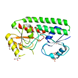
 | | Structure of human Leukotriene C4 synthase mutant R31Q in complex with glutathione | | 分子名称: | DODECYL-BETA-D-MALTOSIDE, GLUTATHIONE, GLYCEROL, ... | | 著者 | Niegowski, D, Martinez-Molina, D, Rinaldo-Matthis, A, Nordlund, P, Haeggstrom, J. | | 登録日 | 2010-01-15 | | 公開日 | 2010-10-27 | | 最終更新日 | 2023-09-06 | | 実験手法 | X-RAY DIFFRACTION (2.1 Å) | | 主引用文献 | Arginine 104 is a key catalytic residue in leukotriene C4 synthase.
J.Biol.Chem., 285, 2010
|
|
4UDO
 | | structure of Mn-bound periplasmic metal binding protein from candidatus liberibacter asiaticus | | 分子名称: | ACETATE ION, GLYCEROL, MANGANESE (II) ION, ... | | 著者 | Sharma, N, Selvakumar, P, Kumar, P, Sharma, A.K. | | 登録日 | 2014-12-10 | | 公開日 | 2015-02-11 | | 最終更新日 | 2023-12-20 | | 実験手法 | X-RAY DIFFRACTION (2.22 Å) | | 主引用文献 | Crystal Structure of a Periplasmic Solute Binding Protein in Metal-Free, Intermediate and Metal-Bound States from Candidatus Liberibacter Asiaticus.
J.Struct.Biol., 189, 2015
|
|
4UDN
 | | structure of metal-free periplasmic metal binding protein from candidatus liberibacter asiaticus | | 分子名称: | ACETATE ION, GLYCEROL, PERIPLASMIC SOLUTE BINDING PROTEIN, ... | | 著者 | Sharma, N, Selvakumar, P, Kumar, P, Sharma, A.K. | | 登録日 | 2014-12-10 | | 公開日 | 2015-02-11 | | 最終更新日 | 2023-12-20 | | 実験手法 | X-RAY DIFFRACTION (2.21 Å) | | 主引用文献 | Crystal Structure of a Periplasmic Solute Binding Protein in Metal-Free, Intermediate and Metal-Bound States from Candidatus Liberibacter Asiaticus.
J.Struct.Biol., 189, 2015
|
|
3G4G
 | |
3IAD
 | |


