4WT0
 
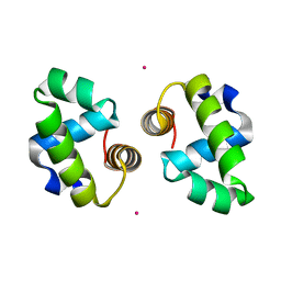 | |
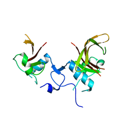
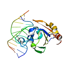
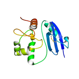
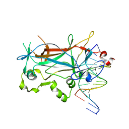
1IRF
 | | INTERFERON REGULATORY FACTOR-2 DNA BINDING DOMAIN, NMR, MINIMIZED AVERAGE STRUCTURE | | 分子名称: | INTERFERON REGULATORY FACTOR-2 | | 著者 | Furui, J, Uegaki, K, Yamazaki, T, Shirakawa, M, Swindells, M.B, Harada, H, Taniguchi, T, Kyogoku, Y. | | 登録日 | 1997-11-24 | | 公開日 | 1998-01-28 | | 最終更新日 | 2024-05-01 | | 実験手法 | SOLUTION NMR | | 主引用文献 | Solution structure of the IRF-2 DNA-binding domain: a novel subgroup of the winged helix-turn-helix family.
Structure, 6, 1998
|
|
8BHV
 | |
8BH3
 | |
8BHY
 | |
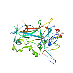
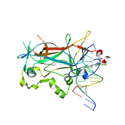
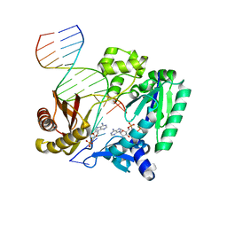
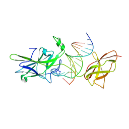
4DUL
 | | ANAC019 NAC domain crystal form IV | | 分子名称: | NAC domain-containing protein 19 | | 著者 | Lo Leggio, L, Helgstrand, C, Welner, D, Olsen, A.N, Skriver, K. | | 登録日 | 2012-02-22 | | 公開日 | 2012-04-11 | | 最終更新日 | 2023-09-13 | | 実験手法 | X-RAY DIFFRACTION (3 Å) | | 主引用文献 | DNA binding by the plant-specific NAC transcription factors in crystal and solution: a firm link to WRKY and GCM transcription factors.
Biochem.J., 444, 2012
|
|
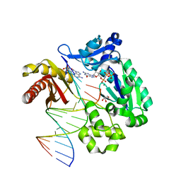
1OZJ
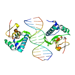 | | Crystal structure of Smad3-MH1 bound to DNA at 2.4 A resolution | | 分子名称: | SMAD 3, Smad binding element, ZINC ION | | 著者 | Chai, J, Wu, J.-W, Yan, N, Massague, J, Pavletich, N.P, Shi, Y. | | 登録日 | 2003-04-09 | | 公開日 | 2004-03-23 | | 最終更新日 | 2024-02-14 | | 実験手法 | X-RAY DIFFRACTION (2.4 Å) | | 主引用文献 | Features of a Smad3 MH1-DNA complex. Roles of water and zinc in DNA binding.
J.Biol.Chem., 278, 2003
|
|
1IRG
 | | INTERFERON REGULATORY FACTOR-2 DNA BINDING DOMAIN, NMR, 20 STRUCTURES | | 分子名称: | INTERFERON REGULATORY FACTOR-2 | | 著者 | Furui, J, Uegaki, K, Yamazaki, T, Shirakawa, M, Swindells, M.B, Harada, H, Taniguchi, T, Kyogoku, Y. | | 登録日 | 1997-11-25 | | 公開日 | 1998-03-18 | | 最終更新日 | 2024-05-01 | | 実験手法 | SOLUTION NMR | | 主引用文献 | Solution structure of the IRF-2 DNA-binding domain: a novel subgroup of the winged helix-turn-helix family.
Structure, 6, 1998
|
|
3ALC
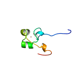 | | ETHANOL REGULON TRANSCRIPTIONAL ACTIVATOR DNA-BINDING DOMAIN FROM ASPERGILLUS NIDULANS | | 分子名称: | PROTEIN (ETHANOL REGULON TRANSCRIPTIONAL ACTIVATOR), ZINC ION | | 著者 | Cerdan, R, Cahuzac, B, Felenbok, B, Guittet, E. | | 登録日 | 1999-03-11 | | 公開日 | 2000-05-15 | | 最終更新日 | 2023-12-27 | | 実験手法 | SOLUTION NMR | | 主引用文献 | NMR solution structure of AlcR (1-60) provides insight in the unusual DNA binding properties of this zinc binuclear cluster protein.
J.Mol.Biol., 295, 2000
|
|
2GPE
 | |
3ZI5
 | | Crystal STRUCTURE OF RESTRICTION ENDONUCLEASE BFII C-TERMINAL RECOGNITION DOMAIN IN COMPLEX WITH COGNATE DNA | | 分子名称: | 5'-D(*AP*GP*CP*AP*CP*TP*GP*GP*GP*TP*CP*GP)-3', 5'-D(*CP*GP*AP*CP*CP*CP*AP*GP*TP*GP*CP*TP)-3', RESTRICTION ENDONUCLEASE | | 著者 | Golovenko, D, Manakova, E, Zakrys, L, Zaremba, M, Sasnauskas, G, Grazulis, S, Siksnys, V. | | 登録日 | 2013-01-03 | | 公開日 | 2014-01-15 | | 最終更新日 | 2023-12-20 | | 実験手法 | X-RAY DIFFRACTION (3.2 Å) | | 主引用文献 | Structural Insight Into the Specificity of the B3 DNA-Binding Domains Provided by the Co-Crystal Structure of the C-Terminal Fragment of Bfii Restriction Enzyme
Nucleic Acids Res., 42, 2014
|
|
2EVH
 | | Structure of a Ndt80-DNA complex (MSE mutant mA7G) | | 分子名称: | 5'-D(*AP*GP*TP*TP*TP*CP*TP*GP*TP*GP*TP*CP*GP*C)-3', 5'-D(*TP*GP*CP*GP*AP*CP*AP*CP*AP*GP*AP*AP*AP*C)-3', NDT80 protein | | 著者 | Lamoureux, J.S, Glover, J.N. | | 登録日 | 2005-10-31 | | 公開日 | 2006-03-21 | | 最終更新日 | 2023-08-23 | | 実験手法 | X-RAY DIFFRACTION (1.989 Å) | | 主引用文献 | Principles of Protein-DNA Recognition Revealed in the Structural Analysis of Ndt80-MSE DNA Complexes.
Structure, 14, 2006
|
|
2EVI
 | | Structure of a Ndt80-DNA complex (MSE mutant mA8T) | | 分子名称: | 5'-D(*AP*GP*TP*TP*AP*TP*TP*GP*TP*GP*TP*CP*GP*C)-3', 5'-D(*TP*GP*CP*GP*AP*CP*AP*CP*AP*AP*TP*AP*AP*C)-3', NDT80 protein | | 著者 | Lamoureux, J.S, Glover, J.N. | | 登録日 | 2005-10-31 | | 公開日 | 2006-03-21 | | 最終更新日 | 2023-08-23 | | 実験手法 | X-RAY DIFFRACTION (1.8 Å) | | 主引用文献 | Principles of Protein-DNA Recognition Revealed in the Structural Analysis of Ndt80-MSE DNA Complexes.
Structure, 14, 2006
|
|
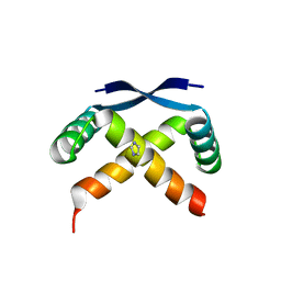
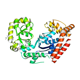
6RZ3
 | | Crystal structure of a complex between the DNA-binding domain of p53 and the carboxyl-terminal conserved region of iASPP | | 分子名称: | Cellular tumor antigen p53, RelA-associated inhibitor, ZINC ION | | 著者 | Chen, S, Ren, J, Jones, E.Y, Lu, X. | | 登録日 | 2019-06-12 | | 公開日 | 2019-10-30 | | 最終更新日 | 2024-01-24 | | 実験手法 | X-RAY DIFFRACTION (4.23 Å) | | 主引用文献 | iASPP mediates p53 selectivity through a modular mechanism fine-tuning DNA recognition.
Proc.Natl.Acad.Sci.USA, 116, 2019
|
|
2EVJ
 | | Structure of an Ndt80-DNA complex (MSE mutant mA9C) | | 分子名称: | 5'-D(*AP*GP*TP*GP*TP*TP*TP*GP*TP*GP*TP*CP*GP*C)-3', 5'-D(*TP*GP*CP*GP*AP*CP*AP*CP*AP*AP*AP*CP*AP*C)-3', NDT80 protein | | 著者 | Lamoureux, J.S, Glover, J.N. | | 登録日 | 2005-10-31 | | 公開日 | 2006-03-21 | | 最終更新日 | 2023-08-23 | | 実験手法 | X-RAY DIFFRACTION (1.89 Å) | | 主引用文献 | Principles of Protein-DNA Recognition Revealed in the Structural Analysis of Ndt80-MSE DNA Complexes.
Structure, 14, 2006
|
|
6VG6
 | | Crystal structure of DPO4 with 8-oxoadenine (oxoA) and dGTP* | | 分子名称: | 2'-deoxy-5'-O-[(R)-hydroxy{[(R)-hydroxy(phosphonooxy)phosphoryl]amino}phosphoryl]guanosine, DNA (5'-D(*GP*GP*GP*GP*GP*AP*AP*GP*GP*AP*TP*TP*C)-3'), DNA (5'-D(*TP*CP*AP*TP*(A38)P*GP*AP*AP*TP*CP*CP*TP*TP*CP*CP*CP*CP*C)-3'), ... | | 著者 | Jung, H, Lee, S. | | 登録日 | 2020-01-07 | | 公開日 | 2021-01-06 | | 最終更新日 | 2023-10-11 | | 実験手法 | X-RAY DIFFRACTION (3.08 Å) | | 主引用文献 | Promutagenic bypass of 7,8-dihydro-8-oxoadenine by translesion synthesis DNA polymerase Dpo4.
Biochem.J., 477, 2020
|
|
6VKP
 | | Crystal structure of DPO4 extension past 8-oxoadenine (oxoA) and dG | | 分子名称: | 2'-deoxy-5'-O-[(R)-hydroxy{[(R)-hydroxy(phosphonooxy)phosphoryl]amino}phosphoryl]adenosine, DNA (5'-D(*GP*GP*GP*GP*GP*AP*AP*AP*GP*AP*TP*TP*CP*G)-3'), DNA (5'-D(*TP*TP*CP*AP*TP*(A38)P*GP*AP*AP*TP*CP*TP*TP*TP*CP*CP*CP*CP*C)-3'), ... | | 著者 | Jung, H, Lee, S. | | 登録日 | 2020-01-21 | | 公開日 | 2021-01-27 | | 最終更新日 | 2023-10-11 | | 実験手法 | X-RAY DIFFRACTION (2.54 Å) | | 主引用文献 | Insights into the mismatch discrimination mechanism of Y-family DNA polymerase Dpo4.
Biochem.J., 478, 2021
|
|
1W0U
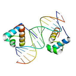 | | hTRF2 DNA-binding domain in complex with telomeric DNA. | | 分子名称: | 5'-D(*CP*TP*AP*AP*CP*CP*CP*TP*AP*AP *CP*CP*CP*TP*AP*GP*A)-3', 5'-D(*TP*CP*TP*AP*GP*GP*GP*TP*TP*AP *GP*GP*GP*TP*TP*AP*G)-3', TELOMERIC REPEAT BINDING FACTOR 2 | | 著者 | Court, R.I, Chapman, L.M, Fairall, L, Rhodes, D. | | 登録日 | 2004-06-11 | | 公開日 | 2004-12-22 | | 最終更新日 | 2024-05-08 | | 実験手法 | X-RAY DIFFRACTION (1.8 Å) | | 主引用文献 | How the Human Telomeric Proteins Trf1 and Trf2 Recognize Telomeric DNA: A View from High-Resolution Crystal Structures
Embo Rep., 6, 2005
|
|
3C94
 | | ExoI/SSB-Ct complex | | 分子名称: | Exodeoxyribonuclease I, MAGNESIUM ION, Single-stranded DNA-binding C-terminal tail peptide | | 著者 | Lu, D, Keck, J.L. | | 登録日 | 2008-02-15 | | 公開日 | 2008-07-08 | | 最終更新日 | 2023-08-30 | | 実験手法 | X-RAY DIFFRACTION (2.7 Å) | | 主引用文献 | Structural basis of Escherichia coli single-stranded DNA-binding protein stimulation of exonuclease I.
Proc.Natl.Acad.Sci.USA, 105, 2008
|
|
8HSG
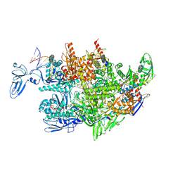 | | Thermus thermophilus RNA polymerase elongation complex | | 分子名称: | DNA (27-MER), DNA (37-MER), DNA-directed RNA polymerase subunit alpha, ... | | 著者 | Murayama, Y, Ehara, H, Sekine, S. | | 登録日 | 2022-12-19 | | 公開日 | 2023-05-03 | | 最終更新日 | 2024-07-03 | | 実験手法 | ELECTRON MICROSCOPY (3.2 Å) | | 主引用文献 | Structural basis of the transcription termination factor Rho engagement with transcribing RNA polymerase from Thermus thermophilus.
Sci Adv, 9, 2023
|
|
5LLQ
 | |
1XJY
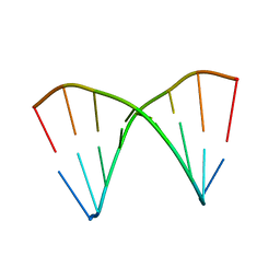 | | The crystal structures of the DNA binding sites of the RUNX1 transcription factor | | 分子名称: | 5'-D(*TP*CP*TP*GP*CP*GP*GP*TP*C)-3', 5'-D(*TP*GP*AP*CP*CP*GP*CP*AP*G)-3' | | 著者 | Kitayner, M, Rozenberg, H, Rabinovich, D, Shakked, Z. | | 登録日 | 2004-09-26 | | 公開日 | 2005-03-15 | | 最終更新日 | 2024-04-03 | | 実験手法 | X-RAY DIFFRACTION (2 Å) | | 主引用文献 | Structures of the DNA-binding site of Runt-domain transcription regulators.
Acta Crystallogr.,Sect.D, 61, 2005
|
|
1OWR
 | |
1XJX
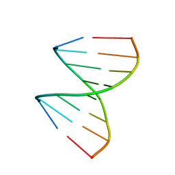 | | The crystal structures of the DNA binding sites of the RUNX1 transcription factor | | 分子名称: | 5'-D(*TP*CP*TP*GP*CP*GP*GP*TP*C)-3', 5'-D(*TP*GP*AP*CP*CP*GP*CP*AP*G)-3' | | 著者 | Kitayner, M, Rozenberg, H, Rabinovich, D, Shakked, Z. | | 登録日 | 2004-09-26 | | 公開日 | 2005-03-15 | | 最終更新日 | 2024-04-03 | | 実験手法 | X-RAY DIFFRACTION (1.7 Å) | | 主引用文献 | Structures of the DNA-binding site of Runt-domain transcription regulators.
Acta Crystallogr.,Sect.D, 61, 2005
|
|
2EVF
 | | Structure of a Ndt80-DNA complex (MSE mutant mA6T) | | 分子名称: | 5'-D(*AP*GP*TP*TP*TP*TP*AP*GP*TP*GP*TP*CP*GP*C)-3', 5'-D(*TP*GP*CP*GP*AP*CP*AP*CP*TP*AP*AP*AP*AP*C)-3', NDT80 protein | | 著者 | Lamoureux, J.S, Glover, J.N. | | 登録日 | 2005-10-31 | | 公開日 | 2006-03-21 | | 最終更新日 | 2023-08-23 | | 実験手法 | X-RAY DIFFRACTION (1.56 Å) | | 主引用文献 | Principles of Protein-DNA Recognition Revealed in the Structural Analysis of Ndt80-MSE DNA Complexes.
Structure, 14, 2006
|
|


