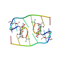
4MQR
 
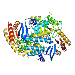 | | Mycobaterium tuberculosis transaminase BioA complexed with E)-5-hydroxy-4-(((Z)-isonicotinoyldiazenyl)methylene)-6-methyl-1,4-dihydropyridin-3-yl)methyl phosphate | | 分子名称: | 1,2-ETHANEDIOL, Adenosylmethionine-8-amino-7-oxononanoate aminotransferase, [(4Z)-5-hydroxy-6-methyl-4-{[(E)-(pyridin-4-ylcarbonyl)diazenyl]methylidene}-1,4-dihydropyridin-3-yl]methyl dihydrogen phosphate | | 著者 | Finzel, B.C, Dai, R. | | 登録日 | 2013-09-16 | | 公開日 | 2014-03-05 | | 最終更新日 | 2023-09-20 | | 実験手法 | X-RAY DIFFRACTION (2.1 Å) | | 主引用文献 | Inhibition of Mycobacterium tuberculosis Transaminase BioA by Aryl Hydrazines and Hydrazides.
Chembiochem, 15, 2014
|
|
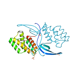
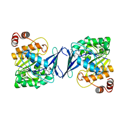
4ILU
 | | Crystal structure of Mycobacterium tuberculosis CarD | | 分子名称: | RNA polymerase-binding transcription factor CarD, SULFATE ION | | 著者 | Thakur, K.G, Kaur, G. | | 登録日 | 2013-01-01 | | 公開日 | 2013-10-30 | | 最終更新日 | 2024-05-29 | | 実験手法 | X-RAY DIFFRACTION (2.3 Å) | | 主引用文献 | Crystal structure of Mycobacterium tuberculosis CarD, an essential RNA polymerase binding protein, reveals a quasidomain-swapped dimeric structural architecture.
Proteins, 82, 2014
|
|
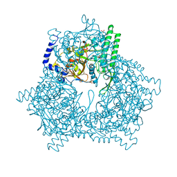
3ZKD
 | | CRYSTAL STRUCTURE OF THE ATPASE REGION OF Mycobacterium tuberculosis GyrB WITH AMPPNP | | 分子名称: | DNA GYRASE SUBUNIT B, MAGNESIUM ION, PHOSPHOAMINOPHOSPHONIC ACID-ADENYLATE ESTER | | 著者 | Agrawal, A, Roue, M, Spitzfaden, C, Petrella, S, Aubry, A, Volker, C, Mossakowska, D, Hann, M, Bax, B, Mayer, C. | | 登録日 | 2013-01-22 | | 公開日 | 2013-09-18 | | 最終更新日 | 2023-12-20 | | 実験手法 | X-RAY DIFFRACTION (2.95 Å) | | 主引用文献 | Mycobacterium Tuberculosis DNA Gyrase ATPase Domain Structures Suggest a Dissociative Mechanism that Explains How ATP Hydrolysis is Coupled to Domain Motion.
Biochem.J., 456, 2013
|
|
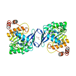
4MFR
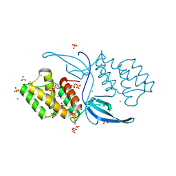 | | Crystal structure of Mycobacterium tuberculosis CarD | | 分子名称: | GLYCEROL, IODIDE ION, RNA polymerase-binding transcription factor CarD, ... | | 著者 | Kaur, G, Thakur, K.G. | | 登録日 | 2013-08-28 | | 公開日 | 2013-11-06 | | 最終更新日 | 2024-05-29 | | 実験手法 | X-RAY DIFFRACTION (2.5 Å) | | 主引用文献 | Crystal structure of Mycobacterium tuberculosis CarD, an essential RNA polymerase binding protein, reveals a quasidomain-swapped dimeric structural architecture.
Proteins, 82, 2014
|
|
4HEC
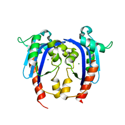 | |
3ZM7
 | | CRYSTAL STRUCTURE OF THE ATPASE REGION OF Mycobacterium tuberculosis GyrB WITH AMPPCP | | 分子名称: | DNA GYRASE SUBUNIT B, MAGNESIUM ION, PHOSPHOMETHYLPHOSPHONIC ACID ADENYLATE ESTER | | 著者 | Agrawal, A, Roue, M, Spitzfaden, C, Petrella, S, Aubry, A, Volker, C, Mossakowska, D, Hann, M, Bax, B, Mayer, C. | | 登録日 | 2013-02-05 | | 公開日 | 2013-09-18 | | 最終更新日 | 2023-12-20 | | 実験手法 | X-RAY DIFFRACTION (3.3 Å) | | 主引用文献 | Mycobacterium Tuberculosis DNA Gyrase ATPase Domain Structures Suggest a Dissociative Mechanism that Explains How ATP Hydrolysis is Coupled to Domain Motion.
Biochem.J., 456, 2013
|
|
1FJA
 | |
1GCB
 | | GAL6, YEAST BLEOMYCIN HYDROLASE DNA-BINDING PROTEASE (THIOL) | | 分子名称: | GAL6 HG (EMTS) DERIVATIVE, GLYCEROL, MERCURY (II) ION, ... | | 著者 | Joshua-Tor, L, Xu, H.E, Johnston, S.A, Rees, D.C. | | 登録日 | 1995-07-18 | | 公開日 | 1995-10-15 | | 最終更新日 | 2024-02-07 | | 実験手法 | X-RAY DIFFRACTION (2.2 Å) | | 主引用文献 | Crystal structure of a conserved protease that binds DNA: the bleomycin hydrolase, Gal6.
Science, 269, 1995
|
|
3ZKB
 | | CRYSTAL STRUCTURE OF THE ATPASE REGION OF Mycobacterium tuberculosis GyrB WITH AMPPNP | | 分子名称: | DNA GYRASE SUBUNIT B, MAGNESIUM ION, PHOSPHOAMINOPHOSPHONIC ACID-ADENYLATE ESTER | | 著者 | Agrawal, A, Roue, M, Spitzfaden, C, Petrella, S, Aubry, A, Volker, C, Mossakowska, D, Hann, M, Bax, B, Mayer, C. | | 登録日 | 2013-01-22 | | 公開日 | 2013-09-18 | | 最終更新日 | 2024-05-08 | | 実験手法 | X-RAY DIFFRACTION (2.9 Å) | | 主引用文献 | Mycobacterium Tuberculosis DNA Gyrase ATPase Domain Structures Suggest a Dissociative Mechanism that Explains How ATP Hydrolysis is Coupled to Domain Motion.
Biochem.J., 456, 2013
|
|
6C9S
 | | Mycobacterium tuberculosis adenosine kinase bound to (2R,3R,4S,5R)-2-(6-([1,1'-biphenyl]-4-ylethynyl)-9H-purin-9-yl)-5-(hydroxymethyl)tetrahydrofuran-3,4-diol | | 分子名称: | 6-[([1,1'-biphenyl]-4-yl)ethynyl]-9-beta-D-ribofuranosyl-9H-purine, Adenosine kinase, SODIUM ION, ... | | 著者 | Crespo, R.A, TB Structural Genomics Consortium (TBSGC) | | 登録日 | 2018-01-28 | | 公開日 | 2019-05-01 | | 最終更新日 | 2023-10-04 | | 実験手法 | X-RAY DIFFRACTION (2.23 Å) | | 主引用文献 | Structure-Guided Drug Design of 6-Substituted Adenosine Analogues as Potent Inhibitors of Mycobacterium tuberculosis Adenosine Kinase.
J.Med.Chem., 62, 2019
|
|
6C9R
 | | Mycobacterium tuberculosis adenosine kinase bound to (2R,3S,4R,5R)-2-(hydroxymethyl)-5-(6-(thiophen-3-yl)-9H-purin-9-yl)tetrahydrofuran-3,4-diol | | 分子名称: | 9-beta-D-ribofuranosyl-6-(thiophen-3-yl)-9H-purine, Adenosine kinase, GLYCEROL, ... | | 著者 | Crespo, R.A, TB Structural Genomics Consortium (TBSGC) | | 登録日 | 2018-01-28 | | 公開日 | 2019-05-01 | | 最終更新日 | 2023-10-04 | | 実験手法 | X-RAY DIFFRACTION (2.1 Å) | | 主引用文献 | Structure-Guided Drug Design of 6-Substituted Adenosine Analogues as Potent Inhibitors of Mycobacterium tuberculosis Adenosine Kinase.
J.Med.Chem., 62, 2019
|
|
6C9Q
 | |
6C9P
 | |
4RPI
 | | Crystal Structure of Nicotinate Mononucleotide Adenylyltransferase from Mycobacterium tuberculosis | | 分子名称: | 4-(2-HYDROXYETHYL)-1-PIPERAZINE ETHANESULFONIC ACID, DIMETHYL SULFOXIDE, nicotinate-nucleotide adenylyltransferase | | 著者 | Rodionova, I, Zuccola, H, Sorci, L, Aleshin, A.E, Kazanov, M, Sergienko, E, Rubin, E, Locher, C, Osterman, A. | | 登録日 | 2014-10-30 | | 公開日 | 2015-01-28 | | 最終更新日 | 2024-02-28 | | 実験手法 | X-RAY DIFFRACTION (2.417 Å) | | 主引用文献 | Mycobacterial nicotinate mononucleotide adenylyltransferase: structure, mechanism, and implications for drug discovery.
J. Biol. Chem., 290, 2015
|
|
4IDY
 | | Mycobacterium Tuberculosis Methionine aminopeptidase Type 1c in complex with 2-hydroxyethyl disulfide | | 分子名称: | 2-HYDROXYETHYL DISULFIDE, Methionine aminopeptidase 2, POTASSIUM ION | | 著者 | Reddi, R, Gumpena, R, Kishor, C, Addlagatta, A. | | 登録日 | 2012-12-13 | | 公開日 | 2013-12-18 | | 最終更新日 | 2024-11-13 | | 実験手法 | X-RAY DIFFRACTION (2 Å) | | 主引用文献 | Selective targeting of the conserved active site cysteine of Mycobacterium tuberculosis methionine aminopeptidase with electrophilic reagents
Febs J., 281, 2014
|
|
4IF7
 | | Mycobacterium Tuberculosis Methionine aminopeptidase Type 1c in complex with homocysteine-methyl disulfide | | 分子名称: | (2S)-2-amino-4-(methyldisulfanyl)butanoic acid, COBALT (II) ION, Methionine aminopeptidase 2 | | 著者 | Reddi, R, Gumpena, R, Kishor, C, Addlagatta, A. | | 登録日 | 2012-12-14 | | 公開日 | 2013-12-18 | | 最終更新日 | 2025-03-26 | | 実験手法 | X-RAY DIFFRACTION (2 Å) | | 主引用文献 | Selective targeting of the conserved active site cysteine of Mycobacterium tuberculosis methionine aminopeptidase with electrophilic reagents
Febs J., 281, 2014
|
|
4Q6U
 | |
2VZW
 | | X-ray structure of the heme-bound GAF domain of sensory histidine kinase DosT of Mycobacterium tuberculosis | | 分子名称: | ACETATE ION, OXYGEN MOLECULE, PROBABLE HISTIDINE KINASE RESPONSE REGULATOR, ... | | 著者 | Podust, L.M, Ioanoviciu, A, Ortiz de Montellano, P.R. | | 登録日 | 2008-08-06 | | 公開日 | 2009-03-17 | | 最終更新日 | 2024-05-08 | | 実験手法 | X-RAY DIFFRACTION (2.3 Å) | | 主引用文献 | 2.3 A X-Ray Structure of the Heme-Bound Gaf Domain of Sensory Histidine Kinase Dost of Mycobacterium Tuberculosis.
Biochemistry, 47, 2008
|
|
6A2Q
 | | Mycobacterium tuberculosis LexA C-domain I | | 分子名称: | GLYCEROL, LexA repressor | | 著者 | Chandran, A.V, Srikalaivani, R, Paul, A, Vijayan, M. | | 登録日 | 2018-06-12 | | 公開日 | 2019-01-23 | | 最終更新日 | 2024-10-30 | | 実験手法 | X-RAY DIFFRACTION (1.48 Å) | | 主引用文献 | Biochemical characterization of Mycobacterium tuberculosis LexA and structural studies of its C-terminal segment.
Acta Crystallogr D Struct Biol, 75, 2019
|
|
6A2T
 | | Mycobacterium tuberculosis LexA C-domain K197A | | 分子名称: | ACRYLIC ACID, LexA repressor | | 著者 | Chandran, A.V, Srikalaivani, R, Paul, A, Vijayan, M. | | 登録日 | 2018-06-12 | | 公開日 | 2019-01-23 | | 最終更新日 | 2024-10-30 | | 実験手法 | X-RAY DIFFRACTION (1.9 Å) | | 主引用文献 | Biochemical characterization of Mycobacterium tuberculosis LexA and structural studies of its C-terminal segment.
Acta Crystallogr D Struct Biol, 75, 2019
|
|
2WM4
 | | X-ray structure of Mycobacterium tuberculosis cytochrome P450 CYP124 in complex with phytanic acid | | 分子名称: | (3R,7S,11S)-3,7,11,15-tetramethylhexadecanoic acid, CALCIUM ION, PROTOPORPHYRIN IX CONTAINING FE, ... | | 著者 | Johnston, J.B, Kells, P.M, Podust, L.M, Ortiz de Montellano, P.R. | | 登録日 | 2009-06-30 | | 公開日 | 2009-10-06 | | 最終更新日 | 2023-12-13 | | 実験手法 | X-RAY DIFFRACTION (2.11 Å) | | 主引用文献 | Biochemical and structural characterization of CYP124: a methyl-branched lipid omega-hydroxylase from Mycobacterium tuberculosis.
Proc. Natl. Acad. Sci. U.S.A., 106, 2009
|
|
3ZQJ
 | | Mycobacterium tuberculosis UvrA | | 分子名称: | UVRABC SYSTEM PROTEIN A, ZINC ION | | 著者 | Rossi, F, Khanduja, J.S, Bortoluzzi, A, Houghton, J, Sander, P, Guthlein, C, Davis, E.O, Springer, B, Bottger, E.C, Relini, A, Penco, A, Muniyappa, K, Rizzi, M. | | 登録日 | 2011-06-09 | | 公開日 | 2011-06-22 | | 最終更新日 | 2023-12-20 | | 実験手法 | X-RAY DIFFRACTION (3.4 Å) | | 主引用文献 | The Biological and Structural Characterization of Mycobacterium Tuberculosis Uvra Provides Novel Insights Into its Mechanism of Action
Nucleic Acids Res., 39, 2011
|
|
2YW6
 | | Structural studies of N terminal deletion mutant of Dps from Mycobacterium smegmatis | | 分子名称: | DNA protection during starvation protein | | 著者 | Roy, S, Saraswathi, R, Gupta, S, Sekar, K, Chatterji, D, Vijayan, M. | | 登録日 | 2007-04-19 | | 公開日 | 2007-07-17 | | 最終更新日 | 2023-10-25 | | 実験手法 | X-RAY DIFFRACTION (2.53 Å) | | 主引用文献 | Role of N and C-terminal Tails in DNA Binding and Assembly in Dps: Structural Studies of Mycobacterium smegmatis Dps Deletion Mutants
J.Mol.Biol., 370, 2007
|
|
2WM5
 | | X-ray structure of the substrate-free Mycobacterium tuberculosis cytochrome P450 CYP124 | | 分子名称: | PROTOPORPHYRIN IX CONTAINING FE, PUTATIVE CYTOCHROME P450 124 | | 著者 | Johnston, J.B, Kells, P.M, Podust, L.M, Ortiz de Montellano, P.R. | | 登録日 | 2009-06-30 | | 公開日 | 2009-10-06 | | 最終更新日 | 2024-05-01 | | 実験手法 | X-RAY DIFFRACTION (1.5 Å) | | 主引用文献 | Biochemical and structural characterization of CYP124: a methyl-branched lipid omega-hydroxylase from Mycobacterium tuberculosis.
Proc. Natl. Acad. Sci. U.S.A., 106, 2009
|
|
6AX8
 | | Mycobacterium tuberculosis methionyl-tRNA synthetase in complex with methionyl-adenylate | | 分子名称: | Methionine-tRNA ligase, [[(2R,3S,4R,5R)-5-(6-aminopurin-9-yl)-3,4-dihydroxy-oxolan-2-yl]methoxy-hydroxy-phosphoryl] (2S)-2-azanyl-4-methylsulfanyl-butanoate | | 著者 | Barros-Alvarez, X, Hol, W.G.J. | | 登録日 | 2017-09-06 | | 公開日 | 2018-04-11 | | 最終更新日 | 2024-03-13 | | 実験手法 | X-RAY DIFFRACTION (2.6 Å) | | 主引用文献 | The crystal structure of the drug target Mycobacterium tuberculosis methionyl-tRNA synthetase in complex with a catalytic intermediate.
Acta Crystallogr F Struct Biol Commun, 74, 2018
|
|


