3N7T
 
 | |
6B1X
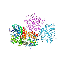
 | | Crystal structure KPC-2 beta-lactamase complexed with WCK 5153 by soaking | | 分子名称: | (2S,5R)-1-formyl-N'-[(3R)-pyrrolidine-3-carbonyl]-5-[(sulfooxy)amino]piperidine-2-carbohydrazide, 1,2-ETHANEDIOL, CITRIC ACID, ... | | 著者 | van den Akker, F, Nguyen, N.Q. | | 登録日 | 2017-09-19 | | 公開日 | 2018-08-01 | | 最終更新日 | 2024-11-13 | | 実験手法 | X-RAY DIFFRACTION (1.45 Å) | | 主引用文献 | Strategic Approaches to Overcome Resistance against Gram-Negative Pathogens Using beta-Lactamase Inhibitors and beta-Lactam Enhancers: Activity of Three Novel Diazabicyclooctanes WCK 5153, Zidebactam (WCK 5107), and WCK 4234.
J. Med. Chem., 61, 2018
|
|
4FSN
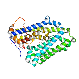
 | | Crystal Structure of the CHK1 | | 分子名称: | 4-(6-{[(4-METHYLCYCLOHEXYL)AMINO]METHYL}-1,4-DIHYDROINDENO[1,2-C]PYRAZOL-3-YL)BENZOIC ACID, ISOPROPYL ALCOHOL, SULFATE ION, ... | | 著者 | Kang, Y.N, Stuckey, J.A, Chang, P, Russell, A.J. | | 登録日 | 2012-06-27 | | 公開日 | 2012-08-22 | | 最終更新日 | 2023-11-29 | | 実験手法 | X-RAY DIFFRACTION (2.1 Å) | | 主引用文献 | Crystal Structure of the CHK1
To be Published
|
|
2ZPK
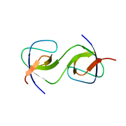
 | | Crystal structure of P20.1 Fab fragment in complex with its antigen peptide | | 分子名称: | GLYCEROL, IgG1-lambda P20.1 Fab (heavy chain), IgG1-lambda P20.1 Fab (light chain), ... | | 著者 | Nogi, T, Sanagawa, T, Takagi, J. | | 登録日 | 2008-07-16 | | 公開日 | 2008-12-23 | | 最終更新日 | 2024-11-20 | | 実験手法 | X-RAY DIFFRACTION (1.8 Å) | | 主引用文献 | Novel affinity tag system using structurally defined antibody-tag interaction: application to single-step protein purification
Protein Sci., 17, 2008
|
|
7NKS
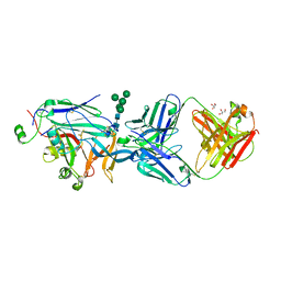 | | Structure of the Hantaan virus Gn glycoprotein ectodomain in complex with Fab HTN-Gn1 | | 分子名称: | 2-acetamido-2-deoxy-beta-D-glucopyranose, Envelope polyprotein, Fab HTN-Gn1 heavy chain, ... | | 著者 | Rissanen, I, Bowden, T.A, Huiskonen, J.T, Stass, R. | | 登録日 | 2021-02-18 | | 公開日 | 2021-06-23 | | 最終更新日 | 2024-10-23 | | 実験手法 | X-RAY DIFFRACTION (3.5 Å) | | 主引用文献 | Structural Basis for a Neutralizing Antibody Response Elicited by a Recombinant Hantaan Virus Gn Immunogen.
Mbio, 12, 2021
|
|
4PE0
 | | Crystal Structure of Calcium-loaded S100B bound to SBi4434 | | 分子名称: | 2-[(2-hydroxyethyl)sulfanyl]naphthalene-1,4-dione, CALCIUM ION, Protein S100-B | | 著者 | Cavalier, M.C, Pierce, P.D, Wilder, P.T, Neau, D, Toth, E.A, Weber, D.J. | | 登録日 | 2014-04-22 | | 公開日 | 2014-11-05 | | 最終更新日 | 2024-11-06 | | 実験手法 | X-RAY DIFFRACTION (1.08 Å) | | 主引用文献 | Covalent Small Molecule Inhibitors of Ca(2+)-Bound S100B.
Biochemistry, 53, 2014
|
|
9DZU
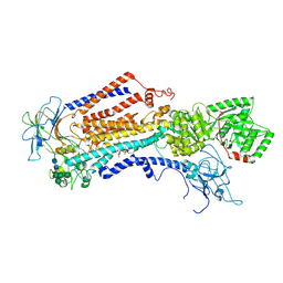 | | Cryo-EM structure of the C. neoformans lipid flippase Apt1-Cdc50 bound with butyrolactol A in the E2P state | | 分子名称: | (3R,4R,5S)-3,4-dihydroxy-5-[(1R,2R,3S,4S,5R,6R,8E,10E,14E,16Z)-1,2,3,4,5-pentahydroxy-6,20,20-trimethylhenicosa-8,10,14,16-tetraen-1-yl]oxolan-2-one, 2-acetamido-2-deoxy-beta-D-glucopyranose, 2-acetamido-2-deoxy-beta-D-glucopyranose-(1-4)-2-acetamido-2-deoxy-beta-D-glucopyranose, ... | | 著者 | Duan, H.D, Li, H. | | 登録日 | 2024-10-17 | | 公開日 | 2025-02-05 | | 最終更新日 | 2025-10-22 | | 実験手法 | ELECTRON MICROSCOPY (2.72 Å) | | 主引用文献 | Butyrolactol A potentiates caspofungin efficacy against resistant fungi via phospholipid flippase inhibition.
Biorxiv, 2025
|
|
6LC7
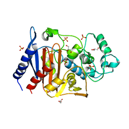 | | Crystal structure of AmpC Ent385 free form | | 分子名称: | 1,4-DIETHYLENE DIOXIDE, Beta-lactamase, GLYCEROL, ... | | 著者 | Kawai, A, Doi, Y. | | 登録日 | 2019-11-18 | | 公開日 | 2020-04-22 | | 最終更新日 | 2023-11-22 | | 実験手法 | X-RAY DIFFRACTION (1.4 Å) | | 主引用文献 | Structural Basis of Reduced Susceptibility to Ceftazidime-Avibactam and Cefiderocol inEnterobacter cloacaeDue to AmpC R2 Loop Deletion.
Antimicrob.Agents Chemother., 64, 2020
|
|
3IEA
 | |
1YVJ
 | | Crystal structure of the Jak3 kinase domain in complex with a staurosporine analogue | | 分子名称: | (2S,3S)-1,4-DIMERCAPTOBUTANE-2,3-DIOL, 1,2,3,4-TETRAHYDROGEN-STAUROSPORINE, Tyrosine-protein kinase JAK3 | | 著者 | Boggon, T.J, Li, Y, Manley, P.W, Eck, M.J. | | 登録日 | 2005-02-15 | | 公開日 | 2005-05-24 | | 最終更新日 | 2024-11-20 | | 実験手法 | X-RAY DIFFRACTION (2.55 Å) | | 主引用文献 | Crystal structure of the Jak3 kinase domain in complex with a staurosporine analog
Blood, 106, 2005
|
|
5PPL
 | | PanDDA analysis group deposition -- Crystal Structure of BRD1 after initial refinement with no ligand modelled (structure 22) | | 分子名称: | 1,2-ETHANEDIOL, Bromodomain-containing protein 1, SODIUM ION | | 著者 | Pearce, N.M, Krojer, T, Talon, R, Bradley, A.R, Fairhead, M, Sethi, R, Wright, N, MacLean, E, Collins, P, Brandao-Neto, J, Douangamath, A, Renjie, Z, Dias, A, Ng, J, Brennan, P.E, Cox, O, Bountra, C, Arrowsmith, C.H, Edwards, A, von Delft, F. | | 登録日 | 2017-02-07 | | 公開日 | 2017-03-29 | | 最終更新日 | 2024-03-06 | | 実験手法 | X-RAY DIFFRACTION (1.63 Å) | | 主引用文献 | A multi-crystal method for extracting obscured crystallographic states from conventionally uninterpretable electron density.
Nat Commun, 8, 2017
|
|
6B88
 | | E. coli LepB in complex with GNE0775 ((4S,7S,10S)-10-((S)-4-amino-2-(2-(4-(tert-butyl)phenyl)-4-methylpyrimidine-5-carboxamido)-N-methylbutanamido)-16,26-bis(2-aminoethoxy)-N-(2-iminoethyl)-7-methyl-6,9-dioxo-5,8-diaza-1,2(1,3)-dibenzenacyclodecaphane-4-carboxamide) | | 分子名称: | (8S,11S,14S)-14-{[(2S)-4-amino-2-{[2-(4-tert-butylphenyl)-4-methylpyrimidine-5-carbonyl]amino}butanoyl](methyl)amino}-3,18-bis(2-aminoethoxy)-N-[(2Z)-2-iminoethyl]-11-methyl-10,13-dioxo-9,12-diazatricyclo[13.3.1.1~2,6~]icosa-1(19),2(20),3,5,15,17-hexaene-8-carboxamide, PENTAETHYLENE GLYCOL, Signal peptidase I | | 著者 | Murray, J.M, Rouge, L. | | 登録日 | 2017-10-05 | | 公開日 | 2018-10-10 | | 最終更新日 | 2024-11-06 | | 実験手法 | X-RAY DIFFRACTION (2.407 Å) | | 主引用文献 | Optimized arylomycins are a new class of Gram-negative antibiotics.
Nature, 561, 2018
|
|
1YWM
 | | Crystal structure of the N-terminal domain of group B Streptococcus alpha C protein | | 分子名称: | (2R,3S)-1,4-DIMERCAPTOBUTANE-2,3-DIOL, C protein alpha-antigen, GLYCEROL | | 著者 | Auperin, T.C, Bolduc, G.R, Baron, M.J, Heroux, A, Filman, D.J, Madoff, L.C, Hogle, J.M. | | 登録日 | 2005-02-18 | | 公開日 | 2005-03-08 | | 最終更新日 | 2024-04-03 | | 実験手法 | X-RAY DIFFRACTION (1.86 Å) | | 主引用文献 | Crystal structure of the N-terminal domain of the group B streptococcus alpha C protein.
J.Biol.Chem., 280, 2005
|
|
1NJX
 | |
5PPY
 | | PanDDA analysis group deposition -- Crystal Structure of BRD1 after initial refinement with no ligand modelled (structure 35) | | 分子名称: | 1,2-ETHANEDIOL, Bromodomain-containing protein 1, SODIUM ION | | 著者 | Pearce, N.M, Krojer, T, Talon, R, Bradley, A.R, Fairhead, M, Sethi, R, Wright, N, MacLean, E, Collins, P, Brandao-Neto, J, Douangamath, A, Renjie, Z, Dias, A, Ng, J, Brennan, P.E, Cox, O, Bountra, C, Arrowsmith, C.H, Edwards, A, von Delft, F. | | 登録日 | 2017-02-07 | | 公開日 | 2017-03-29 | | 最終更新日 | 2024-03-06 | | 実験手法 | X-RAY DIFFRACTION (1.45 Å) | | 主引用文献 | A multi-crystal method for extracting obscured crystallographic states from conventionally uninterpretable electron density.
Nat Commun, 8, 2017
|
|
1NK6
 | |
5PQ6
 | | PanDDA analysis group deposition -- Crystal Structure of BRD1 after initial refinement with no ligand modelled (structure 43) | | 分子名称: | 1,2-ETHANEDIOL, Bromodomain-containing protein 1, SODIUM ION | | 著者 | Pearce, N.M, Krojer, T, Talon, R, Bradley, A.R, Fairhead, M, Sethi, R, Wright, N, MacLean, E, Collins, P, Brandao-Neto, J, Douangamath, A, Renjie, Z, Dias, A, Ng, J, Brennan, P.E, Cox, O, Bountra, C, Arrowsmith, C.H, Edwards, A, von Delft, F. | | 登録日 | 2017-02-07 | | 公開日 | 2017-03-29 | | 最終更新日 | 2024-03-06 | | 実験手法 | X-RAY DIFFRACTION (1.64 Å) | | 主引用文献 | A multi-crystal method for extracting obscured crystallographic states from conventionally uninterpretable electron density.
Nat Commun, 8, 2017
|
|
5PQI
 | | PanDDA analysis group deposition -- Crystal Structure of BRD1 after initial refinement with no ligand modelled (structure 55) | | 分子名称: | 1,2-ETHANEDIOL, Bromodomain-containing protein 1, SODIUM ION | | 著者 | Pearce, N.M, Krojer, T, Talon, R, Bradley, A.R, Fairhead, M, Sethi, R, Wright, N, MacLean, E, Collins, P, Brandao-Neto, J, Douangamath, A, Renjie, Z, Dias, A, Ng, J, Brennan, P.E, Cox, O, Bountra, C, Arrowsmith, C.H, Edwards, A, von Delft, F. | | 登録日 | 2017-02-07 | | 公開日 | 2017-03-29 | | 最終更新日 | 2024-03-06 | | 実験手法 | X-RAY DIFFRACTION (1.33 Å) | | 主引用文献 | A multi-crystal method for extracting obscured crystallographic states from conventionally uninterpretable electron density.
Nat Commun, 8, 2017
|
|
5PQY
 | | PanDDA analysis group deposition -- Crystal Structure of BRD1 after initial refinement with no ligand modelled (structure 71) | | 分子名称: | 1,2-ETHANEDIOL, Bromodomain-containing protein 1, SODIUM ION | | 著者 | Pearce, N.M, Krojer, T, Talon, R, Bradley, A.R, Fairhead, M, Sethi, R, Wright, N, MacLean, E, Collins, P, Brandao-Neto, J, Douangamath, A, Renjie, Z, Dias, A, Ng, J, Brennan, P.E, Cox, O, Bountra, C, Arrowsmith, C.H, Edwards, A, von Delft, F. | | 登録日 | 2017-02-07 | | 公開日 | 2017-03-29 | | 最終更新日 | 2024-03-06 | | 実験手法 | X-RAY DIFFRACTION (1.89 Å) | | 主引用文献 | A multi-crystal method for extracting obscured crystallographic states from conventionally uninterpretable electron density.
Nat Commun, 8, 2017
|
|
5PRE
 | | PanDDA analysis group deposition -- Crystal Structure of BRD1 after initial refinement with no ligand modelled (structure 86) | | 分子名称: | 1,2-ETHANEDIOL, Bromodomain-containing protein 1, SODIUM ION | | 著者 | Pearce, N.M, Krojer, T, Talon, R, Bradley, A.R, Fairhead, M, Sethi, R, Wright, N, MacLean, E, Collins, P, Brandao-Neto, J, Douangamath, A, Renjie, Z, Dias, A, Ng, J, Brennan, P.E, Cox, O, Bountra, C, Arrowsmith, C.H, Edwards, A, von Delft, F. | | 登録日 | 2017-02-07 | | 公開日 | 2017-03-29 | | 最終更新日 | 2024-03-06 | | 実験手法 | X-RAY DIFFRACTION (1.73 Å) | | 主引用文献 | A multi-crystal method for extracting obscured crystallographic states from conventionally uninterpretable electron density.
Nat Commun, 8, 2017
|
|
5PRT
 | | PanDDA analysis group deposition -- Crystal Structure of BRD1 after initial refinement with no ligand modelled (structure 100) | | 分子名称: | 1,2-ETHANEDIOL, Bromodomain-containing protein 1, SODIUM ION | | 著者 | Pearce, N.M, Krojer, T, Talon, R, Bradley, A.R, Fairhead, M, Sethi, R, Wright, N, MacLean, E, Collins, P, Brandao-Neto, J, Douangamath, A, Renjie, Z, Dias, A, Ng, J, Brennan, P.E, Cox, O, Bountra, C, Arrowsmith, C.H, Edwards, A, von Delft, F. | | 登録日 | 2017-02-07 | | 公開日 | 2017-03-29 | | 最終更新日 | 2024-03-06 | | 実験手法 | X-RAY DIFFRACTION (1.89 Å) | | 主引用文献 | A multi-crystal method for extracting obscured crystallographic states from conventionally uninterpretable electron density.
Nat Commun, 8, 2017
|
|
3OM8
 | | The crystal structure of a hydrolase from Pseudomonas aeruginosa PA01 | | 分子名称: | 1,2-ETHANEDIOL, 2-(N-MORPHOLINO)-ETHANESULFONIC ACID, Probable hydrolase | | 著者 | Tan, K, Chhor, G, Buck, K, Joachimiak, A, Midwest Center for Structural Genomics (MCSG) | | 登録日 | 2010-08-26 | | 公開日 | 2010-09-22 | | 最終更新日 | 2024-10-16 | | 実験手法 | X-RAY DIFFRACTION (2.25 Å) | | 主引用文献 | The crystal structure of a hydrolase from Pseudomonas aeruginosa PA01
To be Published
|
|
3OYV
 | |
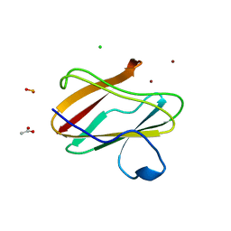
5QU5
 | | Domain Swap in the first SH3 domain of human Nck1 | | 分子名称: | Cytoplasmic protein NCK1 | | 著者 | Burger, D, Ruf, A, Benz, J, Schlatter, D, Rudolph, M.G. | | 登録日 | 2019-12-13 | | 公開日 | 2020-02-12 | | 最終更新日 | 2024-04-03 | | 実験手法 | X-RAY DIFFRACTION (1.11 Å) | | 主引用文献 | Small molecule AX-024 reduces T cell proliferation independently of CD3ε/Nck1 interaction, which is governed by a domain swap in the Nck1-SH3.1 domain.
J.Biol.Chem., 295, 2020
|
|
1WL5
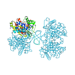 | | Human cytosolic acetoacetyl-CoA thiolase | | 分子名称: | GLYCEROL, SULFATE ION, acetyl-Coenzyme A acetyltransferase 2 | | 著者 | Kursula, P, Fukao, T, Kondo, N, Wierenga, R.K. | | 登録日 | 2004-06-20 | | 公開日 | 2005-03-01 | | 最終更新日 | 2024-10-30 | | 実験手法 | X-RAY DIFFRACTION (2.26 Å) | | 主引用文献 | High Resolution Crystal Structures of Human Cytosolic Thiolase (CT): A Comparison of the Active Sites of Human CT, Bacterial Thiolase, and Bacterial KAS I
J.Mol.Biol., 347, 2005
|
|


