2RP1
 
 | |
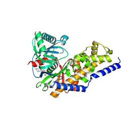
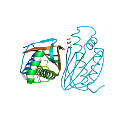
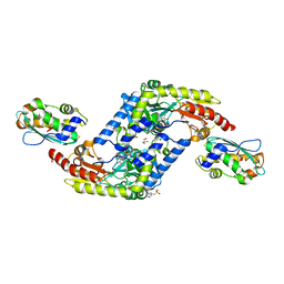
4KZM
 | | Crystal Structure of TR3 LBD S553A Mutant | | 分子名称: | GLYCEROL, Nuclear receptor subfamily 4 group A member 1 | | 著者 | Li, F, Zhang, Q, Li, A, Tian, X, Cai, Q, Wang, W, Wang, Y, Chen, H, Xing, Y, Wu, Q, Lin, T. | | 登録日 | 2013-05-30 | | 公開日 | 2013-12-18 | | 最終更新日 | 2024-03-20 | | 実験手法 | X-RAY DIFFRACTION (2.3 Å) | | 主引用文献 | Orphan nuclear receptor TR3 acts in autophagic cell death via mitochondrial signaling pathway.
Nat.Chem.Biol., 10, 2014
|
|
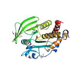
2H1A
 | | ResA C74A Variant | | 分子名称: | 1,2-ETHANEDIOL, Thiol-disulfide oxidoreductase resA | | 著者 | Lewin, A, Crow, A, Oubrie, A, Le Brun, N.E. | | 登録日 | 2006-05-16 | | 公開日 | 2006-09-19 | | 最終更新日 | 2024-02-14 | | 実験手法 | X-RAY DIFFRACTION (2.4 Å) | | 主引用文献 | Molecular Basis for Specificity of the Extracytoplasmic Thioredoxin ResA.
J.Biol.Chem., 281, 2006
|
|
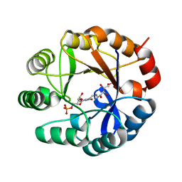
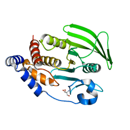
2RI1
 | | Crystal Structure of glucosamine 6-phosphate deaminase (NagB) with GlcN6P from S. mutans | | 分子名称: | 2-[BIS-(2-HYDROXY-ETHYL)-AMINO]-2-HYDROXYMETHYL-PROPANE-1,3-DIOL, 2-amino-2-deoxy-6-O-phosphono-alpha-D-glucopyranose, Glucosamine-6-phosphate deaminase | | 著者 | Liu, C, Li, D, Su, X.D. | | 登録日 | 2007-10-10 | | 公開日 | 2008-03-25 | | 最終更新日 | 2024-03-13 | | 実験手法 | X-RAY DIFFRACTION (2.03 Å) | | 主引用文献 | Ring-opening mechanism revealed by crystal structures of NagB and its ES intermediate complex
J.Mol.Biol., 379, 2008
|
|
1YCK
 | |
3S41
 | | Glucokinase in complex with activator and glucose | | 分子名称: | Glucokinase, N,N-dimethyl-5-({2-methyl-6-[(5-methylpyrazin-2-yl)carbamoyl]-1-benzofuran-4-yl}oxy)pyrimidine-2-carboxamide, SODIUM ION, ... | | 著者 | Liu, S. | | 登録日 | 2011-05-18 | | 公開日 | 2011-09-14 | | 最終更新日 | 2023-09-13 | | 実験手法 | X-RAY DIFFRACTION (2.18 Å) | | 主引用文献 | Designing glucokinase activators with reduced hypoglycemia risk: discovery of N,N-dimethyl-5-(2-methyl-6-((5-methylpyrazin-2-yl)-carbamoyl)benzofuran-4-yloxy)pyrimidine-2-carboxamide as a clinical candidate for the treatment of type 2 diabetes mellitus
MEDCHEMCOMM, 2, 2011
|
|
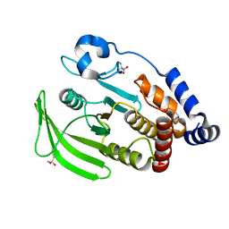
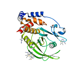
2RI0
 | | Crystal Structure of glucosamine 6-phosphate deaminase (NagB) from S. mutans | | 分子名称: | 2-[BIS-(2-HYDROXY-ETHYL)-AMINO]-2-HYDROXYMETHYL-PROPANE-1,3-DIOL, Glucosamine-6-phosphate deaminase, SODIUM ION | | 著者 | Li, D, Liu, C, Li, L.F, Su, X.D. | | 登録日 | 2007-10-10 | | 公開日 | 2008-03-25 | | 最終更新日 | 2024-03-13 | | 実験手法 | X-RAY DIFFRACTION (1.6 Å) | | 主引用文献 | Ring-opening mechanism revealed by crystal structures of NagB and its ES intermediate complex
J.Mol.Biol., 379, 2008
|
|
2HJO
 | | Crystal structure of V224H design intermediate for GFP metal ion reporter | | 分子名称: | 1,2-ETHANEDIOL, Green Fluorescent Protein, MAGNESIUM ION | | 著者 | Barondeau, D.P, Tubbs, J.L, Tainer, J.A, Getzoff, E.D. | | 登録日 | 2006-06-30 | | 公開日 | 2008-04-08 | | 最終更新日 | 2024-11-13 | | 実験手法 | X-RAY DIFFRACTION (1.25 Å) | | 主引用文献 | Iterative Structure-Based Design of a Green
Fluorescent Protein Metal Ion Reporter
To be Published
|
|
7GSH
 | |
2Y88
 | | CRYSTAL STRUCTURE OF MYCOBACTERIUM TUBERCULOSIS PHOSPHORIBOSYL ISOMERASE (VARIANT D11N) WITH BOUND PRFAR | | 分子名称: | PHOSPHORIBOSYL ISOMERASE A, [(2R,3S,4R,5R)-5-[4-AMINOCARBONYL-5-[[(Z)-[(3R,4R)-3,4-DIHYDROXY-2-OXO-5-PHOSPHONOOXY-PENTYL]IMINOMETHYL]AMINO]IMIDAZOL-1-YL]-3,4-DIHYDROXY-OXOLAN-2-YL]METHYL DIHYDROGEN PHOSPHATE | | 著者 | Kuper, J, Due, A.V, Geerlof, A, Wilmanns, M. | | 登録日 | 2011-02-03 | | 公開日 | 2011-03-02 | | 最終更新日 | 2023-12-20 | | 実験手法 | X-RAY DIFFRACTION (1.33 Å) | | 主引用文献 | Bisubstrate Specificity in Histidine/Tryptophan Biosynthesis Isomerase from Mycobacterium Tuberculosis by Active Site Metamorphosis.
Proc.Natl.Acad.Sci.USA, 108, 2011
|
|
7GT8
 | |
3H4Y
 | |
7GTX
 | |
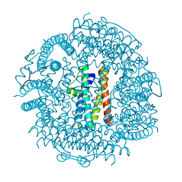
3WIR
 | | Crystal structure of kojibiose phosphorylase complexed with glucose | | 分子名称: | GLYCEROL, Kojibiose phosphorylase, PHOSPHATE ION, ... | | 著者 | Okada, S, Yamamoto, T, Watanabe, H, Nishimoto, T, Chaen, H, Fukuda, S, Wakagi, T, Fushinobu, S. | | 登録日 | 2013-09-24 | | 公開日 | 2014-02-05 | | 最終更新日 | 2024-11-20 | | 実験手法 | X-RAY DIFFRACTION (2.05 Å) | | 主引用文献 | Structural and mutational analysis of substrate recognition in kojibiose phosphorylase
Febs J., 281, 2014
|
|
7GS8
 | | PanDDA Analysis group deposition -- Crystal structure of PTP1B in complex with FMOPL000466a | | 分子名称: | 2-AMINO-2-HYDROXYMETHYL-PROPANE-1,3-DIOL, Tyrosine-protein phosphatase non-receptor type 1, ~{N},~{N},5,6-tetramethylthieno[2,3-d]pyrimidin-4-amine | | 著者 | Mehlman, T, Ginn, H.M, Keedy, D.A. | | 登録日 | 2024-01-03 | | 公開日 | 2024-01-24 | | 最終更新日 | 2024-04-24 | | 実験手法 | X-RAY DIFFRACTION (1.67 Å) | | 主引用文献 | An expanded view of ligandability in the allosteric enzyme PTP1B from computational reanalysis of large-scale crystallographic data.
Biorxiv, 2024
|
|
7GSI
 | |
4EB5
 | | A. fulgidus IscS-IscU complex structure | | 分子名称: | 4-(2-HYDROXYETHYL)-1-PIPERAZINE ETHANESULFONIC ACID, FE2/S2 (INORGANIC) CLUSTER, GLYCEROL, ... | | 著者 | Marinoni, E.N, de Oliveira, J.S, Nicolet, Y, Raulfs, E.C, Amara, P, Dean, D.R, Fontecilla-Camps, J.C. | | 登録日 | 2012-03-23 | | 公開日 | 2012-05-02 | | 最終更新日 | 2024-02-28 | | 実験手法 | X-RAY DIFFRACTION (2.53 Å) | | 主引用文献 | (IscS-IscU)2 complex structures provide insights into Fe2S2 biogenesis and transfer.
Angew.Chem.Int.Ed.Engl., 51, 2012
|
|
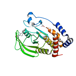
3T9J
 | | Crystal structure HP-NAP from strain YS39 in apo form | | 分子名称: | 1,2-ETHANEDIOL, Neutrophil-activating protein | | 著者 | Tsuruta, O, Yokoyama, H, Fujii, S. | | 登録日 | 2011-08-03 | | 公開日 | 2012-02-08 | | 最終更新日 | 2023-11-01 | | 実験手法 | X-RAY DIFFRACTION (2.2 Å) | | 主引用文献 | A new crystal lattice structure of Helicobacter pylori neutrophil-activating protein (HP-NAP)
Acta Crystallogr.,Sect.F, 68, 2012
|
|
7GTW
 | |
7GU0
 | |
7GT7
 | |
3GJU
 | |
7GTY
 | |
7GTP
 | |
5RWF
 | | INPP5D PanDDA analysis group deposition -- Crystal Structure of the phosphatase and C2 domains of SHIP1 in complex with Z1983897532 | | 分子名称: | 5-fluoro-N-[(2-methyl-1,3-thiazol-5-yl)methyl]pyridin-3-amine, DIMETHYL SULFOXIDE, Phosphatidylinositol 3,4,5-trisphosphate 5-phosphatase 1 | | 著者 | Bradshaw, W.J, Newman, J.A, von Delft, F, Arrowsmith, C.H, Edwards, A.M, Bountra, C, Gileadi, O. | | 登録日 | 2020-10-30 | | 公開日 | 2020-11-11 | | 最終更新日 | 2024-02-14 | | 実験手法 | X-RAY DIFFRACTION (1.35 Å) | | 主引用文献 | Regulation of inositol 5-phosphatase activity by the C2 domain of SHIP1 and SHIP2.
Structure, 2024
|
|


