3DM6
 
 | | Beta-secretase 1 complexed with statine-based inhibitor | | 分子名称: | 5-[[(2S)-2-[[(3R,4S)-5-(3,5-difluorophenoxy)-3-hydroxy-4-[[3-(methyl-methylsulfonyl-amino)-5-[[(1R)-1-phenylethyl]carbamoyl]phenyl]carbonylamino]pentanoyl]amino]-3-methyl-butanoyl]amino]benzene-1,3-dicarboxylic acid, Beta-secretase 1, ISOPROPYL ALCOHOL | | 著者 | Lindberg, J, Borkakoti, N, Nystrom, S. | | 登録日 | 2008-06-30 | | 公開日 | 2008-12-16 | | 最終更新日 | 2024-11-13 | | 実験手法 | X-RAY DIFFRACTION (2.6 Å) | | 主引用文献 | Design, synthesis and SAR of potent statine-based BACE-1 inhibitors: exploration of P1 phenoxy and benzyloxy residues
Bioorg.Med.Chem., 16, 2008
|
|
7JJC
 | |
3II3
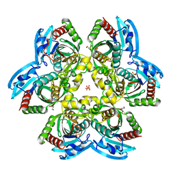
 | | Structure of ORF157 from Acidianus filamentous Virus 1 | | 分子名称: | GLYCEROL, NICKEL (II) ION, Putative uncharacterized protein | | 著者 | Goulet, A, Redder, P, Pina, M, Prangishvili, D, van Tilbeurgh, H, Cambillau, C, Campanacci, V. | | 登録日 | 2009-07-31 | | 公開日 | 2010-03-23 | | 最終更新日 | 2023-11-01 | | 実験手法 | X-RAY DIFFRACTION (2.7 Å) | | 主引用文献 | ORF157 from the archaeal virus Acidianus filamentous virus 1 defines a new class of nuclease
J.Virol., 84, 2010
|
|
3I9G
 | |
4GN8
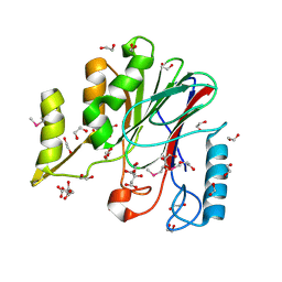
 | | mouse SMP30/GNL-1,5-AG complex | | 分子名称: | 1,5-anhydro-D-glucitol, CALCIUM ION, Regucalcin, ... | | 著者 | Aizawa, S, Senda, M, Harada, A, Maruyama, N, Ishida, T, Aigaki, T, Ishigami, A, Senda, T. | | 登録日 | 2012-08-17 | | 公開日 | 2013-04-10 | | 最終更新日 | 2023-11-08 | | 実験手法 | X-RAY DIFFRACTION (1.7 Å) | | 主引用文献 | Structural basis of the gamma-lactone-ring formation in ascorbic acid biosynthesis by the senescence marker protein-30/gluconolactonase
Plos One, 8, 2013
|
|
3GUX
 | |
3S25
 | |
1MG1
 | | HTLV-1 GP21 ECTODOMAIN/MALTOSE-BINDING PROTEIN CHIMERA | | 分子名称: | CHLORIDE ION, PROTEIN (HTLV-1 GP21 ECTODOMAIN/MALTOSE-BINDING PROTEIN CHIMERA), alpha-D-glucopyranose-(1-4)-alpha-D-glucopyranose | | 著者 | Kobe, B, Center, R.J, Kemp, B.E, Poumbourios, P. | | 登録日 | 1999-03-01 | | 公開日 | 1999-04-06 | | 最終更新日 | 2024-11-20 | | 実験手法 | X-RAY DIFFRACTION (2.5 Å) | | 主引用文献 | Crystal structure of human T cell leukemia virus type 1 gp21 ectodomain crystallized as a maltose-binding protein chimera reveals structural evolution of retroviral transmembrane proteins.
Proc.Natl.Acad.Sci.USA, 96, 1999
|
|
1Y0M
 | |
3EGH
 | | Crystal structure of a complex between Protein Phosphatase 1 alpha (PP1), the PP1 binding and PDZ domains of Spinophilin and the small natural molecular toxin Nodularin-R | | 分子名称: | GLYCEROL, MANGANESE (II) ION, Serine/threonine-protein phosphatase PP1-alpha catalytic subunit, ... | | 著者 | Ragusa, M.J, Page, R, Peti, W. | | 登録日 | 2008-09-10 | | 公開日 | 2010-03-23 | | 最終更新日 | 2023-11-15 | | 実験手法 | X-RAY DIFFRACTION (2 Å) | | 主引用文献 | Spinophilin directs protein phosphatase 1 specificity by blocking substrate binding sites.
Nat.Struct.Mol.Biol., 17, 2010
|
|
7FE2
 | | Crystal structure of the mutant E494Q of GH92 alpha-1,2-mannosidase from Enterococcus faecalis ATCC 10100 in complex with alpha-1,2-mannobiose | | 分子名称: | 1,2-ETHANEDIOL, ACETATE ION, Alpha-1,2-mannosidase, ... | | 著者 | Miyazaki, T, Alonso-Gil, S. | | 登録日 | 2021-07-19 | | 公開日 | 2022-02-02 | | 最終更新日 | 2023-11-29 | | 実験手法 | X-RAY DIFFRACTION (1.75 Å) | | 主引用文献 | Unlocking the Hydrolytic Mechanism of GH92 alpha-1,2-Mannosidases: Computation Inspires the use of C-Glycosides as Michaelis Complex Mimics.
Chemistry, 28, 2022
|
|
3E2M
 | | LFA-1 I domain bound to inhibitors | | 分子名称: | Integrin alpha-L, cis-4-{[2-({4-[(1E)-3-morpholin-4-yl-3-oxoprop-1-en-1-yl]-2,3-bis(trifluoromethyl)phenyl}sulfanyl)phenoxy]methyl}cyclohexanecarboxylic acid | | 著者 | Silvian, L.F. | | 登録日 | 2008-08-05 | | 公開日 | 2008-08-19 | | 最終更新日 | 2023-08-30 | | 実験手法 | X-RAY DIFFRACTION (2 Å) | | 主引用文献 | Structure-activity relationship of ortho- and meta-phenol based LFA-1 ICAM inhibitors
Bioorg.Med.Chem.Lett., 18, 2008
|
|
1RUV
 | | RIBONUCLEASE A-URIDINE VANADATE COMPLEX: HIGH RESOLUTION RESOLUTION X-RAY STRUCTURE (1.3 A) | | 分子名称: | RIBONUCLEASE A, TERTIARY-BUTYL ALCOHOL, URIDINE-2',3'-VANADATE | | 著者 | Ladner, J.E, Wladkowski, B, Svensson, L.A, Sjolin, L, Gilliland, G.L. | | 登録日 | 1995-07-27 | | 公開日 | 1997-04-01 | | 最終更新日 | 2024-11-06 | | 実験手法 | X-RAY DIFFRACTION (1.25 Å) | | 主引用文献 | X-ray structure of a ribonuclease A-uridine vanadate complex at 1.3 A resolution.
Acta Crystallogr.,Sect.D, 53, 1997
|
|
5OX4
 | | Glycogen Phosphorylase in complex with CK900 | | 分子名称: | (2~{S},3~{R},4~{R},5~{S},6~{R})-2-[5-(4-aminophenyl)-4~{H}-1,2,4-triazol-3-yl]-6-(hydroxymethyl)oxane-3,4,5-triol, Glycogen phosphorylase, muscle form, ... | | 著者 | Kyriakis, E, Stravodimos, G.A, Kantsadi, A.L, Chatzileontiadou, D.S.M, Leonidas, D.D. | | 登録日 | 2017-09-05 | | 公開日 | 2018-02-28 | | 最終更新日 | 2025-04-09 | | 実験手法 | X-RAY DIFFRACTION (1.8 Å) | | 主引用文献 | Probing the beta-pocket of the active site of human liver glycogen phosphorylase with 3-(C-beta-d-glucopyranosyl)-5-(4-substituted-phenyl)-1, 2, 4-triazole inhibitors.
Bioorg. Chem., 77, 2018
|
|
7F0M
 | | Crystal Structure of human Pin1 complexed with a potent covalent inhibitor | | 分子名称: | 3,6,9,12,15,18-HEXAOXAICOSANE-1,20-DIOL, 8-(2-chloranylethanoyl)-4-[(5-naphthalen-1-ylfuran-2-yl)methyl]-1-thia-4,8-diazaspiro[4.5]decan-3-one, Peptidyl-prolyl cis-trans isomerase NIMA-interacting 1 | | 著者 | Liu, L, Li, J. | | 登録日 | 2021-06-05 | | 公開日 | 2022-02-16 | | 最終更新日 | 2024-10-23 | | 実験手法 | X-RAY DIFFRACTION (1.9 Å) | | 主引用文献 | Computational and Structure-Based Development of High Potent Cell-Active Covalent Inhibitor Targeting the Peptidyl-Prolyl Isomerase NIMA-Interacting-1 (Pin1).
J.Med.Chem., 65, 2022
|
|
5OMO
 | | CRYSTAL STRUCTURE OF RAT PEROXISOMAL MULTIFUNCTIONAL ENZYME TYPE-1 (RPMFE1) COMPLEXED WITH WITH 3S-HYDROXY-DECANOYL-COA AND 3-KETO-DECANOYL-COA | | 分子名称: | (S)-3-HYDROXYDECANOYL-COA, 3-KETO-DECANOYL-COA, GLYCEROL, ... | | 著者 | Kasaragod, P, Kiema, T.-R, Schmitz, W, Hiltunen, J.K, Wierenga, R.K. | | 登録日 | 2017-08-01 | | 公開日 | 2017-09-06 | | 最終更新日 | 2025-03-19 | | 実験手法 | X-RAY DIFFRACTION (2.49 Å) | | 主引用文献 | Crystallographic binding studies of rat peroxisomal multifunctional enzyme type 1 with 3-ketodecanoyl-CoA: capturing active and inactive states of its hydratase and dehydrogenase catalytic sites.
Acta Crystallogr D Struct Biol, 76, 2020
|
|
4I06
 | | Crystal structure of human Arginase-2 complexed with inhibitor 14 | | 分子名称: | Arginase-2, mitochondrial, BENZAMIDINE, ... | | 著者 | Cousido-Siah, A, Mitschler, A, Ruiz, F.X, Whitehouse, D.L, Golebiowski, A, Ji, M, Zhang, M, Beckett, P, Sheeler, R, Andreoli, M, Conway, B, Mahboubi, K, Schroeter, H, Van Zandt, M.C, Podjarny, A. | | 登録日 | 2012-11-16 | | 公開日 | 2013-03-20 | | 最終更新日 | 2023-09-20 | | 実験手法 | X-RAY DIFFRACTION (1.8 Å) | | 主引用文献 | Discovery of (R)-2-Amino-6-borono-2-(2-(piperidin-1-yl)ethyl)hexanoic Acid and Congeners As Highly Potent Inhibitors of Human Arginases I and II for Treatment of Myocardial Reperfusion Injury.
J.Med.Chem., 56, 2013
|
|
1M0N
 | | Structure of Dialkylglycine Decarboxylase Complexed with 1-Aminocyclopentanephosphonate | | 分子名称: | 1-[((1E)-{3-HYDROXY-2-METHYL-5-[(PHOSPHONOOXY)METHYL]PYRIDIN-4-YL}METHYLENE)AMINO]CYCLOPENTYLPHOSPHONIC ACID, 2,2-Dialkylglycine decarboxylase, POTASSIUM ION, ... | | 著者 | Liu, W, Rogers, C.J, Fisher, A.J, Toney, M.D. | | 登録日 | 2002-06-13 | | 公開日 | 2002-10-23 | | 最終更新日 | 2024-02-14 | | 実験手法 | X-RAY DIFFRACTION (2.2 Å) | | 主引用文献 | Aminophosphonate Inhibitors of Dialkylglycine Decarboxylase: Structural Basis for Slow Binding Inhibition
Biochemistry, 41, 2002
|
|
4R2W
 | | X-ray structure of uridine phosphorylase from Shewanella oneidensis MR-1 in complex with uridine at 1.6 A resolution | | 分子名称: | GLYCEROL, SULFATE ION, URIDINE, ... | | 著者 | Safonova, T.N, Mordkovich, N.N, Manuvera, V.A, Veiko, V.P, Popov, V.O, Polyakov, K.P. | | 登録日 | 2014-08-13 | | 公開日 | 2014-12-10 | | 最終更新日 | 2023-09-20 | | 実験手法 | X-RAY DIFFRACTION (1.6 Å) | | 主引用文献 | High-syn conformation of uridine and asymmetry of the hexameric molecule revealed in the high-resolution structures of Shewanella oneidensis MR-1 uridine phosphorylase in the free form and in complex with uridine.
Acta Crystallogr.,Sect.D, 70, 2014
|
|
1TIV
 | |
1Z9T
 | |
3UL5
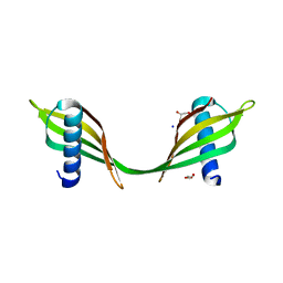 | | Saccharum officinarum canecystatin-1 in space group C2221 | | 分子名称: | Canecystatin-1, GLYCEROL, SODIUM ION | | 著者 | Valadares, N.F, Pereira, H.M, Oliveira-Silva, R, Garratt, R.C. | | 登録日 | 2011-11-10 | | 公開日 | 2012-11-28 | | 最終更新日 | 2023-09-13 | | 実験手法 | X-RAY DIFFRACTION (2.3 Å) | | 主引用文献 | X-ray crystallography and NMR studies of domain-swapped canecystatin-1.
Febs J., 280, 2013
|
|
3ILZ
 | | Structure of TR-alfa bound to selective thyromimetic GC-1 in P212121 space group | | 分子名称: | Thyroid hormone receptor, alpha isoform 1 variant, {4-[4-hydroxy-3-(1-methylethyl)benzyl]-3,5-dimethylphenoxy}acetic acid | | 著者 | Aparicio, R, Polikarpov, L, Bleicher, L. | | 登録日 | 2009-08-07 | | 公開日 | 2010-04-28 | | 最終更新日 | 2024-10-16 | | 実験手法 | X-RAY DIFFRACTION (1.85 Å) | | 主引用文献 | Structural basis of GC-1 selectivity for thyroid hormone receptor isoforms.
Bmc Struct.Biol., 8, 2008
|
|
4EYR
 | | Crystal structure of multidrug-resistant clinical isolate 769 HIV-1 protease in complex with ritonavir | | 分子名称: | HIV-1 PROTEASE, RITONAVIR | | 著者 | Liu, Z, Yedidi, R.S, Wang, Y, Brunzelle, J.S, Kovari, I.A, Kovari, L.C. | | 登録日 | 2012-05-01 | | 公開日 | 2013-01-30 | | 最終更新日 | 2024-02-28 | | 実験手法 | X-RAY DIFFRACTION (1.8 Å) | | 主引用文献 | Insights into the mechanism of drug resistance: X-ray structure analysis of multi-drug resistant HIV-1 protease ritonavir complex.
Biochem.Biophys.Res.Commun., 431, 2013
|
|
4A9L
 | | N-TERMINAL BROMODOMAIN OF HUMAN BRD4 WITH 1,3-DIMETHYL-6-(MORPHOLINE- 4-SULFONYL)-1,2,3,4-TETRAHYDROQUINAZOLIN-2-ONE | | 分子名称: | 1,2-ETHANEDIOL, 1,3-dimethyl-6-(morpholin-4-ylsulfonyl)-3,4-dihydroquinazolin-2(1H)-one, BROMODOMAIN-CONTAINING PROTEIN 4, ... | | 著者 | Chung, C.W, Bamborough, P. | | 登録日 | 2011-11-26 | | 公開日 | 2012-01-11 | | 最終更新日 | 2024-05-08 | | 実験手法 | X-RAY DIFFRACTION (1.6 Å) | | 主引用文献 | Fragment-Based Discovery of Bromodomain Inhibitors Part 1: Inhibitor Binding Modes and Implications for Lead Discovery.
J.Med.Chem., 55, 2012
|
|


