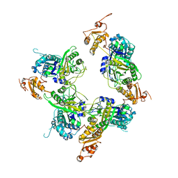
3KVT
 
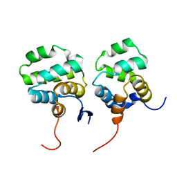
 | | TETRAMERIZATION DOMAIN FROM AKV3.1 (SHAW-SUBFAMILY) VOLTAGE-GATED POTASSIUM CHANNEL | | 分子名称: | POTASSIUM CHANNEL PROTEIN SHAW, ZINC ION | | 著者 | Bixby, K.A, Nanao, M.H, Shen, N.V, Kreusch, A, Bellamy, H, Pfaffinger, P.J, Choe, S. | | 登録日 | 1998-09-25 | | 公開日 | 1999-01-13 | | 最終更新日 | 2024-04-03 | | 実験手法 | X-RAY DIFFRACTION (2 Å) | | 主引用文献 | Zn2+-binding and molecular determinants of tetramerization in voltage-gated K+ channels.
Nat.Struct.Biol., 6, 1999
|
|
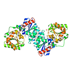
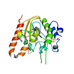
1J5D
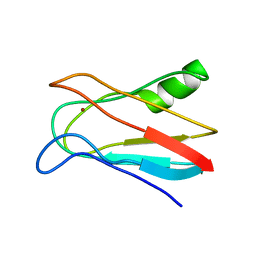
 | | SOLUTION STRUCTURE OF OXIDIZED PARAMAGNETIC CU(II) PLASTOCYANIN FROM SYNECHOCYSTIS PCC6803-MINIMIZED AVERAGE STRUCTURE | | 分子名称: | COPPER (II) ION, PLASTOCYANIN | | 著者 | Bertini, I, Ciurli, S, Dikiy, A, Fernandez, C.O, Luchinat, C, Safarov, N, Shumilin, S, Vila, A.J. | | 登録日 | 2002-04-02 | | 公開日 | 2002-04-10 | | 最終更新日 | 2023-12-27 | | 実験手法 | SOLUTION NMR | | 主引用文献 | The first solution structure of a paramagnetic copper(II) protein: the case of oxidized plastocyanin from the cyanobacterium Synechocystis PCC6803.
J.Am.Chem.Soc., 123, 2001
|
|
8G4E
 | |
5DMJ
 | |
5KXI
 | | X-ray structure of the human Alpha4Beta2 nicotinic receptor | | 分子名称: | (S)-3-(1-METHYLPYRROLIDIN-2-YL)PYRIDINE, 2-acetamido-2-deoxy-beta-D-glucopyranose, Neuronal acetylcholine receptor subunit alpha-4, ... | | 著者 | Morales-Perez, C.L, Noviello, C.M, Hibbs, R.E. | | 登録日 | 2016-07-20 | | 公開日 | 2016-09-28 | | 最終更新日 | 2024-04-03 | | 実験手法 | X-RAY DIFFRACTION (3.941 Å) | | 主引用文献 | X-ray structure of the human alpha 4 beta 2 nicotinic receptor.
Nature, 538, 2016
|
|
8GYE
 | |
8YKY
 | | Structure of human class T GPCR TAS2R14-Ggustducin complex with agonist 28.1 | | 分子名称: | 4-methyl-N-[(2M)-2-(1H-tetrazol-5-yl)phenyl]-6-(trifluoromethyl)pyrimidin-2-amine, CHOLESTEROL, G alpha gustducin protein, ... | | 著者 | Hu, X.L, Wu, L.J, Hua, T, Liu, Z.J. | | 登録日 | 2024-03-05 | | 公開日 | 2024-07-10 | | 実験手法 | ELECTRON MICROSCOPY (2.99 Å) | | 主引用文献 | Bitter taste TAS2R14 activation by intracellular tastants and cholesterol.
Nature, 2024
|
|
7YK4
 | | ox40-antibody | | 分子名称: | 2-acetamido-2-deoxy-beta-D-glucopyranose, Tumor necrosis factor receptor superfamily member 4, antibody-H, ... | | 著者 | Zhou, A. | | 登録日 | 2022-07-21 | | 公開日 | 2023-08-09 | | 実験手法 | X-RAY DIFFRACTION (2.7 Å) | | 主引用文献 | Structural Basis of a Novel Agonistic Anti-OX40 Antibody.
Biomolecules, 12, 2022
|
|
5DMI
 | |
5KVK
 | | Crystal structure of the Competence-Damaged Protein (CinA) Superfamily Protein KP700603 from Klebsiella pneumoniae 700603 | | 分子名称: | Protein KP700603 | | 著者 | Stogios, P.J, Wawrzak, Z, Evdokimova, E, Di Leo, R, Grimshaw, S, Savchenko, A, Anderson, W.F, Center for Structural Genomics of Infectious Diseases (CSGID) | | 登録日 | 2016-07-14 | | 公開日 | 2016-08-03 | | 最終更新日 | 2023-10-04 | | 実験手法 | X-RAY DIFFRACTION (1.66 Å) | | 主引用文献 | To be published
To Be Published
|
|
6R8F
 | | Cryo-EM structure of the Human BRISC-SHMT2 complex | | 分子名称: | BRISC and BRCA1-A complex member 2,BRCC45 (BRE, BRISC and BRCA1-A complex member 2), BRISC complex subunit Abraxas 2, ... | | 著者 | Walden, M, Hesketh, E, Tian, L, Ranson, N.A, Greenberg, R.A, Zeqiraj, E. | | 登録日 | 2019-04-01 | | 公開日 | 2019-06-05 | | 最終更新日 | 2024-05-22 | | 実験手法 | ELECTRON MICROSCOPY (3.8 Å) | | 主引用文献 | Metabolic control of BRISC-SHMT2 assembly regulates immune signalling.
Nature, 570, 2019
|
|
1AF6
 | | MALTOPORIN SUCROSE COMPLEX | | 分子名称: | MAGNESIUM ION, MALTOPORIN, beta-D-fructofuranose-(2-1)-alpha-D-glucopyranose | | 著者 | Dutzler, R, Schirmer, T. | | 登録日 | 1997-03-21 | | 公開日 | 1998-03-25 | | 最終更新日 | 2023-08-02 | | 実験手法 | X-RAY DIFFRACTION (2.4 Å) | | 主引用文献 | Channel specificity: structural basis for sugar discrimination and differential flux rates in maltoporin.
J.Mol.Biol., 272, 1997
|
|
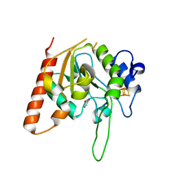
1J5C
 | | SOLUTION STRUCTURE OF OXIDIZED PARAMAGNETIC CU(II) PLASTOCYANIN FROM SYNECHOCYSTIS PCC6803 | | 分子名称: | COPPER (II) ION, PLASTOCYANIN | | 著者 | Bertini, I, Ciurli, S, Dikiy, A, Fernandez, C.O, Luchinat, C, Safarov, N, Shumilin, S, Vila, A.J. | | 登録日 | 2002-04-02 | | 公開日 | 2002-04-10 | | 最終更新日 | 2023-12-27 | | 実験手法 | SOLUTION NMR | | 主引用文献 | The first solution structure of a paramagnetic copper(II) protein: the case of oxidized plastocyanin from the cyanobacterium Synechocystis PCC6803.
J.Am.Chem.Soc., 123, 2001
|
|
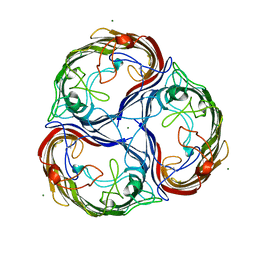
6AEZ
 | | Crystal structure of human CCL5 trimer | | 分子名称: | C-C motif chemokine 5, SULFATE ION | | 著者 | Chen, Y.C, Li, K.M, Chen, P.J, Zarivach, R, Sun, Y.J, Sue, S.C. | | 登録日 | 2018-08-07 | | 公開日 | 2019-08-07 | | 最終更新日 | 2023-11-22 | | 実験手法 | X-RAY DIFFRACTION (1.63 Å) | | 主引用文献 | Integrative Model to Coordinate the Oligomerization and Aggregation Mechanisms of CCL5.
J.Mol.Biol., 432, 2020
|
|
6RZU
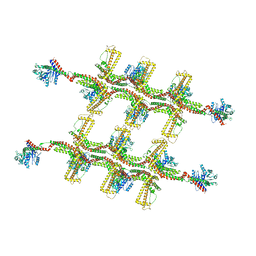 | | Structure of s-Mgm1 decorating the outer surface of tubulated lipid membranes in the GTPgammaS bound state | | 分子名称: | Putative mitochondrial dynamin protein | | 著者 | Faelber, K, Dietrich, L, Noel, J.K, Sanchez, R, Kudryashev, M, Kuelbrandt, W, Daumke, O. | | 登録日 | 2019-06-13 | | 公開日 | 2019-07-24 | | 最終更新日 | 2020-11-18 | | 実験手法 | ELECTRON MICROSCOPY (14.7 Å) | | 主引用文献 | Structure and assembly of the mitochondrial membrane remodelling GTPase Mgm1.
Nature, 571, 2019
|
|
6RZV
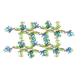 | | Structure of s-Mgm1 decorating the inner surface of tubulated lipid membranes | | 分子名称: | Putative mitochondrial dynamin protein | | 著者 | Faelber, K, Dietrich, L, Noel, J.K, Sanchez, R, Kudryashev, M, Kuelbrandt, W, Daumke, O. | | 登録日 | 2019-06-13 | | 公開日 | 2019-07-24 | | 最終更新日 | 2020-11-18 | | 実験手法 | ELECTRON MICROSCOPY (20.6 Å) | | 主引用文献 | Structure and assembly of the mitochondrial membrane remodelling GTPase Mgm1.
Nature, 571, 2019
|
|
5ERB
 | |
5ELP
 | |
5ENZ
 | | S. aureus MnaA-UDP co-structure | | 分子名称: | 2-AMINO-2-HYDROXYMETHYL-PROPANE-1,3-DIOL, SULFATE ION, TETRAETHYLENE GLYCOL, ... | | 著者 | Fischmann, T.O. | | 登録日 | 2015-11-09 | | 公開日 | 2016-04-27 | | 最終更新日 | 2023-09-27 | | 実験手法 | X-RAY DIFFRACTION (1.91 Å) | | 主引用文献 | Chemical Genetic Analysis and Functional Characterization of Staphylococcal Wall Teichoic Acid 2-Epimerases Reveals Unconventional Antibiotic Drug Targets.
Plos Pathog., 12, 2016
|
|
1U5S
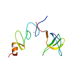 | | NMR structure of the complex between Nck-2 SH3 domain and PINCH-1 LIM4 domain | | 分子名称: | Cytoplasmic protein NCK2, PINCH protein, ZINC ION | | 著者 | Vaynberg, J, Fukuda, T, Vinogradova, O, Velyvis, A, Ng, L, Wu, C, Qin, J. | | 登録日 | 2004-07-28 | | 公開日 | 2005-04-05 | | 最終更新日 | 2024-05-22 | | 実験手法 | SOLUTION NMR | | 主引用文献 | Structure of an ultraweak protein-protein complex and its crucial role in regulation of cell morphology and motility.
Mol.Cell, 17, 2005
|
|
7YXU
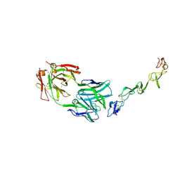 | | Crystal structure of agonistic antibody 1618 fab domain bound to human 4-1BB. | | 分子名称: | MANGANESE (II) ION, Tumor necrosis factor receptor superfamily member 9, heavy chain of Fab, ... | | 著者 | Hakansson, M, Rose, N, Petersson, J, Enell Smith, K, Thorolfsson, M, von Schantz, L. | | 登録日 | 2022-02-16 | | 公開日 | 2023-01-25 | | 最終更新日 | 2024-02-07 | | 実験手法 | X-RAY DIFFRACTION (2.31 Å) | | 主引用文献 | The Bispecific Tumor Antigen-Conditional 4-1BB x 5T4 Agonist, ALG.APV-527, Mediates Strong T-Cell Activation and Potent Antitumor Activity in Preclinical Studies.
Mol.Cancer Ther., 22, 2023
|
|
7CSQ
 | | Solution structure of the complex between p75NTR-DD and TRADD-DD | | 分子名称: | Tumor necrosis factor receptor superfamily member 16, Tumor necrosis factor receptor type 1-associated DEATH domain protein | | 著者 | Lin, Z, Zhang, N. | | 登録日 | 2020-08-16 | | 公開日 | 2021-08-25 | | 最終更新日 | 2024-05-15 | | 実験手法 | SOLUTION NMR | | 主引用文献 | Structural basis of NF-kappa B signaling by the p75 neurotrophin receptor interaction with adaptor protein TRADD through their respective death domains.
J.Biol.Chem., 297, 2021
|
|
6AJO
 | | Complex form of Uracil DNA glycosylase X and uracil-DNA. | | 分子名称: | DNA (5'-D(P*(ORP)P*TP*T)-3'), IRON/SULFUR CLUSTER, PHOSPHATE ION, ... | | 著者 | Ahn, W.C, Aroli, S, Varshney, U, Woo, E.J. | | 登録日 | 2018-08-28 | | 公開日 | 2019-05-29 | | 最終更新日 | 2024-03-27 | | 実験手法 | X-RAY DIFFRACTION (2.269 Å) | | 主引用文献 | Covalent binding of uracil DNA glycosylase UdgX to abasic DNA upon uracil excision.
Nat.Chem.Biol., 15, 2019
|
|
6AJR
 | | Complex form of Uracil DNA glycosylase X and uracil | | 分子名称: | IRON/SULFUR CLUSTER, URACIL, Uracil DNA glycosylase superfamily protein | | 著者 | Ahn, W.C, Aroli, S, Varshney, U, Woo, E.J. | | 登録日 | 2018-08-28 | | 公開日 | 2019-05-29 | | 最終更新日 | 2024-03-27 | | 実験手法 | X-RAY DIFFRACTION (1.341 Å) | | 主引用文献 | Covalent binding of uracil DNA glycosylase UdgX to abasic DNA upon uracil excision.
Nat.Chem.Biol., 15, 2019
|
|
3RHN
 | | HISTIDINE TRIAD NUCLEOTIDE-BINDING PROTEIN (HINT) FROM RABBIT COMPLEXED WITH GMP | | 分子名称: | GUANOSINE-5'-MONOPHOSPHATE, HISTIDINE TRIAD NUCLEOTIDE-BINDING PROTEIN | | 著者 | Brenner, C, Garrison, P, Gilmour, J, Peisach, D, Ringe, D, Petsko, G.A, Lowenstein, J.M. | | 登録日 | 1997-02-11 | | 公開日 | 1997-06-16 | | 最終更新日 | 2024-02-21 | | 実験手法 | X-RAY DIFFRACTION (2.1 Å) | | 主引用文献 | Crystal structures of HINT demonstrate that histidine triad proteins are GalT-related nucleotide-binding proteins.
Nat.Struct.Biol., 4, 1997
|
|


