4IWQ
 
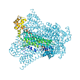
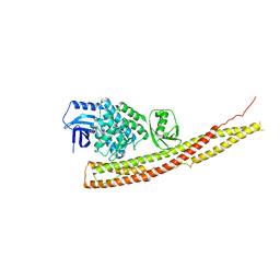 | | Crystal structure and mechanism of activation of TBK1 | | 分子名称: | N-{3-[(5-cyclopropyl-2-{[3-(morpholin-4-ylmethyl)phenyl]amino}pyrimidin-4-yl)amino]propyl}cyclobutanecarboxamide, Serine/threonine-protein kinase TBK1 | | 著者 | Panne, D, Larabi, A. | | 登録日 | 2013-01-24 | | 公開日 | 2013-03-13 | | 最終更新日 | 2024-02-28 | | 実験手法 | X-RAY DIFFRACTION (3 Å) | | 主引用文献 | Crystal structure and mechanism of activation of TANK-binding kinase 1.
Cell Rep, 3, 2013
|
|
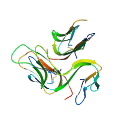
4A2W
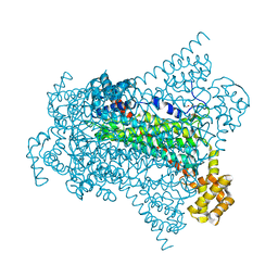
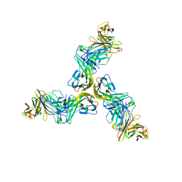
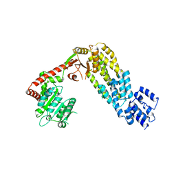 | | Structure of full-length duck RIG-I | | 分子名称: | RETINOIC ACID INDUCIBLE PROTEIN I | | 著者 | Kowalinski, E, Lunardi, T, McCarthy, A.A, Cusack, S. | | 登録日 | 2011-09-29 | | 公開日 | 2011-10-19 | | 最終更新日 | 2024-05-01 | | 実験手法 | X-RAY DIFFRACTION (3.7 Å) | | 主引用文献 | Structural Basis for the Activation of Innate Immune Pattern Recognition Receptor Rig-I by Viral RNA.
Cell(Cambridge,Mass.), 147, 2011
|
|
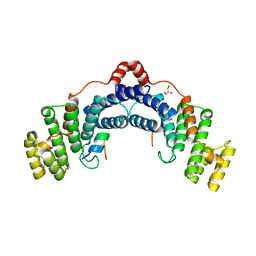
4JLC
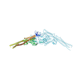 | | Crystal structure of mouse TBK1 bound to SU6668 | | 分子名称: | 3-{2,4-dimethyl-5-[(Z)-(2-oxo-1,2-dihydro-3H-indol-3-ylidene)methyl]-1H-pyrrol-3-yl}propanoic acid, Serine/threonine-protein kinase TBK1 | | 著者 | Li, P. | | 登録日 | 2013-03-12 | | 公開日 | 2013-06-19 | | 最終更新日 | 2023-09-20 | | 実験手法 | X-RAY DIFFRACTION (3 Å) | | 主引用文献 | Structural Insights into the Functions of TBK1 in Innate Antimicrobial Immunity.
Structure, 21, 2013
|
|
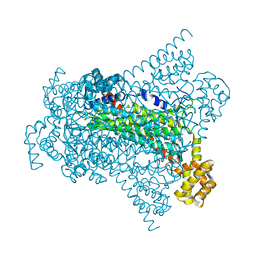
4JBM
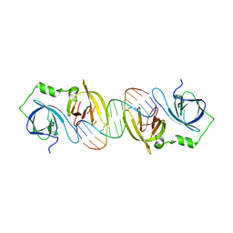
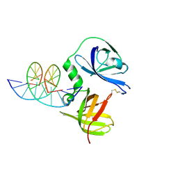 | | Structure of murine DNA binding protein bound with ds DNA | | 分子名称: | DNA (5'-D(*GP*GP*CP*GP*CP*GP*CP*GP*CP*GP*CP*C)-3'), Interferon-inducible protein AIM2 | | 著者 | Ru, H, Ni, X, Crowley, C, Zhao, L, Ding, W, Hung, L.-W, Shaw, N, Cheng, G, Liu, Z.-J. | | 登録日 | 2013-02-19 | | 公開日 | 2013-06-26 | | 最終更新日 | 2023-11-08 | | 実験手法 | X-RAY DIFFRACTION (2.218 Å) | | 主引用文献 | Structural basis for termination of AIM2-mediated signaling by p202
Cell Res., 23, 2013
|
|
3U3Y
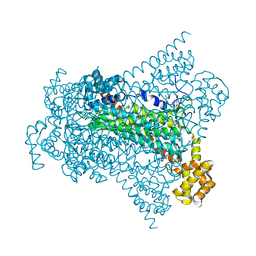
 | | Mouse TREX1 D200H mutant | | 分子名称: | 1,4-BUTANEDIOL, 5'-D(*GP*AP*CP*G)-3', CALCIUM ION, ... | | 著者 | Bailey, S.L, Harvey, S, Perrino, F.W, Hollis, T. | | 登録日 | 2011-10-06 | | 公開日 | 2011-12-28 | | 最終更新日 | 2024-02-28 | | 実験手法 | X-RAY DIFFRACTION (2.28 Å) | | 主引用文献 | Defects in DNA degradation revealed in crystal structures of TREX1 exonuclease mutations linked to autoimmune disease.
Dna Repair, 11, 2012
|
|
3U6F
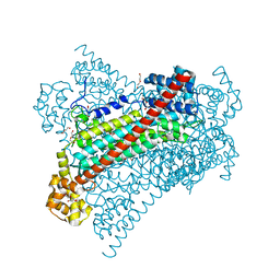
 | | Mouse TREX1 D200N mutant | | 分子名称: | 1,4-BUTANEDIOL, 5'-D(*GP*AP*CP*G)-3', MAGNESIUM ION, ... | | 著者 | Bailey, S.L, Harvey, S, Perrino, F.W, Hollis, T. | | 登録日 | 2011-10-12 | | 公開日 | 2011-12-28 | | 最終更新日 | 2024-02-28 | | 実験手法 | X-RAY DIFFRACTION (2.3 Å) | | 主引用文献 | Defects in DNA degradation revealed in crystal structures of TREX1 exonuclease mutations linked to autoimmune disease.
Dna Repair, 11, 2012
|
|
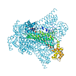
2YVC
 | |
1OJ4
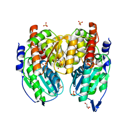
 | | Ternary complex of 4-diphosphocytidyl-2-C-methyl-D-erythritol kinase | | 分子名称: | 4-DIPHOSPHOCYTIDYL-2-C-METHYL-D-ERYTHRITOL, 4-DIPHOSPHOCYTIDYL-2-C-METHYL-D-ERYTHRITOL KINASE, CHLORIDE ION, ... | | 著者 | Miallau, L, Alphey, M.S, Hunter, W.N. | | 登録日 | 2003-06-30 | | 公開日 | 2003-07-31 | | 最終更新日 | 2019-10-16 | | 実験手法 | X-RAY DIFFRACTION (2.01 Å) | | 主引用文献 | Biosynthesis of Isoprenoids: Crystal Structure of 4-Diphosphocytidyl-2C-Methyl-D-Erythritol Kinase
Proc.Natl.Acad.Sci.USA, 100, 2003
|
|
1WUE
 | | Crystal structure of protein GI:29375081, unknown member of enolase superfamily from enterococcus faecalis V583 | | 分子名称: | mandelate racemase/muconate lactonizing enzyme family protein | | 著者 | Fedorov, A.A, Fedorov, E.V, Yew, W.S, Gerlt, J.A, Almo, S.C, Burley, S.K, New York SGX Research Center for Structural Genomics (NYSGXRC) | | 登録日 | 2004-12-05 | | 公開日 | 2004-12-21 | | 最終更新日 | 2024-03-13 | | 実験手法 | X-RAY DIFFRACTION (2.1 Å) | | 主引用文献 | Loss of quaternary structure is associated with rapid sequence divergence in the OSBS family
Proc.Natl.Acad.Sci.USA, 111, 2014
|
|
1WUF
 | | Crystal structure of protein GI:16801725, member of Enolase superfamily from Listeria innocua Clip11262 | | 分子名称: | MAGNESIUM ION, hypothetical protein lin2664 | | 著者 | Fedorov, A.A, Fedorov, E.V, Yew, W.S, Gerlt, J.A, Almo, S.C, Burley, S.K, New York SGX Research Center for Structural Genomics (NYSGXRC) | | 登録日 | 2004-12-07 | | 公開日 | 2004-12-21 | | 最終更新日 | 2024-03-13 | | 実験手法 | X-RAY DIFFRACTION (2.9 Å) | | 主引用文献 | Loss of quaternary structure is associated with rapid sequence divergence in the OSBS family
Proc.Natl.Acad.Sci.USA, 111, 2014
|
|
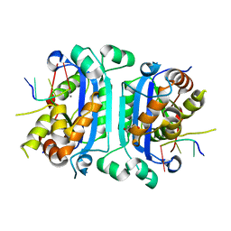
1X9I
 | | Crystal structure of Crystal structure of phosphoglucose/phosphomannose phosphoglucose/phosphomannoseisomerase from Pyrobaculum aerophilum in complex with glucose 6-phosphate | | 分子名称: | GLUCOSE-6-PHOSPHATE, GLYCEROL, glucose-6-phosphate isomerase | | 著者 | Swan, M.K, Hansen, T, Schoenheit, P, Davies, C. | | 登録日 | 2004-08-21 | | 公開日 | 2004-12-07 | | 最終更新日 | 2023-08-23 | | 実験手法 | X-RAY DIFFRACTION (1.16 Å) | | 主引用文献 | Structural basis for phosphomannose isomerase activity in phosphoglucose isomerase from Pyrobaculum aerophilum: a subtle difference between distantly related enzymes.
Biochemistry, 43, 2004
|
|
1X9J
 | |
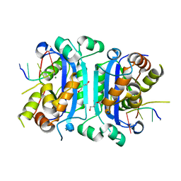
1X9H
 | | Crystal structure of phosphoglucose/phosphomannose isomerase from Pyrobaculum aerophilum in complex with fructose 6-phosphate | | 分子名称: | FRUCTOSE -6-PHOSPHATE, GLYCEROL, SULFATE ION, ... | | 著者 | Swan, M.K, Hansen, T, Schoenheit, P, Davies, C. | | 登録日 | 2004-08-21 | | 公開日 | 2004-12-07 | | 最終更新日 | 2023-08-23 | | 実験手法 | X-RAY DIFFRACTION (1.5 Å) | | 主引用文献 | Structural basis for phosphomannose isomerase activity in phosphoglucose isomerase from Pyrobaculum aerophilum: a subtle difference between distantly related enzymes.
Biochemistry, 43, 2004
|
|
3B94
 | |
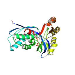
6G3E
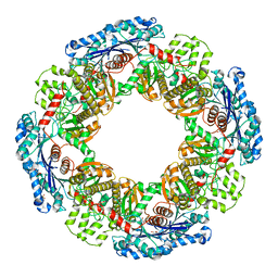
 | | Crystal structure of EDDS lyase in complex with formate | | 分子名称: | Argininosuccinate lyase, FORMIC ACID, SODIUM ION | | 著者 | Poddar, H, Thunnissem, A.M.W.H, Poelarends, G.J. | | 登録日 | 2018-03-25 | | 公開日 | 2018-05-16 | | 最終更新日 | 2024-01-17 | | 実験手法 | X-RAY DIFFRACTION (1.9 Å) | | 主引用文献 | Structural Basis for the Catalytic Mechanism of Ethylenediamine- N, N'-disuccinic Acid Lyase, a Carbon-Nitrogen Bond-Forming Enzyme with a Broad Substrate Scope.
Biochemistry, 57, 2018
|
|
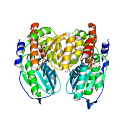
6G3G
 | | Crystal structure of EDDS lyase in complex with succinate | | 分子名称: | Argininosuccinate lyase, DI(HYDROXYETHYL)ETHER, SUCCINIC ACID | | 著者 | Poddar, H, Thunnissem, A.M.W.H, Poelarends, G.J. | | 登録日 | 2018-03-25 | | 公開日 | 2018-05-16 | | 最終更新日 | 2024-01-17 | | 実験手法 | X-RAY DIFFRACTION (2.606 Å) | | 主引用文献 | Structural Basis for the Catalytic Mechanism of Ethylenediamine- N, N'-disuccinic Acid Lyase, a Carbon-Nitrogen Bond-Forming Enzyme with a Broad Substrate Scope.
Biochemistry, 57, 2018
|
|
3B93
 | |
4RQI
 | | Structure of TRF2/RAP1 secondary interaction binding site | | 分子名称: | GLYCEROL, MAGNESIUM ION, Telomeric repeat-binding factor 2, ... | | 著者 | Miron, S, Guimaraes, B, Gaullier, G, Giraud-Panis, M.-J, Gilson, E, Le Du, M.-H. | | 登録日 | 2014-11-03 | | 公開日 | 2016-02-10 | | 最終更新日 | 2024-02-28 | | 実験手法 | X-RAY DIFFRACTION (2.4405 Å) | | 主引用文献 | A higher-order entity formed by the flexible assembly of RAP1 with TRF2.
Nucleic Acids Res., 44, 2016
|
|
6G3D
 | |
6FXN
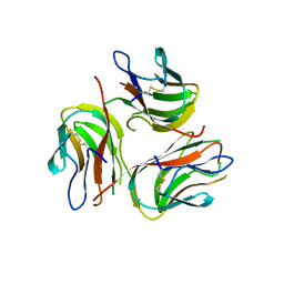
 | | Crystal structure of human BAFF in complex with Fab fragment of anti-BAFF antibody belimumab | | 分子名称: | Tumor necrosis factor ligand superfamily member 13B, belimumab heavy chain, belimumab light chain | | 著者 | Lammens, A, Maskos, K, Willen, L, Jiang, X, Schneider, P. | | 登録日 | 2018-03-09 | | 公開日 | 2018-04-04 | | 最終更新日 | 2024-01-17 | | 実験手法 | X-RAY DIFFRACTION (2.9 Å) | | 主引用文献 | A loop region of BAFF controls B cell survival and regulates recognition by different inhibitors.
Nat Commun, 9, 2018
|
|
1ZLP
 | | Petal death protein PSR132 with cysteine-linked glutaraldehyde forming a thiohemiacetal adduct | | 分子名称: | 5-HYDROXYPENTANAL, MAGNESIUM ION, petal death protein | | 著者 | Teplyakov, A, Liu, S, Lu, Z, Howard, A, Dunaway-Mariano, D, Herzberg, O. | | 登録日 | 2005-05-08 | | 公開日 | 2006-01-03 | | 最終更新日 | 2011-07-13 | | 実験手法 | X-RAY DIFFRACTION (2.7 Å) | | 主引用文献 | Crystal Structure of the Petal Death Protein from Carnation Flower.
Biochemistry, 44, 2005
|
|
6G3I
 | | Crystal structure of EDDS lyase in complex with N-(2-aminoethyl)aspartic acid (AEAA) | | 分子名称: | (2~{S})-2-(2-azanylethylamino)butanedioic acid, Argininosuccinate lyase, FUMARIC ACID | | 著者 | Poddar, H, Thunnissem, A.M.W.H, Poelarends, G.J. | | 登録日 | 2018-03-25 | | 公開日 | 2018-05-16 | | 最終更新日 | 2024-01-17 | | 実験手法 | X-RAY DIFFRACTION (2.41 Å) | | 主引用文献 | Structural Basis for the Catalytic Mechanism of Ethylenediamine- N, N'-disuccinic Acid Lyase, a Carbon-Nitrogen Bond-Forming Enzyme with a Broad Substrate Scope.
Biochemistry, 57, 2018
|
|
6G3F
 | | Crystal structure of EDDS lyase in complex with fumarate | | 分子名称: | Argininosuccinate lyase, DI(HYDROXYETHYL)ETHER, FUMARIC ACID | | 著者 | Poddar, H, Thunnissem, A.M.W.H, Poelarends, G.J. | | 登録日 | 2018-03-25 | | 公開日 | 2018-05-16 | | 最終更新日 | 2024-01-17 | | 実験手法 | X-RAY DIFFRACTION (2.222 Å) | | 主引用文献 | Structural Basis for the Catalytic Mechanism of Ethylenediamine- N, N'-disuccinic Acid Lyase, a Carbon-Nitrogen Bond-Forming Enzyme with a Broad Substrate Scope.
Biochemistry, 57, 2018
|
|
6G3H
 | | Crystal structure of EDDS lyase in complex with SS-EDDS | | 分子名称: | (2~{S})-2-[2-[[(2~{S})-1,4-bis(oxidanyl)-1,4-bis(oxidanylidene)butan-2-yl]amino]ethylamino]butanedioic acid, Argininosuccinate lyase | | 著者 | Poddar, H, Thunnissem, A.M.W.H, Poelarends, G.J. | | 登録日 | 2018-03-25 | | 公開日 | 2018-05-16 | | 最終更新日 | 2024-01-17 | | 実験手法 | X-RAY DIFFRACTION (2.269 Å) | | 主引用文献 | Structural Basis for the Catalytic Mechanism of Ethylenediamine- N, N'-disuccinic Acid Lyase, a Carbon-Nitrogen Bond-Forming Enzyme with a Broad Substrate Scope.
Biochemistry, 57, 2018
|
|
4JBK
 | | Molecular basis for abrogation of activation of pro-inflammatory cytokines | | 分子名称: | DNA (5'-D(P*GP*GP*AP*AP*TP*TP*AP*TP*AP*AP*TP*TP*CP*C)-3'), Interferon-activable protein 202 | | 著者 | Ru, H, Ni, X, Crowley, C, Zhao, L, Ding, W, Hung, L.-W, Shaw, N, Cheng, G, Liu, Z.-J. | | 登録日 | 2013-02-19 | | 公開日 | 2013-06-26 | | 最終更新日 | 2023-11-08 | | 実験手法 | X-RAY DIFFRACTION (2.963 Å) | | 主引用文献 | Structural basis for termination of AIM2-mediated signaling by p202
Cell Res., 23, 2013
|
|


