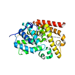
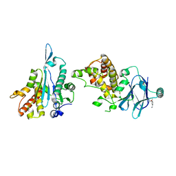
3HZU
 
 | |
6PW6
 | | The HIV-1 Envelope Glycoprotein Clone BG505 SOSIP.664 in Complex with Three Copies of the Bovine Broadly Neutralizing Antibody, NC-Cow1, Fragment Antigen Binding Domain | | 分子名称: | 2-acetamido-2-deoxy-beta-D-glucopyranose, 2-acetamido-2-deoxy-beta-D-glucopyranose-(1-4)-2-acetamido-2-deoxy-beta-D-glucopyranose, Broadly Neutralizing Antibody NC-Cow1 Heavy Chain, ... | | 著者 | Berndsen, Z.T, Ward, A.B. | | 登録日 | 2019-07-22 | | 公開日 | 2020-06-24 | | 最終更新日 | 2024-10-23 | | 実験手法 | ELECTRON MICROSCOPY (4.5 Å) | | 主引用文献 | Structural basis of broad HIV neutralization by a vaccine-induced cow antibody.
Sci Adv, 6, 2020
|
|
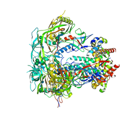
7EG0
 | | Cryo-EM structure of anagrelide-induced PDE3A-SLFN12 complex | | 分子名称: | 6,7-bis(chloranyl)-3,5-dihydro-1H-imidazo[2,1-b]quinazolin-2-one, MAGNESIUM ION, Schlafen family member 12, ... | | 著者 | Liu, N, Chen, J, Wang, X.D, Wang, H.W. | | 登録日 | 2021-03-23 | | 公開日 | 2021-09-29 | | 最終更新日 | 2025-07-02 | | 実験手法 | ELECTRON MICROSCOPY (3.4 Å) | | 主引用文献 | Structure of PDE3A-SLFN12 complex and structure-based design for a potent apoptosis inducer of tumor cells.
Nat Commun, 12, 2021
|
|
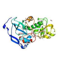
5SK4
 | | CRYSTAL STRUCTURE OF HUMAN PHOSPHODIESTERASE 10 IN COMPLEX WITH c1ccc(cc1N4N=C(c2ccnn2c3ccccc3)C(=O)C=C4C)OC(F)(F)F, micromolar IC50=0.181240 | | 分子名称: | 6-methyl-3-(1-phenyl-1H-pyrazol-5-yl)-1-[3-(trifluoromethoxy)phenyl]pyridazin-4(1H)-one, MAGNESIUM ION, ZINC ION, ... | | 著者 | Joseph, C, Benz, J, Flohr, A, Rogers-Evans, M, Rudolph, M.G. | | 登録日 | 2022-02-01 | | 公開日 | 2022-10-12 | | 最終更新日 | 2024-10-16 | | 実験手法 | X-RAY DIFFRACTION (2 Å) | | 主引用文献 | Crystal Structure of a human phosphodiesterase 10 complex
To be published
|
|
5SKL
 | | CRYSTAL STRUCTURE OF HUMAN PHOSPHODIESTERASE 10 IN COMPLEX WITH c4(c(C(=O)Nc2cc1nc(cn1cc2C#N)c3ccccc3)n(C)nc4)C(=O)N5CCOCC5, micromolar IC50=0.000990 | | 分子名称: | MAGNESIUM ION, N-[(4R)-6-cyano-2-phenylimidazo[1,2-a]pyridin-7-yl]-1-methyl-4-(morpholine-4-carbonyl)-1H-pyrazole-5-carboxamide, ZINC ION, ... | | 著者 | Joseph, C, Benz, J, Flohr, A, Peters, J, Rudolph, M.G. | | 登録日 | 2022-02-01 | | 公開日 | 2022-10-12 | | 最終更新日 | 2024-10-16 | | 実験手法 | X-RAY DIFFRACTION (2.2 Å) | | 主引用文献 | Crystal Structure of a human phosphodiesterase 10 complex
To be published
|
|
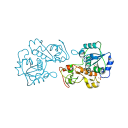
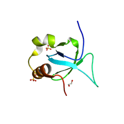
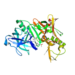
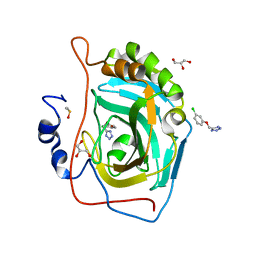
3HY7
 | | Crystal Structure of the Catalytic Domain of ADAMTS-5 in Complex with Marimastat | | 分子名称: | (2S,3R)-N~4~-[(1S)-2,2-dimethyl-1-(methylcarbamoyl)propyl]-N~1~,2-dihydroxy-3-(2-methylpropyl)butanediamide, A disintegrin and metalloproteinase with thrombospondin motifs 5, CALCIUM ION, ... | | 著者 | Shieh, H.-S, Williams, J.M, Caspers, N, Mathis, K.J, Tortorella, M.D, Tomasselli, A. | | 登録日 | 2009-06-22 | | 公開日 | 2009-07-07 | | 最終更新日 | 2024-11-06 | | 実験手法 | X-RAY DIFFRACTION (1.69 Å) | | 主引用文献 | Structural and inhibition analysis reveals the mechanism of selectivity of a series of aggrecanase inhibitors
J.Biol.Chem., 284, 2009
|
|
7HG4
 | | PanDDA analysis group deposition -- Crystal structure of HRP-2 PWWP domain in complex with Z1696844792 | | 分子名称: | 1,2-ETHANEDIOL, 1-(diphenylmethyl)azetidin-3-ol, Hepatoma-derived growth factor-related protein 2, ... | | 著者 | Vantieghem, T, Osipov, E, Fearon, D, Douangamath, A, von Delft, F, Strelkov, S. | | 登録日 | 2024-08-29 | | 公開日 | 2024-11-06 | | 実験手法 | X-RAY DIFFRACTION (1.57 Å) | | 主引用文献 | Rational fragment-based design of compounds targeting the PWWP domain of the HRP family.
Eur.J.Med.Chem., 280, 2024
|
|
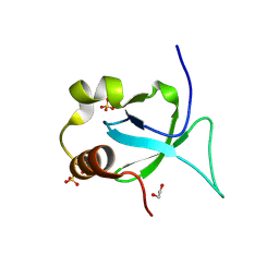
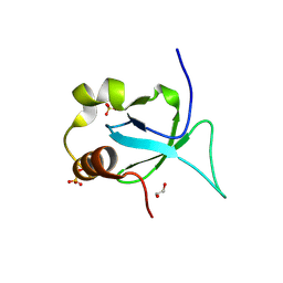
4F2Q
 | | Quisqualate bound to the D655A mutant of the ligand binding domain of GluA3 | | 分子名称: | (S)-2-AMINO-3-(3,5-DIOXO-[1,2,4]OXADIAZOLIDIN-2-YL)-PROPIONIC ACID, Glutamate receptor 3, ZINC ION | | 著者 | Ahmed, A.H, Oswald, R.E. | | 登録日 | 2012-05-08 | | 公開日 | 2012-05-30 | | 最終更新日 | 2024-11-06 | | 実験手法 | X-RAY DIFFRACTION (2.202 Å) | | 主引用文献 | The loss of an electrostatic contact unique to AMPA receptor ligand binding domain 2 slows channel activation.
Biochemistry, 51, 2012
|
|
7HG0
 | | PanDDA analysis group deposition -- Crystal structure of HRP-2 PWWP domain in complex with Z46169212 | | 分子名称: | 1,2-ETHANEDIOL, Hepatoma-derived growth factor-related protein 2, N-(4-acetylphenyl)-2-(pyrrolidin-1-yl)acetamide, ... | | 著者 | Vantieghem, T, Osipov, E, Fearon, D, Douangamath, A, von Delft, F, Strelkov, S. | | 登録日 | 2024-08-29 | | 公開日 | 2024-11-06 | | 実験手法 | X-RAY DIFFRACTION (1.58 Å) | | 主引用文献 | Rational fragment-based design of compounds targeting the PWWP domain of the HRP family.
Eur.J.Med.Chem., 280, 2024
|
|
6D6W
 | | Bacteroides uniformis beta-glucuronidase 1 bound to glucuronate | | 分子名称: | Beta-galactosidase/beta-glucuronidase, CHLORIDE ION, GLYCEROL, ... | | 著者 | Walton, W.G, Pellock, S.J, Redinbo, M.R. | | 登録日 | 2018-04-23 | | 公開日 | 2018-10-17 | | 最終更新日 | 2024-03-13 | | 実験手法 | X-RAY DIFFRACTION (1.8 Å) | | 主引用文献 | Three structurally and functionally distinct beta-glucuronidases from the human gut microbeBacteroides uniformis.
J. Biol. Chem., 293, 2018
|
|
7XIJ
 | | Crystal structure of CBP bromodomain liganded with Y08175 | | 分子名称: | 3-[(1-ethanoyl-5-methoxy-indol-3-yl)carbonylamino]-4-fluoranyl-5-(1-methylpyrazol-4-yl)benzoic acid, DIMETHYL SULFOXIDE, GLYCEROL, ... | | 著者 | Xiang, Q, Wang, C, Wu, T, Zhang, Y, Zhang, C, Luo, G, Wu, X, Shen, H, Xu, Y. | | 登録日 | 2022-04-13 | | 公開日 | 2022-06-08 | | 最終更新日 | 2023-11-29 | | 実験手法 | X-RAY DIFFRACTION (1.82 Å) | | 主引用文献 | Crystal structure of CBP bromodomain liganded with Y08175
To Be Published
|
|
4MSS
 | | Crystal structure of Burkholderia cenocepacia family 3 glycoside hydrolase (NagZ) bound to (3S,4R,5R,6S)-3-acetamido-4,5,6-trihydroxyazepane | | 分子名称: | Beta-hexosaminidase 1, GLYCEROL, N-[(3S,4R,5R,6S)-4,5,6-trihydroxyazepan-3-yl]acetamide | | 著者 | Vadlamani, G, Mark, B.L. | | 登録日 | 2013-09-18 | | 公開日 | 2013-10-30 | | 最終更新日 | 2023-09-20 | | 実験手法 | X-RAY DIFFRACTION (1.8 Å) | | 主引用文献 | Selective trihydroxyazepane NagZ inhibitors increase sensitivity of Pseudomonas aeruginosa to beta-lactams.
Chem.Commun.(Camb.), 49, 2013
|
|
5STJ
 | | PanDDA analysis group deposition -- Aar2/RNaseH in complex with fragment P02H11 from the F2X-Universal Library | | 分子名称: | 1-(3-methoxy-4-methylphenyl)methanamine, A1 cistron-splicing factor AAR2, Pre-mRNA-splicing factor 8 | | 著者 | Barthel, T, Wollenhaupt, J, Lima, G.M.A, Wahl, M.C, Weiss, M.S. | | 登録日 | 2022-08-26 | | 公開日 | 2022-11-02 | | 最終更新日 | 2024-05-22 | | 実験手法 | X-RAY DIFFRACTION (1.76 Å) | | 主引用文献 | Large-Scale Crystallographic Fragment Screening Expedites Compound Optimization and Identifies Putative Protein-Protein Interaction Sites.
J.Med.Chem., 65, 2022
|
|
4Q99
 | |
4QG8
 | | crystal structure of PKM2-K305Q mutant | | 分子名称: | GLYCEROL, MAGNESIUM ION, MALONATE ION, ... | | 著者 | Wang, P, Sun, C, Zhu, T, Xu, Y. | | 登録日 | 2014-05-22 | | 公開日 | 2015-02-25 | | 最終更新日 | 2024-05-29 | | 実験手法 | X-RAY DIFFRACTION (2.3 Å) | | 主引用文献 | Structural insight into mechanisms for dynamic regulation of PKM2.
Protein Cell, 6, 2015
|
|
3X00
 | | Crystal structure of the core streptavidin mutant V212 (Y22S/N23D/S27D/S45N/Y83S/R84K/E101D/R103K/E116N) complexed with bis iminobiotin long tail (Bis-IMNtail) at 1.3 A resolution | | 分子名称: | 6-({5-[(2E,3aS,4S,6aR)-2-iminohexahydro-1H-thieno[3,4-d]imidazol-4-yl]pentanoyl}amino)hexanoic acid, ETHANE-1,2-DIAMINE, Streptavidin | | 著者 | Kawato, T, Mizohata, E, Shimizu, Y, Meshizuka, T, Yamamoto, T, Takasu, N, Matsuoka, M, Matsumura, H, Kodama, T, Kanai, M, Doi, H, Inoue, T, Sugiyama, A. | | 登録日 | 2014-10-09 | | 公開日 | 2015-01-21 | | 最終更新日 | 2023-11-08 | | 実験手法 | X-RAY DIFFRACTION (1.3 Å) | | 主引用文献 | Structure-based design and synthesis of a bivalent iminobiotin analog showing strong affinity toward a low immunogenic streptavidin mutant.
Biosci.Biotechnol.Biochem., 79, 2015
|
|
7HGZ
 | | PanDDA analysis group deposition -- Crystal structure of HRP-2 PWWP domain in complex with Z1509882419 | | 分子名称: | 1,2-ETHANEDIOL, 1-(propan-2-yl)-1H-imidazole-4-sulfonamide, Hepatoma-derived growth factor-related protein 2, ... | | 著者 | Vantieghem, T, Osipov, E, Fearon, D, Douangamath, A, von Delft, F, Strelkov, S. | | 登録日 | 2024-08-29 | | 公開日 | 2024-11-06 | | 実験手法 | X-RAY DIFFRACTION (1.65 Å) | | 主引用文献 | Rational fragment-based design of compounds targeting the PWWP domain of the HRP family.
Eur.J.Med.Chem., 280, 2024
|
|
3TPR
 | | Crystal structure of BACE1 complexed with an inhibitor | | 分子名称: | Beta-secretase 1, CHLORIDE ION, N-[(1S,2R)-1-BENZYL-3-(CYCLOPROPYLAMINO)-2-HYDROXYPROPYL]-5-[METHYL(METHYLSULFONYL)AMINO]-N'-[(1R)-1-PHENYLETHYL]ISOPHTHALAMIDE | | 著者 | Xu, Y.C, Li, M.J, Greenblatt, H, Chen, T.T, Silman, I, Sussman, J.L. | | 登録日 | 2011-09-08 | | 公開日 | 2011-11-23 | | 最終更新日 | 2024-11-06 | | 実験手法 | X-RAY DIFFRACTION (2.55 Å) | | 主引用文献 | Flexibility of the flap in the active site of BACE1 as revealed by crystal structures and molecular dynamics simulations
Acta Crystallogr.,Sect.D, 68, 2012
|
|
7HHI
 | | PanDDA analysis group deposition -- Crystal structure of HRP-2 PWWP domain in complex with Z805551440 | | 分子名称: | 1,2-ETHANEDIOL, Hepatoma-derived growth factor-related protein 2, SULFATE ION, ... | | 著者 | Vantieghem, T, Osipov, E, Fearon, D, Douangamath, A, von Delft, F, Strelkov, S. | | 登録日 | 2024-08-29 | | 公開日 | 2024-11-06 | | 実験手法 | X-RAY DIFFRACTION (2.11 Å) | | 主引用文献 | Rational fragment-based design of compounds targeting the PWWP domain of the HRP family.
Eur.J.Med.Chem., 280, 2024
|
|
3LQW
 | |
7HG1
 | | PanDDA analysis group deposition -- Crystal structure of HRP-2 PWWP domain in complex with Z1079512010 | | 分子名称: | 1,2-ETHANEDIOL, 6-methyl-2-[(3-methyl-1,2-oxazol-5-yl)methyl]pyridazin-3(2H)-one, Hepatoma-derived growth factor-related protein 2, ... | | 著者 | Vantieghem, T, Osipov, E, Fearon, D, Douangamath, A, von Delft, F, Strelkov, S. | | 登録日 | 2024-08-29 | | 公開日 | 2024-11-06 | | 実験手法 | X-RAY DIFFRACTION (1.81 Å) | | 主引用文献 | Rational fragment-based design of compounds targeting the PWWP domain of the HRP family.
Eur.J.Med.Chem., 280, 2024
|
|
5FNH
 | | Native state mass spectrometry, surface plasmon resonance and X-ray crystallography correlate strongly as a fragment screening combination | | 分子名称: | 5-[(3-chloranylphenoxy)methyl]-1,2,4-triaza-3-azanidacyclopenta-1,4-diene, CARBONIC ANHYDRASE 2, DIMETHYL SULFOXIDE, ... | | 著者 | Woods, L.A, Dolezal, O, Ren, B, Ryan, J.H, Peat, T.S, Poulsen, S.A. | | 登録日 | 2015-11-15 | | 公開日 | 2016-03-02 | | 最終更新日 | 2024-01-10 | | 実験手法 | X-RAY DIFFRACTION (1.66 Å) | | 主引用文献 | Native State Mass Spectrometry, Surface Plasmon Resonance and X-Ray Crystallography Correlate Strongly as a Fragment Screening Combination.
J.Med.Chem., 59, 2016
|
|
4IDC
 | | Structure of the Fragaria x ananassa enone oxidoreductase in complex with NADPH and HDMF | | 分子名称: | (2R)-4-hydroxy-2,5-dimethylfuran-3(2H)-one, 1,2-ETHANEDIOL, NADPH DIHYDRO-NICOTINAMIDE-ADENINE-DINUCLEOTIDE PHOSPHATE, ... | | 著者 | Schiefner, A, Skerra, A. | | 登録日 | 2012-12-12 | | 公開日 | 2013-04-17 | | 最終更新日 | 2023-11-08 | | 実験手法 | X-RAY DIFFRACTION (1.4 Å) | | 主引用文献 | Structural basis for the enzymatic formation of the key strawberry flavor compound 4-hydroxy-2,5-dimethyl-3(2H)-furanone
J.Biol.Chem., 288, 2013
|
|
3LHY
 | | Crystal structure of the mutant I199A of orotidine 5'-monophosphate decarboxylase from Methanobacterium thermoautotrophicum complexed with inhibitor BMP | | 分子名称: | 1-(5'-PHOSPHO-BETA-D-RIBOFURANOSYL)BARBITURIC ACID, Orotidine 5'-phosphate decarboxylase | | 著者 | Fedorov, A.A, Fedorov, E.V, Wood, B.M, Gerlt, J.A, Almo, S.C. | | 登録日 | 2010-01-23 | | 公開日 | 2010-06-16 | | 最終更新日 | 2023-09-06 | | 実験手法 | X-RAY DIFFRACTION (1.4 Å) | | 主引用文献 | Conformational changes in orotidine 5'-monophosphate decarboxylase: "remote" residues that stabilize the active conformation.
Biochemistry, 49, 2010
|
|
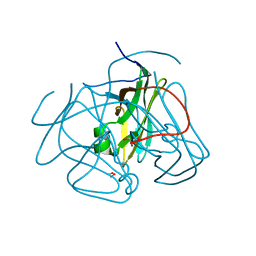
3ZUH
 | | Negative stain EM Map of the AAA protein CbbX, a red-type Rubisco activase from R. sphaeroides | | 分子名称: | ADENOSINE-5'-DIPHOSPHATE, PROTEIN CBBX, RIBULOSE-1,5-DIPHOSPHATE | | 著者 | Mueller-Cajar, O, Stotz, M, Wendler, P, Hartl, F.U, Bracher, A, Hayer-Hartl, M. | | 登録日 | 2011-07-19 | | 公開日 | 2011-11-09 | | 最終更新日 | 2024-05-08 | | 実験手法 | ELECTRON MICROSCOPY (21 Å) | | 主引用文献 | Structure and Function of the Aaa+ Protein Cbbx, a Red-Type Rubisco Activase.
Nature, 479, 2011
|
|


