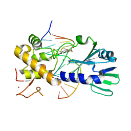
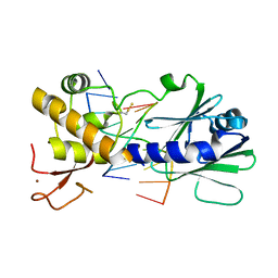
5H6K
 
 | | DNA targeting ADP-ribosyltransferase Pierisin-1 | | 分子名称: | 1,2-ETHANEDIOL, Pierisin-1 | | 著者 | Oda, T, Hirabayashi, H, Shikauchi, G, Takamura, R, Hiraga, K, Minami, H, Hashimoto, H, Yamamoto, M, Wakabayashi, K, Sugimura, T, Shimizu, T, Sato, M. | | 登録日 | 2016-11-14 | | 公開日 | 2017-08-09 | | 最終更新日 | 2024-03-20 | | 実験手法 | X-RAY DIFFRACTION (1.8 Å) | | 主引用文献 | Structural basis of autoinhibition and activation of the DNA-targeting ADP-ribosyltransferase pierisin-1
J. Biol. Chem., 292, 2017
|
|
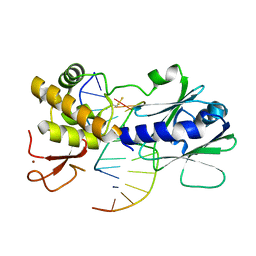
3U6C
 | | MutM set 1 ApGo | | 分子名称: | DNA (5'-D(*GP*GP*TP*AP*GP*AP*TP*CP*CP*TP*GP*AP*C)-3'), DNA (5'-D(P*CP*AP*(8OG)P*GP*AP*(TX)P*CP*TP*AP*C)-3'), Formamidopyrimidine-DNA glycosylase, ... | | 著者 | Sung, R.J, Zhang, M, Qi, Y, Verdine, G.L. | | 登録日 | 2011-10-12 | | 公開日 | 2012-04-25 | | 最終更新日 | 2013-09-25 | | 実験手法 | X-RAY DIFFRACTION (1.8 Å) | | 主引用文献 | Sequence-dependent structural variation in DNA undergoing intrahelical inspection by the DNA glycosylase MutM.
J.Biol.Chem., 287, 2012
|
|
3U6D
 | | MutM set 1 GpGo | | 分子名称: | DNA (5'-D(*A*GP*GP*TP*AP*GP*AP*TP*CP*CP*CP*GP*AP*CP*GP*C)-3'), DNA (5'-D(*TP*GP*CP*GP*TP*CP*GP*(8OG)P*GP*AP*(TX)P*CP*TP*AP*CP*C)-3'), Formamidopyrimidine-DNA glycosylase, ... | | 著者 | Sung, R.J, Zhang, M, Qi, Y, Verdine, G.L. | | 登録日 | 2011-10-12 | | 公開日 | 2012-04-25 | | 最終更新日 | 2013-09-25 | | 実験手法 | X-RAY DIFFRACTION (1.87 Å) | | 主引用文献 | Sequence-dependent structural variation in DNA undergoing intrahelical inspection by the DNA glycosylase MutM.
J.Biol.Chem., 287, 2012
|
|
2QRV
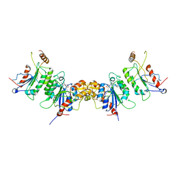 | | Structure of Dnmt3a-Dnmt3L C-terminal domain complex | | 分子名称: | DNA (cytosine-5)-methyltransferase 3-like, DNA (cytosine-5)-methyltransferase 3A, S-ADENOSYL-L-HOMOCYSTEINE | | 著者 | Jia, D, Cheng, X. | | 登録日 | 2007-07-29 | | 公開日 | 2007-12-04 | | 最終更新日 | 2023-08-30 | | 実験手法 | X-RAY DIFFRACTION (2.89 Å) | | 主引用文献 | Structure of Dnmt3a bound to Dnmt3L suggests a model for de novo DNA methylation.
Nature, 449, 2007
|
|
3U6L
 | | MutM set 2 CpGo | | 分子名称: | DNA (5'-D(*AP*GP*GP*TP*AP*GP*AP*CP*CP*GP*GP*AP*CP*GP*C)-3'), DNA (5'-D(*TP*GP*CP*GP*TP*CP*CP*(8OG)P*GP*TP*(CX2)P*TP*AP*CP*C)-3'), Formamidopyrimidine-DNA glycosylase, ... | | 著者 | Sung, R.J, Zhang, M, Qi, Y, Verdine, G.L. | | 登録日 | 2011-10-12 | | 公開日 | 2012-04-25 | | 最終更新日 | 2017-01-11 | | 実験手法 | X-RAY DIFFRACTION (1.97 Å) | | 主引用文献 | Sequence-dependent structural variation in DNA undergoing intrahelical inspection by the DNA glycosylase MutM.
J.Biol.Chem., 287, 2012
|
|
3U6M
 | | Structural effects of sequence context on lesion recognition by MutM | | 分子名称: | DNA (5'-D(*AP*GP*GP*TP*AP*GP*AP*CP*CP*AP*GP*GP*AP*CP*GP*C)-3'), DNA (5'-D(*TP*GP*CP*GP*TP*CP*CP*TP*(8OG)P*GP*TP*(CX2)P*TP*AP*CP*C)-3'), Formamidopyrimidine-DNA glycosylase, ... | | 著者 | Sung, R.J, Zhang, M, Qi, Y, Verdine, G.L. | | 登録日 | 2011-10-12 | | 公開日 | 2012-04-25 | | 最終更新日 | 2013-09-25 | | 実験手法 | X-RAY DIFFRACTION (2.1 Å) | | 主引用文献 | Sequence-dependent structural variation in DNA undergoing intrahelical inspection by the DNA glycosylase MutM.
J.Biol.Chem., 287, 2012
|
|
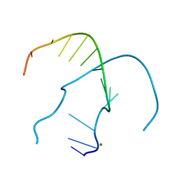
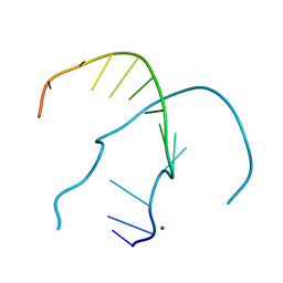
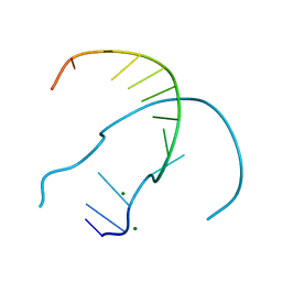
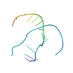
5LME
 | | Specific-DNA binding activity of the cross-brace zinc finger motif of the piggyBac transposase | | 分子名称: | ZINC ION, piggyBac transposase | | 著者 | Morellet, N, Taylor, J.A, Wieninger, S, Moriau, S, Li, X, Lescop, E, Mathy, N, Bischerour, J, Betermier, M, Bardiaux, B, Nilges, M, Craig, N.L, Hickman, A.B, Dyda, F, Guittet, E. | | 登録日 | 2016-07-30 | | 公開日 | 2017-12-20 | | 最終更新日 | 2024-06-19 | | 実験手法 | SOLUTION NMR | | 主引用文献 | Sequence-specific DNA binding activity of the cross-brace zinc finger motif of the piggyBac transposase.
Nucleic Acids Res., 46, 2018
|
|
7JY2
 | | Z-DNA joint X-ray/Neutron | | 分子名称: | Chains: A,B | | 著者 | Harp, J.M, Coates, L, Egli, M. | | 登録日 | 2020-08-28 | | 公開日 | 2021-04-28 | | 最終更新日 | 2024-04-03 | | 実験手法 | NEUTRON DIFFRACTION (1.5 Å), X-RAY DIFFRACTION | | 主引用文献 | Water structure around a left-handed Z-DNA fragment analyzed by cryo neutron crystallography.
Nucleic Acids Res., 49, 2021
|
|
4OCB
 | |
4RP2
 | |
4ROY
 | |
4ROG
 | |
4ROK
 | |
4RON
 | |
4ROO
 | |
4ROZ
 | |
8VMR
 | |
4RP1
 | |
4RNK
 | |
4RO7
 | |
4RO8
 | |
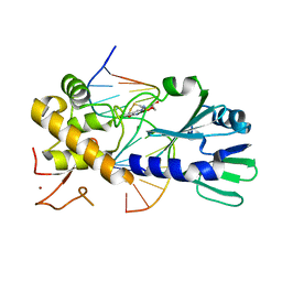
5H6M
 | | DNA targeting ADP-ribosyltransferase Pierisin-1 | | 分子名称: | 1,2-ETHANEDIOL, Pierisin-1 | | 著者 | Oda, T, Hirabayashi, H, Shikauchi, G, Takamura, R, Hiraga, K, Minami, H, Hashimoto, H, Yamamoto, M, Wakabayashi, K, Sugimura, T, Shimizu, T, Sato, M. | | 登録日 | 2016-11-14 | | 公開日 | 2017-08-09 | | 最終更新日 | 2024-03-20 | | 実験手法 | X-RAY DIFFRACTION (1.9 Å) | | 主引用文献 | Structural basis of autoinhibition and activation of the DNA-targeting ADP-ribosyltransferase pierisin-1
J. Biol. Chem., 292, 2017
|
|
2Z43
 | |
3V7K
 | |
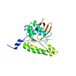
8H5Z
 | | Crystal structure of RadD/ATP analogue complex | | 分子名称: | PHOSPHOTHIOPHOSPHORIC ACID-ADENYLATE ESTER, Putative DNA repair helicase RadD, ZINC ION | | 著者 | Yan, X.X, Tian, L.F. | | 登録日 | 2022-10-14 | | 公開日 | 2023-10-18 | | 実験手法 | X-RAY DIFFRACTION (3.00002718 Å) | | 主引用文献 | Biochemical and Structural Analyses Shed Light on the Mechanisms of RadD DNA Binding and Its ATPase from Escherichia coli.
Int J Mol Sci, 24, 2023
|
|


