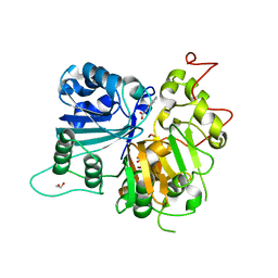
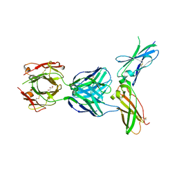
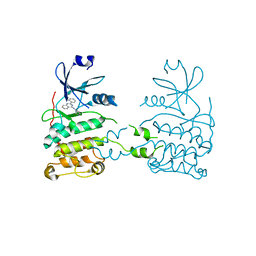
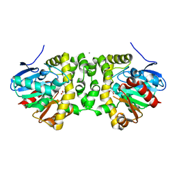
7QUJ
 
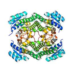 | | Structure of NsNEPS2, a 7S-cis-trans nepetalactone synthase | | 分子名称: | NICOTINAMIDE-ADENINE-DINUCLEOTIDE, NsNEPS2 | | 著者 | Hernandez Lozada, N.J, Hong, B, Wood, J.C, Caputi, L, Basquin, J, Chuang, L, Kunert, M, Rodriguez Lopez, C.R, Langley, C, Zhao, D, Buell, C.R, Lichman, B.R, O'Connor, S.E. | | 登録日 | 2022-01-18 | | 公開日 | 2022-12-28 | | 最終更新日 | 2024-01-31 | | 実験手法 | X-RAY DIFFRACTION (1.85 Å) | | 主引用文献 | Biocatalytic routes to stereo-divergent iridoids.
Nat Commun, 13, 2022
|
|
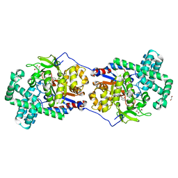
4O4L
 | | Tubulin-Peloruside A-Epothilone A complex | | 分子名称: | 2-(N-MORPHOLINO)-ETHANESULFONIC ACID, CALCIUM ION, EPOTHILONE A, ... | | 著者 | Prota, A.E, Bargsten, K, Northcote, P.T, Marsh, M, Altmann, K.H, Miller, J.H, Diaz, J.F, Steinmetz, M.O. | | 登録日 | 2013-12-18 | | 公開日 | 2014-03-26 | | 最終更新日 | 2024-04-03 | | 実験手法 | X-RAY DIFFRACTION (2.2 Å) | | 主引用文献 | Structural basis of microtubule stabilization by laulimalide and peloruside a.
Angew.Chem.Int.Ed.Engl., 53, 2014
|
|
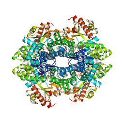
4O4H
 | | Tubulin-Laulimalide complex | | 分子名称: | CALCIUM ION, GLYCEROL, GUANOSINE-5'-DIPHOSPHATE, ... | | 著者 | Prota, A.E, Bargsten, K, Northcote, P.T, Marsh, M, Altmann, K.H, Miller, J.H, Diaz, J.F, Steinmetz, M.O. | | 登録日 | 2013-12-18 | | 公開日 | 2014-03-05 | | 最終更新日 | 2023-09-20 | | 実験手法 | X-RAY DIFFRACTION (2.1 Å) | | 主引用文献 | Structural basis of microtubule stabilization by laulimalide and peloruside a.
Angew.Chem.Int.Ed.Engl., 53, 2014
|
|
5VRK
 | | Crystal structure of SsoPox AsA6 mutant (F46L-C258A-W263M-I280T) - open form | | 分子名称: | 1,2-ETHANEDIOL, Aryldialkylphosphatase, COBALT (II) ION, ... | | 著者 | Hiblot, J, Gotthard, G, Jacquet, P, Daude, D, Bergonzi, C, Chabriere, E, Elias, M. | | 登録日 | 2017-05-10 | | 公開日 | 2018-01-10 | | 最終更新日 | 2023-11-15 | | 実験手法 | X-RAY DIFFRACTION (1.4 Å) | | 主引用文献 | Rational engineering of a native hyperthermostable lactonase into a broad spectrum phosphotriesterase.
Sci Rep, 7, 2017
|
|
6DIM
 | | Crystal structure of Tdp1 catalytic domain in complex with Zenobia fragment ZT1982 from cocktail soak | | 分子名称: | 1,2-ETHANEDIOL, 4-hydroxyquinoline-3-carboxylic acid, Tyrosyl-DNA phosphodiesterase 1 | | 著者 | Lountos, G.T, Zhao, X.Z, Kiselev, E, Tropea, J.E, Needle, D, Burke Jr, T.R, Pommier, Y, Waugh, D.S. | | 登録日 | 2018-05-23 | | 公開日 | 2019-05-29 | | 最終更新日 | 2024-03-13 | | 実験手法 | X-RAY DIFFRACTION (1.81 Å) | | 主引用文献 | Identification of a ligand binding hot spot and structural motifs replicating aspects of tyrosyl-DNA phosphodiesterase I (TDP1) phosphoryl recognition by crystallographic fragment cocktail screening.
Nucleic Acids Res., 47, 2019
|
|
5VD9
 | | Crystal structure of human WEE1 kinase domain in complex with RAC-IV-097, a MK1775 analogue | | 分子名称: | 1,2-ETHANEDIOL, 1-{6-[(1R)-1-hydroxyethyl]pyridin-2-yl}-6-{[4-(4-methylpiperazin-1-yl)phenyl]amino}-2-(prop-2-en-1-yl)-1,2-dihydro-3H-pyrazolo[3,4-d]pyrimidin-3-one, CHLORIDE ION, ... | | 著者 | Zhu, J.-Y, Schonbrunn, E. | | 登録日 | 2017-04-01 | | 公開日 | 2018-04-04 | | 最終更新日 | 2023-10-04 | | 実験手法 | X-RAY DIFFRACTION (1.87 Å) | | 主引用文献 | Structural basis of Wee family kinase inhibition by small molecules
to be published
|
|
4WBX
 | | Conserved hypothetical protein PF1771 from Pyrococcus furiosus solved by sulfur SAD using Swiss Light Source data | | 分子名称: | 2-keto acid:ferredoxin oxidoreductase subunit alpha | | 著者 | Weinert, T, Waltersperger, S, Olieric, V, Panepucci, E, Chen, L, Rose, J.P, Wang, M, Wang, B.C, Southeast Collaboratory for Structural Genomics (SECSG) | | 登録日 | 2014-09-04 | | 公開日 | 2014-12-10 | | 最終更新日 | 2023-12-27 | | 実験手法 | X-RAY DIFFRACTION (2.301 Å) | | 主引用文献 | Fast native-SAD phasing for routine macromolecular structure determination.
Nat.Methods, 12, 2015
|
|
6QHQ
 | | Time resolved structural analysis of the full turnover of an enzyme - 1128 ms | | 分子名称: | Fluoroacetate dehalogenase, fluoroacetic acid | | 著者 | Schulz, E.C, Mehrabi, P, Pai, E.F, Miller, D. | | 登録日 | 2019-01-17 | | 公開日 | 2019-09-25 | | 最終更新日 | 2024-01-24 | | 実験手法 | X-RAY DIFFRACTION (1.735 Å) | | 主引用文献 | Time-resolved crystallography reveals allosteric communication aligned with molecular breathing.
Science, 365, 2019
|
|
6QHW
 | | Time resolved structural analysis of the full turnover of an enzyme - 4512 ms | | 分子名称: | Fluoroacetate dehalogenase, GLYCOLIC ACID, fluoroacetic acid | | 著者 | Schulz, E.C, Mehrabi, P, Pai, E.F, Miller, D. | | 登録日 | 2019-01-17 | | 公開日 | 2019-09-25 | | 最終更新日 | 2023-11-15 | | 実験手法 | X-RAY DIFFRACTION (1.718 Å) | | 主引用文献 | Time-resolved crystallography reveals allosteric communication aligned with molecular breathing.
Science, 365, 2019
|
|
5W06
 | | HUMAN TISSUE FACTOR IN COMPLEX WITH ANTIBODY M1587 | | 分子名称: | GLYCEROL, M1587 FAB HEAVY CHAIN, M1587 FAB LIGHT CHAIN, ... | | 著者 | Teplyakov, A, Obmolova, G, Malia, T.J, Gilliland, G.L. | | 登録日 | 2017-05-30 | | 公開日 | 2017-06-14 | | 最終更新日 | 2023-10-04 | | 実験手法 | X-RAY DIFFRACTION (2.6 Å) | | 主引用文献 | Structural insights into humanization of anti-tissue factor antibody 10H10.
MAbs, 10, 2018
|
|
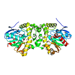
5CSU
 | | Disproportionating enzyme 1 from Arabidopsis - acarviostatin soak | | 分子名称: | 1,2-ETHANEDIOL, 4-alpha-glucanotransferase DPE1, chloroplastic/amyloplastic, ... | | 著者 | O'Neill, E.C, Stevenson, C.E.M, Tantanarat, K, Latousakis, D, Donaldson, M.I, Rejzek, M, Limpaseni, T, Smith, A.M, Field, R.A, Lawson, D.M. | | 登録日 | 2015-07-23 | | 公開日 | 2015-11-04 | | 最終更新日 | 2024-01-10 | | 実験手法 | X-RAY DIFFRACTION (2.53 Å) | | 主引用文献 | Structural Dissection of the Maltodextrin Disproportionation Cycle of the Arabidopsis Plastidial Disproportionating Enzyme 1 (DPE1).
J.Biol.Chem., 290, 2015
|
|
5VKH
 | | Closed conformation of KcsA Y82A-F103A mutant | | 分子名称: | (1S)-2-HYDROXY-1-[(NONANOYLOXY)METHYL]ETHYL MYRISTATE, Antibody Heavy Chain, Antibody Light Chain, ... | | 著者 | Cuello, L.G, Perozo, E, Cortes, D.M. | | 登録日 | 2017-04-21 | | 公開日 | 2017-12-06 | | 最終更新日 | 2020-01-01 | | 実験手法 | X-RAY DIFFRACTION (2.25 Å) | | 主引用文献 | The gating cycle of a K+ channel at atomic resolution.
Elife, 6, 2017
|
|
7LU5
 | | SAMHD1(113-626) H206R D207N R366H | | 分子名称: | 2'-DEOXYGUANOSINE-5'-TRIPHOSPHATE, Deoxynucleoside triphosphate triphosphohydrolase SAMHD1 | | 著者 | Temple, J.T, Bowen, N.E. | | 登録日 | 2021-02-20 | | 公開日 | 2021-11-24 | | 最終更新日 | 2023-10-18 | | 実験手法 | X-RAY DIFFRACTION (3.57 Å) | | 主引用文献 | Structural and functional characterization explains loss of dNTPase activity of the cancer-specific R366C/H mutant SAMHD1 proteins.
J.Biol.Chem., 297, 2021
|
|
7LTW
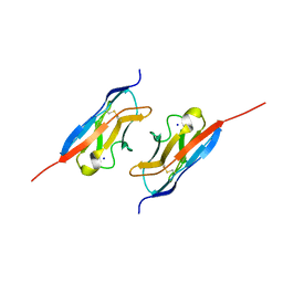 | | Crystal structure of the mouse Kirrel2 D1 homodimer | | 分子名称: | Kin of IRRE-like protein 2, SODIUM ION | | 著者 | Roman, C.A, Pak, J.S, Wang, J, Ozkan, E. | | 登録日 | 2021-02-20 | | 公開日 | 2021-11-24 | | 最終更新日 | 2023-10-18 | | 実験手法 | X-RAY DIFFRACTION (1.8 Å) | | 主引用文献 | Molecular and structural basis of olfactory sensory neuron axon coalescence by Kirrel receptors.
Cell Rep, 37, 2021
|
|
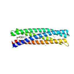
4OH9
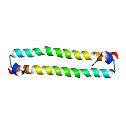 | | Crystal Structure of the human MST2 SARAH homodimer | | 分子名称: | Serine/threonine-protein kinase 3 | | 著者 | Hwang, E, Cheong, H.-K, Ul Mushtaq, A, Kim, H.-Y, Yeo, K.J, Kim, E, Lee, W.C, Hwang, K.Y, Cheong, C, Jeon, Y.H. | | 登録日 | 2014-01-17 | | 公開日 | 2014-07-23 | | 最終更新日 | 2023-09-20 | | 実験手法 | X-RAY DIFFRACTION (1.699 Å) | | 主引用文献 | Structural basis of the heterodimerization of the MST and RASSF SARAH domains in the Hippo signalling pathway.
Acta Crystallogr.,Sect.D, 70, 2014
|
|
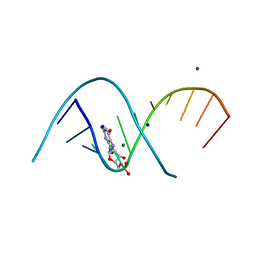
7QSH
 | | 23S ribosomal RNA Sarcin Ricin Loop 27-nt fragment containing a Xanthosine residue at position 2648 | | 分子名称: | 23S ribosomal RNA Sarcin Ricin Loop 27-nucleotide fragment, 9-[(2~{R},3~{R},4~{S},5~{R})-3,4-bis(oxidanyl)-5-[[tris(oxidanyl)-$l^{5}-phosphanyl]oxymethyl]oxolan-2-yl]-2-oxidanyl-1~{H}-purin-6-one, GLYCEROL, ... | | 著者 | Ennifar, E, Micura, R. | | 登録日 | 2022-01-13 | | 公開日 | 2023-01-25 | | 最終更新日 | 2024-02-07 | | 実験手法 | X-RAY DIFFRACTION (0.86 Å) | | 主引用文献 | Towards a comprehensive understanding of RNA deamination: synthesis and properties of xanthosine-modified RNA.
Nucleic Acids Res., 50, 2022
|
|
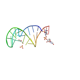
7QUA
 | | Duplex RNA containing Xanthosine-Cytosine base pairs | | 分子名称: | MAGNESIUM ION, RNA (5'-R(*CP*GP*CP*GP*(XAN)P*AP*UP*UP*AP*GP*CP*G)-3'), SODIUM ION | | 著者 | Ennifar, E, Micura, R. | | 登録日 | 2022-01-17 | | 公開日 | 2023-01-25 | | 最終更新日 | 2024-02-07 | | 実験手法 | X-RAY DIFFRACTION (1 Å) | | 主引用文献 | Towards a comprehensive understanding of RNA deamination: synthesis and properties of xanthosine-modified RNA.
Nucleic Acids Res., 50, 2022
|
|
5VP8
 | | I38T mutant of 2009 H1N1 PA Endonuclease | | 分子名称: | 1-[(3R,5S,7R)-1,5,7,9-tetrakis(2-oxopyrrolidin-1-yl)nonan-3-yl]-1,3-dihydro-2H-pyrrol-2-one, MANGANESE (II) ION, Polymerase acidic protein, ... | | 著者 | Kumar, G, White, S.W. | | 登録日 | 2017-05-04 | | 公開日 | 2018-04-11 | | 最終更新日 | 2023-10-04 | | 実験手法 | X-RAY DIFFRACTION (2.2 Å) | | 主引用文献 | Identification of the I38T PA Substitution as a Resistance Marker for Next-Generation Influenza Virus Endonuclease Inhibitors.
MBio, 9, 2018
|
|
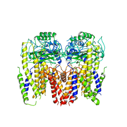
3USG
 | | Crystal structure of LeuT bound to L-leucine in space group C2 from lipid bicelles | | 分子名称: | ACETATE ION, DI(HYDROXYETHYL)ETHER, LEUCINE, ... | | 著者 | Wang, H, Elferich, J, Gouaux, E. | | 登録日 | 2011-11-23 | | 公開日 | 2012-01-11 | | 最終更新日 | 2023-09-13 | | 実験手法 | X-RAY DIFFRACTION (2.502 Å) | | 主引用文献 | Structures of LeuT in bicelles define conformation and substrate binding in a membrane-like context.
Nat.Struct.Mol.Biol., 19, 2012
|
|
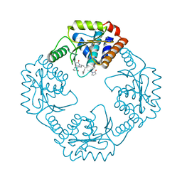
5VJS
 | | De Novo Photosynthetic Reaction Center Protein Equipped with Heme B, a synthetic Zn porphyrin, and Zn(II) cations | | 分子名称: | CHLORIDE ION, PROTOPORPHYRIN IX CONTAINING FE, Reaction Center Maquette, ... | | 著者 | Ennist, N.M, Dutton, P.L, Stayrook, S.E, Moser, C.C. | | 登録日 | 2017-04-19 | | 公開日 | 2018-04-25 | | 最終更新日 | 2024-05-22 | | 実験手法 | X-RAY DIFFRACTION (2 Å) | | 主引用文献 | De novo protein design of photochemical reaction centers.
Nat Commun, 13, 2022
|
|
2XM8
 | | Co-crystal structure of a small molecule inhibitor bound to the kinase domain of Chk2 | | 分子名称: | 2-{4-[(3S)-PYRROLIDIN-3-YLAMINO]QUINAZOLIN-2-YL}PHENOL, SERINE/THREONINE-PROTEIN KINASE CHK2 | | 著者 | Caldwell, J.J, Welsh, E.J, Matijssen, C, Anderson, V.E, Antoni, L, Boxall, K, Urban, F, Hayes, A, Raynaud, F.I, Rigoreau, L.J, Raynham, T, Aherne, G.W, Pearl, L.H, Oliver, A.W, Garrett, M.D, Collins, I. | | 登録日 | 2010-07-26 | | 公開日 | 2011-01-12 | | 最終更新日 | 2023-12-20 | | 実験手法 | X-RAY DIFFRACTION (3.4 Å) | | 主引用文献 | Structure-Based Design of Potent and Selective 2-(Quinazolin-2-Yl)Phenol Inhibitors of Checkpoint Kinase 2.
J.Med.Chem., 54, 2011
|
|
6DU8
 | | Human Polycsytin 2-l1 | | 分子名称: | 2-acetamido-2-deoxy-beta-D-glucopyranose, Polycystic kidney disease 2-like 1 protein | | 著者 | Hulse, R.E, Clapham, D.E, Li, Z, Huang, R.K, Zhang, J. | | 登録日 | 2018-06-20 | | 公開日 | 2018-07-25 | | 最終更新日 | 2020-07-29 | | 実験手法 | ELECTRON MICROSCOPY (3.11 Å) | | 主引用文献 | Cryo-EM structure of the polycystin 2-l1 ion channel.
Elife, 7, 2018
|
|
6QHS
 | | Time resolved structural analysis of the full turnover of an enzyme - 564 ms | | 分子名称: | Fluoroacetate dehalogenase, fluoroacetic acid | | 著者 | Schulz, E.C, Mehrabi, P, Pai, E.F, Miller, D. | | 登録日 | 2019-01-17 | | 公開日 | 2019-09-25 | | 最終更新日 | 2024-01-24 | | 実験手法 | X-RAY DIFFRACTION (1.733 Å) | | 主引用文献 | Time-resolved crystallography reveals allosteric communication aligned with molecular breathing.
Science, 365, 2019
|
|
6QI0
 | | Time resolved structural analysis of the full turnover of an enzyme - 9024 ms | | 分子名称: | CALCIUM ION, Fluoroacetate dehalogenase, GLYCOLIC ACID, ... | | 著者 | Schulz, E.C, Mehrabi, P, Pai, E.F, Miller, D. | | 登録日 | 2019-01-17 | | 公開日 | 2019-09-25 | | 最終更新日 | 2023-11-15 | | 実験手法 | X-RAY DIFFRACTION (1.733 Å) | | 主引用文献 | Time-resolved crystallography reveals allosteric communication aligned with molecular breathing.
Science, 365, 2019
|
|
6QHY
 | | Time resolved structural analysis of the full turnover of an enzyme - 100 ms | | 分子名称: | Fluoroacetate dehalogenase, fluoroacetic acid | | 著者 | Schulz, E.C, Mehrabi, P, Pai, E.F, Miller, D. | | 登録日 | 2019-01-17 | | 公開日 | 2019-09-25 | | 最終更新日 | 2024-01-24 | | 実験手法 | X-RAY DIFFRACTION (1.698 Å) | | 主引用文献 | Time-resolved crystallography reveals allosteric communication aligned with molecular breathing.
Science, 365, 2019
|
|


