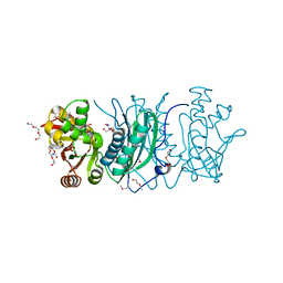
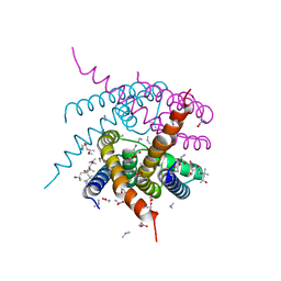
4M9H
 
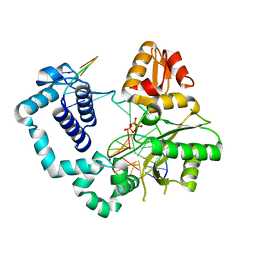 | | DNA Polymerase Beta E295K Soaked with dTTP | | 分子名称: | CHLORIDE ION, DNA Downstream Strand, DNA Primer Strand, ... | | 著者 | Eckenroth, B.E, Doublie, S. | | 登録日 | 2013-08-14 | | 公開日 | 2013-10-16 | | 最終更新日 | 2024-02-28 | | 実験手法 | X-RAY DIFFRACTION (2.394 Å) | | 主引用文献 | The E295K Cancer Variant of Human Polymerase beta Favors the Mismatch Conformational Pathway during Nucleotide Selection.
J.Biol.Chem., 288, 2013
|
|
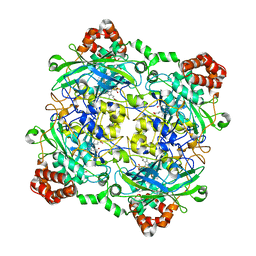
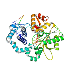
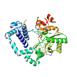
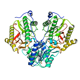
4QOL
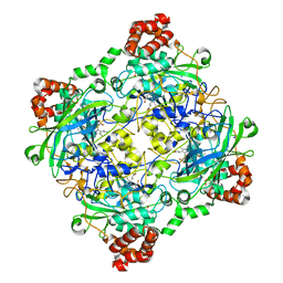
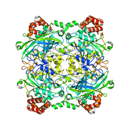 | | Structure of Bacillus pumilus catalase | | 分子名称: | ACETATE ION, CHLORIDE ION, Catalase, ... | | 著者 | Loewen, P.C. | | 登録日 | 2014-06-20 | | 公開日 | 2015-02-25 | | 最終更新日 | 2023-09-20 | | 実験手法 | X-RAY DIFFRACTION (1.65 Å) | | 主引用文献 | Unprecedented access of phenolic substrates to the heme active site of a catalase: Substrate binding and peroxidase-like reactivity of Bacillus pumilus catalase monitored by X-ray crystallography and EPR spectroscopy.
Proteins, 83, 2015
|
|
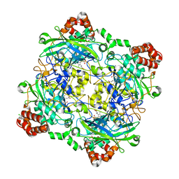
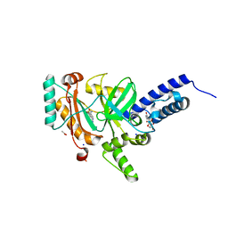
4QOP
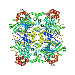 | | Structure of Bacillus pumilus catalase with hydroquinone bound. | | 分子名称: | CHLORIDE ION, Catalase, PROTOPORPHYRIN IX CONTAINING FE, ... | | 著者 | Loewen, P.C. | | 登録日 | 2014-06-20 | | 公開日 | 2015-02-25 | | 最終更新日 | 2024-02-28 | | 実験手法 | X-RAY DIFFRACTION (1.9 Å) | | 主引用文献 | Unprecedented access of phenolic substrates to the heme active site of a catalase: Substrate binding and peroxidase-like reactivity of Bacillus pumilus catalase monitored by X-ray crystallography and EPR spectroscopy.
Proteins, 83, 2015
|
|
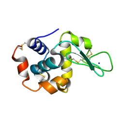
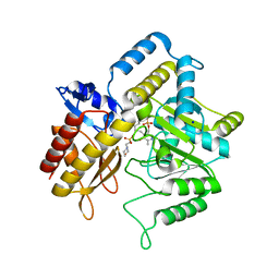
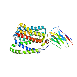
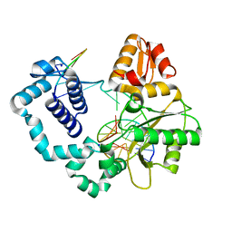
4MFA
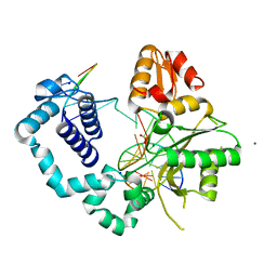 | | Structure of human DNA polymerase beta complexed with nicked DNA containing a mismatched template O6MG and incoming TTP | | 分子名称: | DNA polymerase beta, MAGNESIUM ION, PHOSPHATE ION, ... | | 著者 | Koag, M.C, Min, K, Monzingo, A.F, Lee, S. | | 登録日 | 2013-08-27 | | 公開日 | 2014-08-27 | | 最終更新日 | 2024-02-28 | | 実験手法 | X-RAY DIFFRACTION (2.27 Å) | | 主引用文献 | Structures of human DNA polymerase beta inserting bases opposite templating O6MG
To be Published
|
|
4QOQ
 | | Structure of Bacillus pumilus catalase with guaiacol bound | | 分子名称: | CHLORIDE ION, Catalase, Guaiacol, ... | | 著者 | Loewen, P.C. | | 登録日 | 2014-06-20 | | 公開日 | 2015-02-25 | | 最終更新日 | 2024-02-28 | | 実験手法 | X-RAY DIFFRACTION (1.7 Å) | | 主引用文献 | Unprecedented access of phenolic substrates to the heme active site of a catalase: Substrate binding and peroxidase-like reactivity of Bacillus pumilus catalase monitored by X-ray crystallography and EPR spectroscopy.
Proteins, 83, 2015
|
|
8H8V
 | |
4QOO
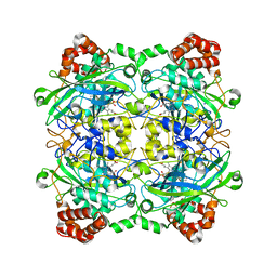 | | Structure of Bacillus pumilus catalase with resorcinol bound. | | 分子名称: | CHLORIDE ION, Catalase, PROTOPORPHYRIN IX CONTAINING FE, ... | | 著者 | Loewen, P.C. | | 登録日 | 2014-06-20 | | 公開日 | 2015-02-25 | | 最終更新日 | 2023-09-20 | | 実験手法 | X-RAY DIFFRACTION (1.75 Å) | | 主引用文献 | Unprecedented access of phenolic substrates to the heme active site of a catalase: Substrate binding and peroxidase-like reactivity of Bacillus pumilus catalase monitored by X-ray crystallography and EPR spectroscopy.
Proteins, 83, 2015
|
|
8H8U
 | |
8H8T
 | |
4QON
 | | Structure of Bacillus pumilus catalase with catechol bound. | | 分子名称: | CATECHOL, CHLORIDE ION, Catalase, ... | | 著者 | Loewen, P.C. | | 登録日 | 2014-06-20 | | 公開日 | 2015-02-25 | | 最終更新日 | 2023-09-20 | | 実験手法 | X-RAY DIFFRACTION (1.8 Å) | | 主引用文献 | Unprecedented access of phenolic substrates to the heme active site of a catalase: Substrate binding and peroxidase-like reactivity of Bacillus pumilus catalase monitored by X-ray crystallography and EPR spectroscopy.
Proteins, 83, 2015
|
|
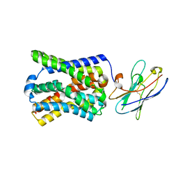
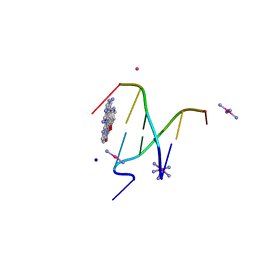
4LVS
 | | DNA polymerase beta mismatched substrate complex with Mn2+, 2.5 min | | 分子名称: | 2'-DEOXYADENOSINE 5'-TRIPHOSPHATE, DNA (5'-D(*CP*CP*GP*AP*CP*GP*GP*CP*GP*CP*AP*TP*CP*AP*GP*C)-3'), DNA (5'-D(*GP*CP*TP*GP*AP*TP*GP*CP*GP*C)-3'), ... | | 著者 | Freudenthal, B.D, Beard, W.A, Shock, D.D, Wilson, S.H. | | 登録日 | 2013-07-26 | | 公開日 | 2013-09-04 | | 最終更新日 | 2023-09-20 | | 実験手法 | X-RAY DIFFRACTION (2.002 Å) | | 主引用文献 | Observing a DNA polymerase choose right from wrong.
Cell(Cambridge,Mass.), 154, 2013
|
|
4QOR
 | | Structure of Bacillus pumilus catalase with chlorophenol bound. | | 分子名称: | 2-CHLOROPHENOL, CHLORIDE ION, Catalase, ... | | 著者 | Loewen, P.C. | | 登録日 | 2014-06-20 | | 公開日 | 2015-02-25 | | 最終更新日 | 2024-02-28 | | 実験手法 | X-RAY DIFFRACTION (1.95 Å) | | 主引用文献 | Unprecedented access of phenolic substrates to the heme active site of a catalase: Substrate binding and peroxidase-like reactivity of Bacillus pumilus catalase monitored by X-ray crystallography and EPR spectroscopy.
Proteins, 83, 2015
|
|
8H8W
 | |
3MCQ
 | |
4R65
 | | Ternary complex crystal structure of R258A mutant of DNA polymerase Beta | | 分子名称: | 2'-DEOXYURIDINE 5'-ALPHA,BETA-IMIDO-TRIPHOSPHATE, CHLORIDE ION, DNA (5'-D(*CP*CP*GP*AP*CP*AP*GP*CP*GP*CP*AP*TP*CP*AP*GP*C)-3'), ... | | 著者 | Batra, V.K, Beard, W.A, Wilson, S.H. | | 登録日 | 2014-08-22 | | 公開日 | 2014-10-08 | | 最終更新日 | 2024-02-28 | | 実験手法 | X-RAY DIFFRACTION (1.95 Å) | | 主引用文献 | Substrate-induced DNA Polymerase beta Activation.
J.Biol.Chem., 289, 2014
|
|
3O69
 | | Structure of the E100A E.coli GDP-mannose hydrolase (yffh) in complex with Mg++ | | 分子名称: | CHLORIDE ION, DI(HYDROXYETHYL)ETHER, GDP-mannose pyrophosphatase nudK, ... | | 著者 | Amzel, L.M, Gabelli, S.B, Boto, A.N. | | 登録日 | 2010-07-28 | | 公開日 | 2011-05-11 | | 最終更新日 | 2023-09-06 | | 実験手法 | X-RAY DIFFRACTION (2.1 Å) | | 主引用文献 | Structural studies of the Nudix GDP-mannose hydrolase from E. coli reveals a new motif for mannose recognition.
Proteins, 79, 2011
|
|
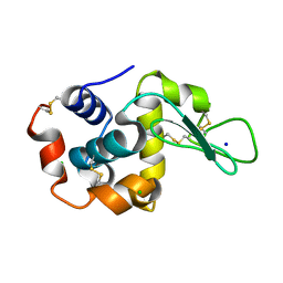
4LH6
 | | Crystal structure of a LigA inhibitor | | 分子名称: | 4-amino-2-bromothieno[3,2-c]pyridine-7-carboxamide, ACETATE ION, BETA-NICOTINAMIDE RIBOSE MONOPHOSPHATE, ... | | 著者 | Benenato, K, Wang, H, Mcguire, H.M, Davis, H, Gao, N, Prince, D.B, Jahic, H, Stokes, S.S, Boriack-Sjodin, P.A. | | 登録日 | 2013-06-30 | | 公開日 | 2013-12-25 | | 最終更新日 | 2024-02-28 | | 実験手法 | X-RAY DIFFRACTION (1.65 Å) | | 主引用文献 | Identification through structure-based methods of a bacterial NAD(+)-dependent DNA ligase inhibitor that avoids known resistance mutations.
Bioorg.Med.Chem.Lett., 24, 2014
|
|
1ZOD
 | |
4R7C
 | | Crystal Structure of CNG mimicking NaK-ETPP mutant cocrystallized with DiMethylammonium | | 分子名称: | (4S)-2-METHYL-2,4-PENTANEDIOL, DIMETHYLAMINE, GLYCINE, ... | | 著者 | De March, M, Napolitano, L.M.R, Onesti, S. | | 登録日 | 2014-08-27 | | 公開日 | 2015-07-01 | | 最終更新日 | 2023-09-20 | | 実験手法 | X-RAY DIFFRACTION (2.3 Å) | | 主引用文献 | A structural, functional, and computational analysis suggests pore flexibility as the base for the poor selectivity of CNG channels.
Proc.Natl.Acad.Sci.USA, 112, 2015
|
|
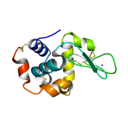
4M9N
 | | DNA Polymerase Beta E295K Soaked with dATP | | 分子名称: | 2'-DEOXYADENOSINE 5'-TRIPHOSPHATE, DNA Downstream Strand, DNA Primer Strand, ... | | 著者 | Eckenroth, B.E, Doublie, S. | | 登録日 | 2013-08-14 | | 公開日 | 2013-10-16 | | 最終更新日 | 2024-02-28 | | 実験手法 | X-RAY DIFFRACTION (2.275 Å) | | 主引用文献 | The E295K Cancer Variant of Human Polymerase beta Favors the Mismatch Conformational Pathway during Nucleotide Selection.
J.Biol.Chem., 288, 2013
|
|
7PQQ
 | |
7PQG
 | |
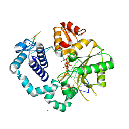
4R60
 | | Crystal Structure of Xaa-Pro dipeptidase from Xanthomonas campestris | | 分子名称: | MANGANESE (II) ION, PHOSPHATE ION, Proline dipeptidase, ... | | 著者 | Kumar, A, Ghosh, B, Are, V.N, Jamdar, S.N, Makde, R.D, Sharma, S.M. | | 登録日 | 2014-08-22 | | 公開日 | 2014-09-17 | | 最終更新日 | 2023-11-08 | | 実験手法 | X-RAY DIFFRACTION (1.83 Å) | | 主引用文献 | Crystal Structure of Xaa-Pro dipeptidase from Xanthomonas campestris
to be published
|
|
4MNB
 | | Crystal Structure of a complex between the marine anticancer drug Variolin B and DNA | | 分子名称: | 5'-D(*CP*GP*TP*AP*CP*G)-3', 9-amino-5-(2-aminopyrimidin-4-yl)pyrido[3',2':4,5]pyrrolo[1,2-c]pyrimidin-4-ol, COBALT (II) ION, ... | | 著者 | Canals, A, Arribas-Bosacoma, R, Alvarez, M, Albericio, F, Aymami, J, Coll, M. | | 登録日 | 2013-09-10 | | 公開日 | 2015-03-11 | | 最終更新日 | 2024-02-28 | | 実験手法 | X-RAY DIFFRACTION (1.4 Å) | | 主引用文献 | Intercalative DNA binding of the marine anticancer drug variolin B.
Sci Rep, 7, 2017
|
|
4R64
 | | Binary complex crystal structure of E295K mutant of DNA polymerase Beta | | 分子名称: | DNA (5'-D(*CP*CP*GP*AP*CP*AP*GP*CP*GP*CP*AP*TP*CP*AP*GP*C)-3'), DNA (5'-D(*GP*CP*TP*GP*AP*TP*GP*CP*GP*C)-3'), DNA (5'-D(P*GP*TP*CP*GP*G)-3'), ... | | 著者 | Batra, V.K, Beard, W.A, Wilson, S.H. | | 登録日 | 2014-08-22 | | 公開日 | 2014-10-08 | | 最終更新日 | 2023-09-20 | | 実験手法 | X-RAY DIFFRACTION (2.2 Å) | | 主引用文献 | Substrate-induced DNA Polymerase beta Activation.
J.Biol.Chem., 289, 2014
|
|


