6D0I
 
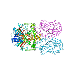
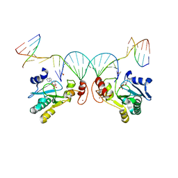
 | | ParT: Prs ADP-ribosylating toxin bound to cognate antitoxin ParS. L48M ParT, SeMet-substituted complex. | | 分子名称: | GLYCEROL, ParS: COG5642 (DUF2384) antitoxin fragment, ParT: COG5654 (RES domain) toxin | | 著者 | Piscotta, F.J, Jeffrey, P.D, Link, A.J. | | 登録日 | 2018-04-10 | | 公開日 | 2019-01-09 | | 最終更新日 | 2024-10-23 | | 実験手法 | X-RAY DIFFRACTION (1.55 Å) | | 主引用文献 | ParST is a widespread toxin-antitoxin module that targets nucleotide metabolism.
Proc. Natl. Acad. Sci. U.S.A., 116, 2019
|
|
6D17
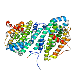
 | | Crystal structure of KPC-2 complexed with compound 3 | | 分子名称: | Carbapenem-hydrolyzing beta-lactamase KPC, GLYCEROL, [(6-oxo-2H,6H-[1,3]dioxolo[4,5-g][1]benzopyran-8-yl)methyl]phosphonic acid | | 著者 | Pemberton, O.A, Chen, Y. | | 登録日 | 2018-04-11 | | 公開日 | 2019-04-17 | | 最終更新日 | 2024-11-06 | | 実験手法 | X-RAY DIFFRACTION (1.45 Å) | | 主引用文献 | Heteroaryl Phosphonates as Noncovalent Inhibitors of Both Serine- and Metallocarbapenemases.
J.Med.Chem., 62, 2019
|
|
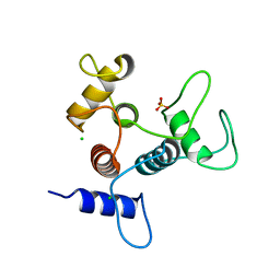
6D1R
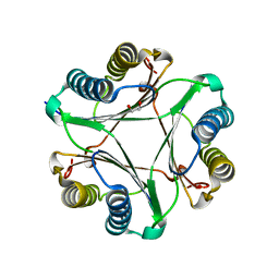
 | | Structure of Staphylococcus aureus RNase P protein at 2.0 angstrom | | 分子名称: | Ribonuclease P protein component | | 著者 | Ha, L, Colquhoun, J, Noinaj, N, Das, C, Dunman, P, Flaherty, D.P. | | 登録日 | 2018-04-12 | | 公開日 | 2018-09-26 | | 最終更新日 | 2024-03-13 | | 実験手法 | X-RAY DIFFRACTION (1.995 Å) | | 主引用文献 | Crystal structure of the ribonuclease-P-protein subunit from Staphylococcus aureus.
Acta Crystallogr F Struct Biol Commun, 74, 2018
|
|
6D20
 | | Crystal structure of Tyrosine-protein kinase receptor in complex with 5-(4-fluorophenyl)thieno[2,3-d]pyrimidin-4(3H)-one and 5-{[2,4-dichloro-5-(pyridin-2-yl)benzene-1-carbonyl]amino}-N-(2-hydroxy-2-methylpropyl)-1-phenyl-1H-pyrazole-3-carboxamide Inhibitors | | 分子名称: | 5-(4-fluorophenyl)thieno[2,3-d]pyrimidin-4(3H)-one, 5-{[2,4-dichloro-5-(pyridin-2-yl)benzene-1-carbonyl]amino}-N-(2-hydroxy-2-methylpropyl)-1-phenyl-1H-pyrazole-3-carboxamide, High affinity nerve growth factor receptor | | 著者 | Greasley, S.E, Johnson, E, Kraus, M.L, Cronin, C.N. | | 登録日 | 2018-04-12 | | 公開日 | 2018-05-02 | | 最終更新日 | 2024-03-13 | | 実験手法 | X-RAY DIFFRACTION (1.94 Å) | | 主引用文献 | Discovery of Allosteric, Potent, Subtype Selective, and Peripherally Restricted TrkA Kinase Inhibitors.
J. Med. Chem., 62, 2019
|
|
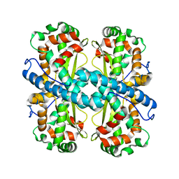
6D3H
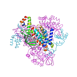
 | | FT_T dioxygenase with bound dichlorprop | | 分子名称: | (2R)-2-(2,4-dichlorophenoxy)propanoic acid, 2-OXOGLUTARIC ACID, CHLORIDE ION, ... | | 著者 | Rydel, T.J, Halls, C.E. | | 登録日 | 2018-04-16 | | 公開日 | 2018-08-15 | | 最終更新日 | 2024-03-13 | | 実験手法 | X-RAY DIFFRACTION (2.03 Å) | | 主引用文献 | Development of enzymes for robust aryloxyphenoxypropionate and synthetic auxin herbicide tolerance traits in maize and soybean crops.
Pest Manag. Sci., 75, 2019
|
|
6CWP
 | |
6CUQ
 | |
6CXT
 | | Crystal structure of FAD-dependent dehydrogenase | | 分子名称: | 1,2-ETHANEDIOL, Alpha/beta hydrolase fold protein, Butyryl-CoA dehydrogenase, ... | | 著者 | Agarwal, V. | | 登録日 | 2018-04-04 | | 公開日 | 2019-01-16 | | 最終更新日 | 2024-11-13 | | 実験手法 | X-RAY DIFFRACTION (1.9 Å) | | 主引用文献 | Insights into Thiotemplated Pyrrole Biosynthesis Gained from the Crystal Structure of Flavin-Dependent Oxidase in Complex with Carrier Protein.
Biochemistry, 58, 2019
|
|
6CVO
 | | Human Aprataxin (Aptx) bound to nicked RNA-DNA, AMP and Zn product complex | | 分子名称: | ADENOSINE MONOPHOSPHATE, Aprataxin, BETA-MERCAPTOETHANOL, ... | | 著者 | Schellenberg, M.J, Tumbale, P.P, Williams, R.S. | | 登録日 | 2018-03-28 | | 公開日 | 2018-08-29 | | 最終更新日 | 2023-10-04 | | 実験手法 | X-RAY DIFFRACTION (2.4 Å) | | 主引用文献 | Mechanism of APTX nicked DNA sensing and pleiotropic inactivation in neurodegenerative disease.
EMBO J., 37, 2018
|
|
6CY4
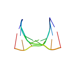 | | RNA octamer containing 2'-OMe, 4'- Cbeta-OMe U. | | 分子名称: | RNA (5'-R(*(CBV)P*GP*AP*AP*(UOB)P*UP*CP*G)-3') | | 著者 | Harp, J.M, Egli, M. | | 登録日 | 2018-04-04 | | 公開日 | 2018-08-29 | | 最終更新日 | 2024-03-13 | | 実験手法 | X-RAY DIFFRACTION (1.851 Å) | | 主引用文献 | Structural basis for the synergy of 4'- and 2'-modifications on siRNA nuclease resistance, thermal stability and RNAi activity.
Nucleic Acids Res., 46, 2018
|
|
6CVZ
 | | Crystal structure of the WD40-repeat of RFWD3 | | 分子名称: | E3 ubiquitin-protein ligase RFWD3, MAGNESIUM ION | | 著者 | DONG, A, LOPPNAU, P, SEITOVA, A, HUTCHINSON, A, TEMPEL, W, WEI, Y, Bountra, C, Arrowsmith, C.H, Edwards, A.M, BROWN, P.J, TONG, Y, Structural Genomics Consortium (SGC) | | 登録日 | 2018-03-29 | | 公開日 | 2018-06-27 | | 最終更新日 | 2024-10-09 | | 実験手法 | X-RAY DIFFRACTION (1.8 Å) | | 主引用文献 | Target highlights in CASP13: Experimental target structures through the eyes of their authors.
Proteins, 87, 2019
|
|
6CWC
 | | Crystal structure of SpaA-SLH | | 分子名称: | CHLORIDE ION, SULFATE ION, Surface (S-) layer glycoprotein | | 著者 | Blackler, R.J, Evans, S.V. | | 登録日 | 2018-03-30 | | 公開日 | 2018-08-15 | | 最終更新日 | 2024-03-13 | | 実験手法 | X-RAY DIFFRACTION (1.9 Å) | | 主引用文献 | Structural basis of cell wall anchoring by SLH domains in Paenibacillus alvei.
Nat Commun, 9, 2018
|
|
6CYP
 | |
6CZQ
 | |
8IIY
 | |
6D18
 | | Crystal structure of KPC-2 complexed with compound 6 | | 分子名称: | Carbapenem-hydrolyzing beta-lactamase KPC, GLYCEROL, [(5,7-dimethyl-2-oxo-2H-1-benzopyran-4-yl)methyl]phosphonic acid | | 著者 | Pemberton, O.A, Chen, Y. | | 登録日 | 2018-04-11 | | 公開日 | 2019-04-17 | | 最終更新日 | 2024-11-20 | | 実験手法 | X-RAY DIFFRACTION (1.35 Å) | | 主引用文献 | Heteroaryl Phosphonates as Noncovalent Inhibitors of Both Serine- and Metallocarbapenemases.
J.Med.Chem., 62, 2019
|
|
8IIZ
 | |
6D47
 | |
8IN1
 | | beta-glucosidase protein from Aplysia kurodai | | 分子名称: | 2-acetamido-2-deoxy-beta-D-glucopyranose, Beta-Glucosidase, alpha-L-fucopyranose-(1-3)-2-acetamido-2-deoxy-beta-D-glucopyranose, ... | | 著者 | Sun, X.M, Ye, Y.X, Kato, K, Yu, J, Yao, M. | | 登録日 | 2023-03-08 | | 公開日 | 2023-11-15 | | 最終更新日 | 2024-11-20 | | 実験手法 | X-RAY DIFFRACTION (2.7 Å) | | 主引用文献 | Structural basis of EHEP-mediated offense against phlorotannin-induced defense from brown algae to protect aku BGL activity.
Elife, 12, 2023
|
|
6CXS
 | | Crystal Structure of Clostridium perfringens beta-glucuronidase bound with a novel, potent inhibitor 4-(8-(piperazin-1-yl)-1,2,3,4-tetrahydro-[1,2,3]triazino[4',5':4,5]thieno[2,3-c]isoquinolin-5-yl)morpholine | | 分子名称: | 4-(8-(piperazin-1-yl)-1,2,3,4-tetrahydro-[1,2,3]triazino[4',5':4,5]thieno[2,3-c]isoquinolin-5-yl)morpholine, Beta-glucuronidase, Maltose/maltodextrin-binding periplasmic protein | | 著者 | Wallace, B.D, Redinbo, M.R. | | 登録日 | 2018-04-04 | | 公開日 | 2019-04-17 | | 最終更新日 | 2023-10-04 | | 実験手法 | X-RAY DIFFRACTION (2.8 Å) | | 主引用文献 | Targeted inhibition of gut bacterial beta-glucuronidase activity enhances anticancer drug efficacy.
Proc.Natl.Acad.Sci.USA, 2020
|
|
6CYO
 | |
6CYS
 | |
6D14
 | |
6D1Z
 | | Crystal structure of Tyrosine-protein kinase receptor in complex with 5-(4-fluorophenyl)thieno[2,3-d]pyrimidin-4(3H)-one Inhibitor | | 分子名称: | 5-(4-fluorophenyl)thieno[2,3-d]pyrimidin-4(3H)-one, 5-{[5-(6-aminopyridin-2-yl)-2-chlorobenzene-1-carbonyl]amino}-1-phenyl-1H-pyrazole-3-carboxamide, GLYCEROL, ... | | 著者 | Greasley, S.E, Johnson, E, Kraus, M.L, Cronin, C.N. | | 登録日 | 2018-04-12 | | 公開日 | 2018-05-02 | | 最終更新日 | 2024-03-13 | | 実験手法 | X-RAY DIFFRACTION (1.87 Å) | | 主引用文献 | Discovery of Allosteric, Potent, Subtype Selective, and Peripherally Restricted TrkA Kinase Inhibitors.
J. Med. Chem., 62, 2019
|
|
1M1J
 | | Crystal structure of native chicken fibrinogen with two different bound ligands | | 分子名称: | 2-acetamido-2-deoxy-alpha-D-glucopyranose, 2-acetamido-2-deoxy-beta-D-glucopyranose, CALCIUM ION, ... | | 著者 | Yang, Z, Kollman, J.M, Pandi, L, Doolittle, R.F. | | 登録日 | 2002-06-19 | | 公開日 | 2002-06-26 | | 最終更新日 | 2024-10-30 | | 実験手法 | X-RAY DIFFRACTION (2.7 Å) | | 主引用文献 | Crystal Structure of Native Chicken Fibrinogen at 2.7 A Resolution
Biochemistry, 40, 2001
|
|


