3MY2
 
 | |
3NDY
 | |
3NDZ
 | |
3KM6
 | |
3KD6
 | |
3NYC
 | |
3NO1
 | |
3B69
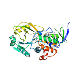
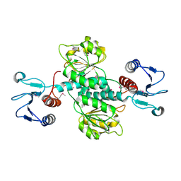
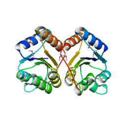
 | | T cruzi Trans-sialidase complex with benzoylated NANA derivative | | 分子名称: | 4-(2-HYDROXYETHYL)-1-PIPERAZINE ETHANESULFONIC ACID, 5-acetamido-9-(benzoylamino)-3,5,9-trideoxy-3-fluoro-D-erythro-alpha-L-manno-non-2-ulopyranosonic acid, CHLORIDE ION, ... | | 著者 | Buschiazzo, A. | | 登録日 | 2007-10-28 | | 公開日 | 2008-05-13 | | 最終更新日 | 2024-11-13 | | 実験手法 | X-RAY DIFFRACTION (1.67 Å) | | 主引用文献 | A new generation of specific Trypanosoma cruzi trans-sialidase inhibitors.
Angew.Chem.Int.Ed.Engl., 47, 2008
|
|
3GU4
 | |
3GU7
 | |
2E40
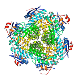
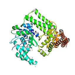
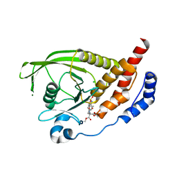
 | | Crystal structure of intracellular family 1 beta-glucosidase BGL1A from the basidiomycete Phanerochaete chrysosporium in complex with gluconolactone | | 分子名称: | Beta-glucosidase, D-glucono-1,5-lactone | | 著者 | Nijikken, Y, Tsukada, T, Igarashi, K, Samejima, M, Fushinobu, S. | | 登録日 | 2006-12-01 | | 公開日 | 2007-03-27 | | 最終更新日 | 2023-10-25 | | 実験手法 | X-RAY DIFFRACTION (1.9 Å) | | 主引用文献 | Crystal structure of intracellular family 1 beta-glucosidase BGL1A from the basidiomycete Phanerochaete chrysosporium
Febs Lett., 581, 2007
|
|
3GU6
 | |
3GUB
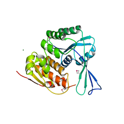
 | | Crystal structure of DAPKL93G complexed with N6-(2-Phenylethyl)adenosine | | 分子名称: | 9-alpha-L-lyxofuranosyl-N-(2-phenylethyl)-9H-purin-6-amine, Death-associated protein kinase 1, SULFATE ION | | 著者 | McNamara, L.K, Schumacher, A.M, Schavocky, J.S, Watterson, D.M, Brunzelle, J.S. | | 登録日 | 2009-03-29 | | 公開日 | 2010-03-09 | | 最終更新日 | 2023-09-06 | | 実験手法 | X-RAY DIFFRACTION (1.71 Å) | | 主引用文献 | Crystal structures of the DAPK gatekeeper mutant complexed with N6-modified adenosine analogs.
To be Published
|
|
3GVX
 | |
2EWF
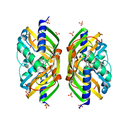
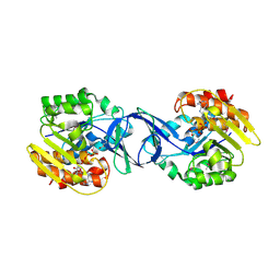
 | | Crystal structure of the site-specific DNA nickase N.BspD6I | | 分子名称: | BROMIDE ION, Nicking endonuclease N.BspD6I | | 著者 | Kachalova, G.S, Bartunik, H.D, Artyukh, R.I, Rogulin, E.A, Perevyazova, T.A, Zheleznaya, L.A, Matvienko, N.I. | | 登録日 | 2005-11-03 | | 公開日 | 2006-11-21 | | 最終更新日 | 2024-02-14 | | 実験手法 | X-RAY DIFFRACTION (1.84 Å) | | 主引用文献 | Structural analysis of the heterodimeric type IIS restriction endonuclease R.BspD6I acting as a complex between a monomeric site-specific nickase and a catalytic subunit.
J.Mol.Biol., 384, 2008
|
|
3HIC
 | |
2F99
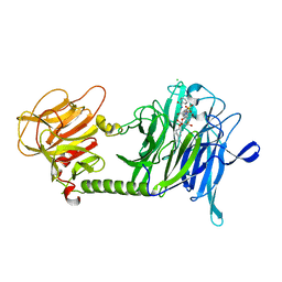
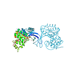
 | | Crystal structure of the polyketide cyclase AknH with bound substrate and product analogue: implications for catalytic mechanism and product stereoselectivity. | | 分子名称: | Aklanonic Acid methyl Ester Cyclase, AknH, SULFATE ION, ... | | 著者 | Kallio, P, Sultana, A, Neimi, J, Mantsala, P, Schneider, G. | | 登録日 | 2005-12-05 | | 公開日 | 2006-02-14 | | 最終更新日 | 2023-08-30 | | 実験手法 | X-RAY DIFFRACTION (1.9 Å) | | 主引用文献 | Crystal structure of the polyketide cyclase AknH with bound substrate and product analogue: implications for catalytic mechanism and product stereoselectivity.
J.Mol.Biol., 357, 2006
|
|
3HEB
 | |
2H03
 | | Structural studies of protein tyrosine phosphatase beta catalytic domain in complex with inhibitors | | 分子名称: | (4-{4-[(TERT-BUTOXYCARBONYL)AMINO]-2,2-BIS(ETHOXYCARBONYL)BUTYL}PHENYL)SULFAMIC ACID, CHLORIDE ION, MAGNESIUM ION, ... | | 著者 | Evdokimov, A.G, Pokross, M.E, Walter, R.L, Mekel, M, Gray, J.L, Peters, K.G, Maier, M.B, Amarasinghe, K.D, Clark, C.M, Nichols, R. | | 登録日 | 2006-05-13 | | 公開日 | 2006-06-13 | | 最終更新日 | 2023-08-30 | | 実験手法 | X-RAY DIFFRACTION (1.65 Å) | | 主引用文献 | Design and synthesis of potent, non-peptidic inhibitors of HPTPbeta.
Bioorg.Med.Chem.Lett., 16, 2006
|
|
3IH0
 | | Crystal structure of an uncharacterized sugar kinase PH1459 from Pyrococcus horikoshii in complex with AMP-PNP | | 分子名称: | GLYCEROL, PHOSPHOAMINOPHOSPHONIC ACID-ADENYLATE ESTER, Uncharacterized sugar kinase PH1459 | | 著者 | Kumar, G, Eswaramoorthy, S, Burley, S.K, Swaminathan, S, New York SGX Research Center for Structural Genomics (NYSGXRC) | | 登録日 | 2009-07-29 | | 公開日 | 2009-09-08 | | 最終更新日 | 2024-11-20 | | 実験手法 | X-RAY DIFFRACTION (1.9 Å) | | 主引用文献 | Crystal structure of an uncharacterized sugar kinase PH1459 from Pyrococcus horikoshii in complex with AMP-PNP
To be Published
|
|
3IKH
 | |
3IE7
 | |
3ILH
 | |
3HKR
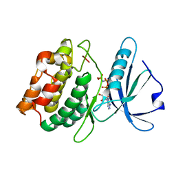
 | | Crystal Structure of Glutathione Transferase Pi Y108V Mutant | | 分子名称: | 2-(N-MORPHOLINO)-ETHANESULFONIC ACID, CALCIUM ION, CARBONATE ION, ... | | 著者 | Parker, L.J. | | 登録日 | 2009-05-25 | | 公開日 | 2009-09-22 | | 最終更新日 | 2023-11-01 | | 実験手法 | X-RAY DIFFRACTION (1.8 Å) | | 主引用文献 | Influence of the H-site residue 108 on human glutathione transferase P1-1 ligand binding: structure-thermodynamic relationships and thermal stability.
Protein Sci., 18, 2009
|
|
2G7F
 | | The 1.95 A crystal structure of Vibrio cholerae extracellular endonuclease I | | 分子名称: | CHLORIDE ION, Endonuclease I, MAGNESIUM ION | | 著者 | Altermark, B, Smalaas, A.O, Willassen, N.P, Helland, R. | | 登録日 | 2006-02-28 | | 公開日 | 2006-10-31 | | 最終更新日 | 2024-10-30 | | 実験手法 | X-RAY DIFFRACTION (1.95 Å) | | 主引用文献 | The structure of Vibrio cholerae extracellular endonuclease I reveals the presence of a buried chloride ion.
Acta Crystallogr.,Sect.D, 62, 2006
|
|


