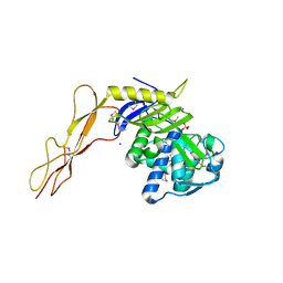
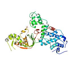
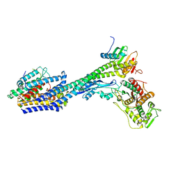
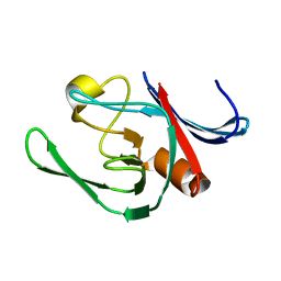
8F3V
 
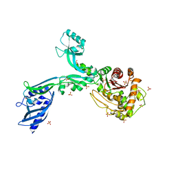 | | Crystal structure of Penicillin Binding Protein 5 (PBP5) PAPAPAP variant apo form from Enterococcus faecium | | 分子名称: | Penicillin binding protein 5, SULFATE ION | | 著者 | Schoenle, M.V, D'Andrea, E.D, Choy, M.S, Peti, W, Page, R. | | 登録日 | 2022-11-10 | | 公開日 | 2023-11-15 | | 実験手法 | X-RAY DIFFRACTION (3.1 Å) | | 主引用文献 | The Molecular Basis for Resistance of E. faecium PBP5 to beta-lactam antibiotics
To Be Published
|
|
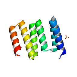
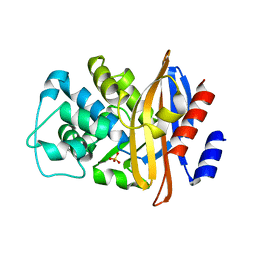
3BRM
 | | Crystal structure of the covalent complex between the Bacillus subtilis glutaminase YbgJ and 5-oxo-L-norleucine formed by reaction of the protein with 6-diazo-5-oxo-L-norleucine | | 分子名称: | 5-OXO-L-NORLEUCINE, Glutaminase 1 | | 著者 | Singer, A.U, Kim, Y, Dementieva, I, Vinokour, E, Joachimiak, A, Savchenko, A, Yakunin, A. | | 登録日 | 2007-12-21 | | 公開日 | 2008-05-20 | | 最終更新日 | 2011-07-13 | | 実験手法 | X-RAY DIFFRACTION (2.29 Å) | | 主引用文献 | Functional and structural characterization of four glutaminases from Escherichia coli and Bacillus subtilis.
Biochemistry, 47, 2008
|
|
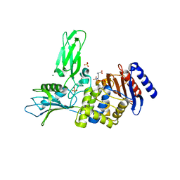
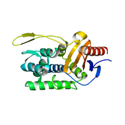
8F3F
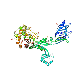 | | Crystal structure of Penicillin Binding Protein 5 (PBP5) T485M variant apo form from Enterococcus faecium | | 分子名称: | Penicillin binding protein 5, SULFATE ION | | 著者 | D'Andrea, E.D, Choy, M.S, Hunashal, Y, Schoenle, M.V, Page, R, Peti, W. | | 登録日 | 2022-11-10 | | 公開日 | 2023-07-05 | | 最終更新日 | 2023-10-25 | | 実験手法 | X-RAY DIFFRACTION (2.84 Å) | | 主引用文献 | The Molecular Basis for Resistance of E. faecium PBP5 to beta-lactam Antibiotics
Nat Commun, 2023
|
|
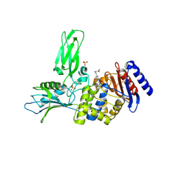
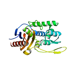
8F3G
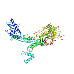 | | Crystal structure of Penicillin Binding Protein 5 (PBP5) T485M variant in the penicillin bound form from Enterococcus faecium | | 分子名称: | OPEN FORM - PENICILLIN G, Penicillin binding protein 5, SULFATE ION | | 著者 | D'Andrea, E.D, Choy, M.S, Schoenle, M.V, Page, R, Peti, W. | | 登録日 | 2022-11-10 | | 公開日 | 2023-07-05 | | 最終更新日 | 2024-03-13 | | 実験手法 | X-RAY DIFFRACTION (3.59 Å) | | 主引用文献 | The Molecular Basis for Resistance of E. faecium PBP5 to beta-lactam Antibiotics
Nat Commun, 2023
|
|
1SAX
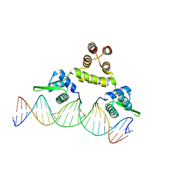 | | Three-dimensional structure of s.aureus methicillin-resistance regulating transcriptional repressor meci in complex with 25-bp ds-DNA | | 分子名称: | 5'-d(CAAAATTACAACTGTAATATCGGAG)-3', 5'-d(GCTCCGATATTACAGTTGTAATTTT)-3', Methicillin resistance regulatory protein mecI, ... | | 著者 | Garcia-Castellanos, R, Mallorqui-Fernandez, G, Marrero, A, Potempa, J, Coll, M, Gomis-Ruth, F.X. | | 登録日 | 2004-02-09 | | 公開日 | 2004-04-27 | | 最終更新日 | 2023-08-23 | | 実験手法 | X-RAY DIFFRACTION (2.8 Å) | | 主引用文献 | On the transcriptional regulation of methicillin resistance: MecI repressor in complex with its operator
J.Biol.Chem., 279, 2004
|
|
1XP4
 | | Crystal structure of a peptidoglycan synthesis regulatory factor (PBP3) from Streptococcus pneumoniae | | 分子名称: | D-alanyl-D-alanine carboxypeptidase, IODIDE ION, SULFATE ION | | 著者 | Morlot, C, Pernot, L, Le Gouellec, A, Di Guilmi, A.M, Vernet, T, Dideberg, O, Dessen, A. | | 登録日 | 2004-10-08 | | 公開日 | 2004-11-09 | | 最終更新日 | 2023-11-15 | | 実験手法 | X-RAY DIFFRACTION (2.8 Å) | | 主引用文献 | Crystal structure of a peptidoglycan synthesis regulatory factor (PBP3) from Streptococcus pneumoniae
J.Biol.Chem., 280, 2005
|
|
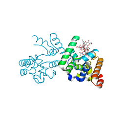
6W5Q
 | | Structure of the globular C-terminal domain of P. aeruginosa LpoP | | 分子名称: | Peptidoglycan synthase activator LpoP, SULFATE ION, TRIETHYLENE GLYCOL | | 著者 | Caveney, N.A, Robb, C.S, Simorre, J.P, Strynadka, N.C.J. | | 登録日 | 2020-03-13 | | 公開日 | 2020-05-06 | | 最終更新日 | 2023-10-18 | | 実験手法 | X-RAY DIFFRACTION (2.2 Å) | | 主引用文献 | Structure of the Peptidoglycan Synthase Activator LpoP in Pseudomonas aeruginosa.
Structure, 28, 2020
|
|
2VGK
 | | Crystal structure of Actinomadura R39 DD-peptidase complexed with a peptidoglycan-mimetic cephalosporin | | 分子名称: | (2R)-2-AMINO-7-{[(1R)-1-CARBOXYETHYL]AMINO}-7-OXOHEPTANOIC ACID, D-ALANYL-D-ALANINE CARBOXYPEPTIDASE, MAGNESIUM ION, ... | | 著者 | Sauvage, E, kerff, F, Herman, R, Charlier, P. | | 登録日 | 2007-11-14 | | 公開日 | 2008-11-25 | | 最終更新日 | 2023-12-13 | | 実験手法 | X-RAY DIFFRACTION (2.25 Å) | | 主引用文献 | Crystal Structures of Complexes of Bacterial Dd-Peptidases with Peptidoglycan-Mimetic Ligands: The Substrate Specificity Puzzle.
J.Mol.Biol., 381, 2008
|
|
2VGJ
 | | Crystal structure of Actinomadura R39 DD-peptidase complexed with a peptidoglycan-mimetic cephalosporin | | 分子名称: | CEPHALOSPORIN, D-ALANYL-D-ALANINE CARBOXYPEPTIDASE, MAGNESIUM ION, ... | | 著者 | Sauvage, E, kerff, F, Herman, R, Charlier, P. | | 登録日 | 2007-11-14 | | 公開日 | 2008-11-25 | | 最終更新日 | 2023-12-13 | | 実験手法 | X-RAY DIFFRACTION (2.4 Å) | | 主引用文献 | Crystal Structures of Complexes of Bacterial Dd-Peptidases with Peptidoglycan-Mimetic Ligands: The Substrate Specificity Puzzle.
J.Mol.Biol., 381, 2008
|
|
4RYE
 | | The crystal structure of D-ALANYL-D-ALANINE CARBOXYPEPTIDASE from Mycobacterium tuberculosis H37Rv | | 分子名称: | D-alanyl-D-alanine carboxypeptidase | | 著者 | Cuff, M, Tan, K, Hatzos-Skintges, C, Jedrzejczak, R, Sacchettini, J, Joachimiak, A, Midwest Center for Structural Genomics (MCSG), Structures of Mtb Proteins Conferring Susceptibility to Known Mtb Inhibitors (MTBI) | | 登録日 | 2014-12-15 | | 公開日 | 2015-01-28 | | 最終更新日 | 2017-11-22 | | 実験手法 | X-RAY DIFFRACTION (1.901 Å) | | 主引用文献 | The crystal structure of D-ALANYL-D-ALANINE CARBOXYPEPTIDASE from Mycobacterium tuberculosis H37Rv
To be Published
|
|
6NTW
 | | Crystal structure of E. coli YcbB | | 分子名称: | (2S,3R,4S)-4-{[(3S,5R)-5-(dimethylcarbamoyl)pyrrolidin-3-yl]sulfanyl}-2-[(2S,3R)-3-hydroxy-1-oxobutan-2-yl]-3-methyl-3,4-dihydro-2H-pyrrole-5-carboxylic acid, Probable L,D-transpeptidase YcbB, SULFATE ION | | 著者 | Caveney, N.A, Strynadka, N.C.J, Caballero, G, Worrall, L.J. | | 登録日 | 2019-01-30 | | 公開日 | 2019-03-20 | | 最終更新日 | 2020-01-08 | | 実験手法 | X-RAY DIFFRACTION (2.76 Å) | | 主引用文献 | Structural insight into YcbB-mediated beta-lactam resistance in Escherichia coli.
Nat Commun, 10, 2019
|
|
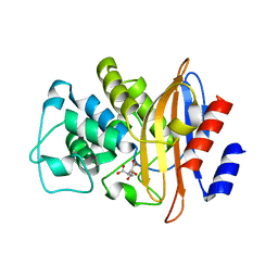
4JMX
 | | Structure of LD transpeptidase LdtMt1 in complex with imipenem | | 分子名称: | (5R)-5-[(1S,2R)-1-formyl-2-hydroxypropyl]-3-[(2-{[(E)-iminomethyl]amino}ethyl)sulfanyl]-4,5-dihydro-1H-pyrrole-2-carboxylic acid, Probable L,D-transpeptidase LdtA | | 著者 | Correale, S, Ruggiero, A, Capparelli, R, Pedone, E, Berisio, R. | | 登録日 | 2013-03-14 | | 公開日 | 2013-10-23 | | 実験手法 | X-RAY DIFFRACTION (2.55 Å) | | 主引用文献 | Structures of free and inhibited forms of the L,D-transpeptidase LdtMt1 from Mycobacterium tuberculosis.
Acta Crystallogr.,Sect.D, 69, 2013
|
|
1ESI
 | |
1ES5
 | |
8P1U
 | | Structure of divisome complex FtsWIQLB | | 分子名称: | Cell division protein FtsB, Cell division protein FtsL, Cell division protein FtsQ, ... | | 著者 | Yang, L, Chang, S, Tang, D, Dong, H. | | 登録日 | 2023-05-12 | | 公開日 | 2024-05-22 | | 最終更新日 | 2024-07-03 | | 実験手法 | ELECTRON MICROSCOPY (3.3 Å) | | 主引用文献 | Structural insights into the activation of the divisome complex FtsWIQLB.
Cell Discov, 10, 2024
|
|
4JMN
 | | Crystal structure of LD transpeptidase LdtMt1 from M. tuberculosis | | 分子名称: | Probable L,D-transpeptidase LdtA | | 著者 | Ruggiero, A, Correale, S, Berisio, R. | | 登録日 | 2013-03-14 | | 公開日 | 2013-10-23 | | 最終更新日 | 2024-02-28 | | 実験手法 | X-RAY DIFFRACTION (2.2 Å) | | 主引用文献 | Structures of free and inhibited forms of the L,D-transpeptidase LdtMt1 from Mycobacterium tuberculosis.
Acta Crystallogr.,Sect.D, 69, 2013
|
|
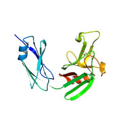
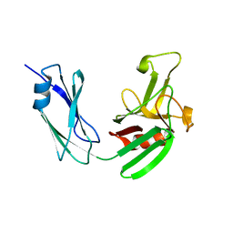
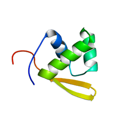
6G5S
 | | Solution structure of the TPR domain of the cell division coordinator, CpoB | | 分子名称: | Cell division coordinator CpoB | | 著者 | Simorre, J.P, Maya Martinez, R.C, Bougault, C, Vollmer, W, Egan, A. | | 登録日 | 2018-03-29 | | 公開日 | 2018-08-08 | | 最終更新日 | 2024-06-19 | | 実験手法 | SOLUTION NMR | | 主引用文献 | Induced conformational changes activate the peptidoglycan synthase PBP1B.
Mol. Microbiol., 110, 2018
|
|
1MKI
 | | Crystal Structure of Bacillus Subtilis Probable Glutaminase, APC1040 | | 分子名称: | 1,2-ETHANEDIOL, FORMIC ACID, Probable Glutaminase ybgJ | | 著者 | Kim, Y, Dementieva, I, Vinokour, E, Joachimiak, A, Midwest Center for Structural Genomics (MCSG) | | 登録日 | 2002-08-29 | | 公開日 | 2003-06-03 | | 最終更新日 | 2017-10-11 | | 実験手法 | X-RAY DIFFRACTION (2 Å) | | 主引用文献 | Functional and structural characterization of four glutaminases from Escherichia coli and Bacillus subtilis.
Biochemistry, 47, 2008
|
|
1W7F
 | |
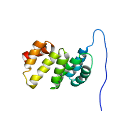
3ZG4
 | | NMR structure of the catalytic domain from E. faecium L,D- transpeptidase | | 分子名称: | ERFK/YBIS/YCFS/YNHG | | 著者 | Lecoq, L, Dubee, V, Triboulet, S, Bougault, C, Hugonnet, J.E, Arthur, M, Simorre, J.P. | | 登録日 | 2012-12-14 | | 公開日 | 2013-04-24 | | 最終更新日 | 2024-06-19 | | 実験手法 | SOLUTION NMR | | 主引用文献 | The Structure of Enterococcus Faecium L,D---Transpeptidase Acylated by Ertapenem Provides Insight Into the Inactivation Mechanism.
Acs Chem.Biol., 8, 2013
|
|
4BLM
 | |
3D3H
 | | Crystal structure of a complex of the peptidoglycan glycosyltransferase domain from Aquifex aeolicus and neryl moenomycin A | | 分子名称: | (2R)-3-{[(S)-{[(2R,3R,4R,5S,6S)-3-{[(2S,3R,4R,5S,6R)-3-(acetylamino)-5-{[(2S,3R,4R,5S,6R)-3-(acetylamino)-5-{[(2R,3R,4S,5R,6S)-6-carbamoyl-3,4,5-trihydroxytetrahydro-2H-pyran-2-yl]oxy}-4-hydroxy-6-methyltetrahydro-2H-pyran-2-yl]oxy}-4-hydroxy-6-({[(2R,3R,4S,5S,6R)-3,4,5-trihydroxy-6-(hydroxymethyl)tetrahydro-2H-pyran-2-yl]oxy}methyl)tetrahydro-2H-pyran-2-yl]oxy}-6-carbamoyl-4-(carbamoyloxy)-5-hydroxy-5-methyltetrahydro-2H-pyran-2-yl]oxy}(hydroxy)phosphoryl]oxy}-2-{[(2Z)-3,7-dimethylocta-2,6-dien-1-yl]oxy}propanoic acid, Penicillin-insensitive transglycosylase | | 著者 | Yuan, Y, Sliz, P, Walker, S. | | 登録日 | 2008-05-11 | | 公開日 | 2008-07-22 | | 最終更新日 | 2023-08-30 | | 実験手法 | X-RAY DIFFRACTION (2.31 Å) | | 主引用文献 | Structural analysis of the contacts anchoring moenomycin to peptidoglycan glycosyltransferases and implications for antibiotic design.
Acs Chem.Biol., 3, 2008
|
|
1U60
 | | MCSG APC5046 Probable glutaminase ybaS | | 分子名称: | 1,2-ETHANEDIOL, FORMIC ACID, Probable glutaminase ybaS | | 著者 | Chang, C, Cuff, M.E, Joachimiak, A, Savchenko, A, Edwards, A, Skarina, T, Midwest Center for Structural Genomics (MCSG) | | 登録日 | 2004-07-28 | | 公開日 | 2004-09-07 | | 最終更新日 | 2024-02-14 | | 実験手法 | X-RAY DIFFRACTION (1.61 Å) | | 主引用文献 | Functional and structural characterization of four glutaminases from Escherichia coli and Bacillus subtilis.
Biochemistry, 47, 2008
|
|
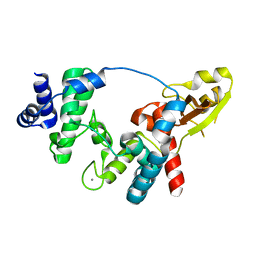
1P6R
 | | Solution structure of the DNA binding domain of the repressor BlaI. | | 分子名称: | Penicillinase repressor | | 著者 | Melckebeke, H.V, Vreuls, C, Gans, P, Llabres, G, Filee, P, Joris, B, Simorre, J.P. | | 登録日 | 2003-04-30 | | 公開日 | 2003-12-09 | | 最終更新日 | 2024-05-22 | | 実験手法 | SOLUTION NMR | | 主引用文献 | Solution structural study of BlaI: implications for the repression of genes involved in beta-lactam antibiotic resistance.
J.Mol.Biol., 333, 2003
|
|
4ANR
 | | Crystal structure of soluble lytic Transglycosylase SltB1 from Pseudomonas aeruginosa | | 分子名称: | CALCIUM ION, SOLUBLE LYTIC TRANSGLYCOSYLASE B | | 著者 | Nikolaidis, I, Izore, T, Job, V, Thielens, N, Breukink, E, Dessen, A. | | 登録日 | 2012-03-22 | | 公開日 | 2012-04-04 | | 最終更新日 | 2023-12-20 | | 実験手法 | X-RAY DIFFRACTION (1.84 Å) | | 主引用文献 | Calcium-Dependent Complex Formation between Pbp2 and Lytic Transglycosylase Sltb1 of Pseudomonas Aeruginosa.
Microb.Drug Resist., 18, 2012
|
|


