7YFU
 
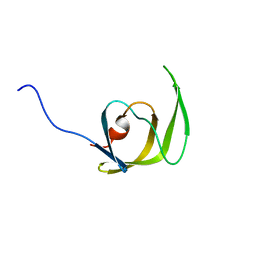
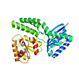
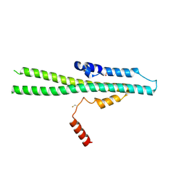
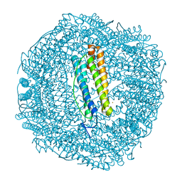
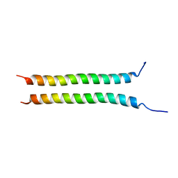 | | Structure of Rpgrip1l CC2 | | 分子名称: | Protein fantom | | 著者 | He, R, Chen, G, Li, Z, Li, J. | | 登録日 | 2022-07-09 | | 公開日 | 2023-05-17 | | 最終更新日 | 2024-05-29 | | 実験手法 | X-RAY DIFFRACTION (1.5 Å) | | 主引用文献 | Structure of the N-terminal coiled-coil domains of the ciliary protein Rpgrip1l.
Iscience, 26, 2023
|
|
6SL3
 | |
8ECC
 | |
6SGI
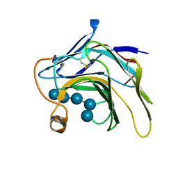
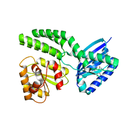
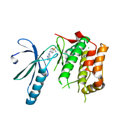 | | Nek2 kinase bound to inhibitor 96 | | 分子名称: | 4-[(6-ethyl-7~{H}-purin-2-yl)amino]benzenesulfonamide, CHLORIDE ION, Serine/threonine-protein kinase Nek2 | | 著者 | Richards, M.W, Mas-Droux, C.P, Bayliss, R. | | 登録日 | 2019-08-05 | | 公開日 | 2020-06-17 | | 最終更新日 | 2024-01-24 | | 実験手法 | X-RAY DIFFRACTION (2.3 Å) | | 主引用文献 | 2-Arylamino-6-ethynylpurines are cysteine-targeting irreversible inhibitors of Nek2 kinase.
Rsc Med Chem, 11, 2020
|
|
6SON
 | |
8EU8
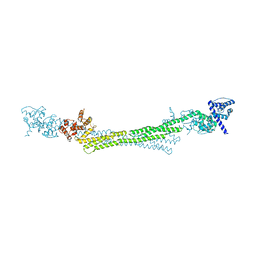
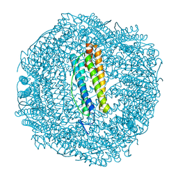
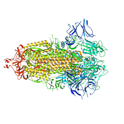
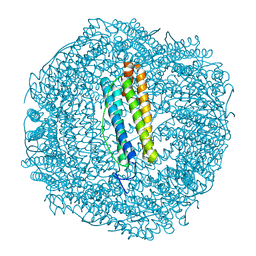
 | | Cryo-EM structure of CH848 10.17DT DS-SOSIP-2P Env | | 分子名称: | 2-acetamido-2-deoxy-beta-D-glucopyranose, 2-acetamido-2-deoxy-beta-D-glucopyranose-(1-4)-2-acetamido-2-deoxy-beta-D-glucopyranose, CH848 10.17DT SOSIP Envelope glycoprotein gp160 | | 著者 | Wrapp, D, Acharya, P, Haynes, B.F. | | 登録日 | 2022-10-18 | | 公開日 | 2023-01-04 | | 最終更新日 | 2023-02-08 | | 実験手法 | ELECTRON MICROSCOPY (3.73 Å) | | 主引用文献 | Structure-Based Stabilization of SOSIP Env Enhances Recombinant Ectodomain Durability and Yield.
J.Virol., 97, 2023
|
|
5UN0
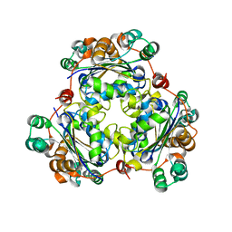
 | | Crystal Structure of Mycobacterium Tuberculosis Proteasome-assembly chaperone homologue Rv2125 | | 分子名称: | proteasome assembly chaperone 2 (PAC2) homologue Rv2125 | | 著者 | Bai, L, Jastrab, J.B, Hu, K, Yu, H, Darwin, K.H, Li, H. | | 登録日 | 2017-01-30 | | 公開日 | 2017-03-01 | | 最終更新日 | 2023-10-04 | | 実験手法 | X-RAY DIFFRACTION (3 Å) | | 主引用文献 | Structural Analysis of Mycobacterium tuberculosis Homologues of the Eukaryotic Proteasome Assembly Chaperone 2 (PAC2).
J. Bacteriol., 199, 2017
|
|
1J9K
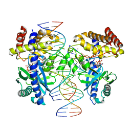
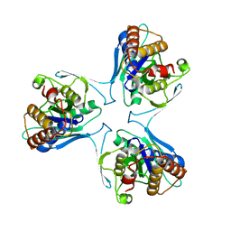
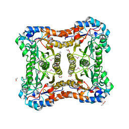
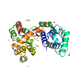
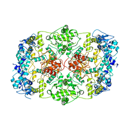
 | | CRYSTAL STRUCTURE OF SURE PROTEIN FROM T.MARITIMA IN COMPLEX WITH TUNGSTATE | | 分子名称: | 4-(2-HYDROXYETHYL)-1-PIPERAZINE ETHANESULFONIC ACID, CALCIUM ION, STATIONARY PHASE SURVIVAL PROTEIN, ... | | 著者 | Suh, S.W, Lee, J.Y, Kwak, J.E, Moon, J. | | 登録日 | 2001-05-27 | | 公開日 | 2001-09-12 | | 最終更新日 | 2023-10-25 | | 実験手法 | X-RAY DIFFRACTION (2.1 Å) | | 主引用文献 | Crystal structure and functional analysis of the SurE protein identify a novel phosphatase family.
Nat.Struct.Biol., 8, 2001
|
|
1Q9P
 | | Solution structure of the mature HIV-1 protease monomer | | 分子名称: | HIV-1 Protease | | 著者 | Ishima, R, Torchia, D.A, Lynch, S.M, Gronenborn, A.M, Louis, J.M. | | 登録日 | 2003-08-25 | | 公開日 | 2004-03-02 | | 最終更新日 | 2024-05-22 | | 実験手法 | SOLUTION NMR | | 主引用文献 | Solution structure of the mature HIV-1 protease monomer: Insight into the tertiary fold and stability of a precursor
J.Biol.Chem., 278, 2003
|
|
3E49
 | |
4HH2
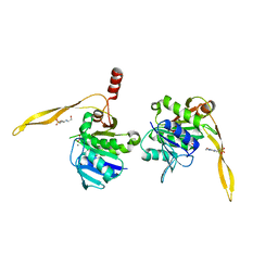
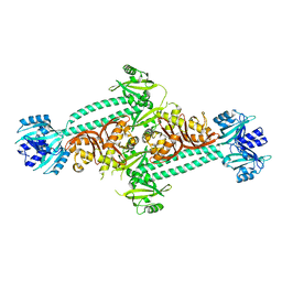 | | Structure of PpsR without the HTH motif from Rb. sphaeroides | | 分子名称: | Transcriptional regulator, PpsR | | 著者 | Winkler, A, Heintz, U, Lindner, R, Reinstein, J, Shoeman, R, Schlichting, I. | | 登録日 | 2012-10-09 | | 公開日 | 2013-06-05 | | 最終更新日 | 2024-02-28 | | 実験手法 | X-RAY DIFFRACTION (2.8 Å) | | 主引用文献 | A ternary AppA-PpsR-DNA complex mediates light regulation of photosynthesis-related gene expression.
Nat.Struct.Mol.Biol., 20, 2013
|
|
7MSV
 | |
4HH0
 | | Dark-state structure of AppA C20S without the Cys-rich region from Rb. sphaeroides | | 分子名称: | AppA protein, CHLORIDE ION, FLAVIN MONONUCLEOTIDE | | 著者 | Winkler, A, Heintz, U, Lindner, R, Reinstein, J, Shoeman, R, Schlichting, I. | | 登録日 | 2012-10-09 | | 公開日 | 2013-06-05 | | 最終更新日 | 2024-02-28 | | 実験手法 | X-RAY DIFFRACTION (2.6 Å) | | 主引用文献 | A ternary AppA-PpsR-DNA complex mediates light regulation of photosynthesis-related gene expression.
Nat.Struct.Mol.Biol., 20, 2013
|
|
5DZE
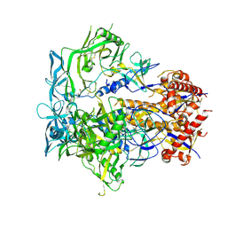
 | | Crystal Structure of the catalytic nucleophile mutant of VvEG16 in complex with cellotetraose | | 分子名称: | beta-D-glucopyranose, beta-D-glucopyranose-(1-4)-beta-D-glucopyranose-(1-4)-beta-D-glucopyranose-(1-4)-alpha-D-glucopyranose, endo-glucanase | | 著者 | McGregor, N.G.S, Tung, C.C, Van Petegem, F, Brumer, H. | | 登録日 | 2015-09-25 | | 公開日 | 2016-09-21 | | 最終更新日 | 2020-07-29 | | 実験手法 | X-RAY DIFFRACTION (0.97 Å) | | 主引用文献 | Crystallographic insight into the evolutionary origins of xyloglucan endotransglycosylases and endohydrolases.
Plant J., 89, 2017
|
|
7MK0
 | |
6V4C
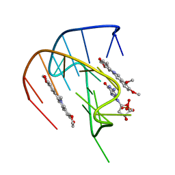
 | | Culex quinquefasciatus D7 long form 1- CxD7L1 in complex with ADP | | 分子名称: | 1,2-ETHANEDIOL, 2-ETHOXYETHANOL, ADENOSINE-5'-DIPHOSPHATE, ... | | 著者 | Calvo, E, Garboczi, D.N, Martin-Martin, I, Gittis, A.G. | | 登録日 | 2019-11-27 | | 公開日 | 2020-06-24 | | 最終更新日 | 2023-10-11 | | 実験手法 | X-RAY DIFFRACTION (1.97 Å) | | 主引用文献 | ADP binding by the Culex quinquefasciatus mosquito D7 salivary protein enhances blood feeding on mammals.
Nat Commun, 11, 2020
|
|
7LYL
 | |
7LYO
 | |
7LYP
 | |
7LYN
 | |
7LHL
 | |
4HH1
 | | Dark-state structure of AppA wild-type without the Cys-rich region from Rb. sphaeroides | | 分子名称: | AppA protein, FLAVIN MONONUCLEOTIDE | | 著者 | Winkler, A, Heintz, U, Lindner, R, Reinstein, J, Shoeman, R, Schlichting, I. | | 登録日 | 2012-10-09 | | 公開日 | 2013-06-05 | | 最終更新日 | 2024-02-28 | | 実験手法 | X-RAY DIFFRACTION (3.501 Å) | | 主引用文献 | A ternary AppA-PpsR-DNA complex mediates light regulation of photosynthesis-related gene expression.
Nat.Struct.Mol.Biol., 20, 2013
|
|
5IJH
 | |
6SOM
 | |
6SOQ
 | |


