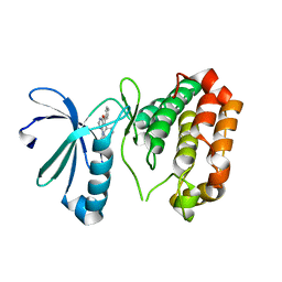
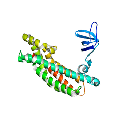
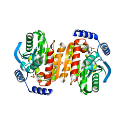
3BSQ
 
 | | Crystal structure of human kallikrein 7 produced as a secretion protein in E.coli | | 分子名称: | Kallikrein-7, SULFATE ION | | 著者 | Fernandez, I.S, Standker, L, Magert, H.J, Forssmann, W.G, Gimenez-Gallego, G, Romero, A. | | 登録日 | 2007-12-26 | | 公開日 | 2008-04-29 | | 最終更新日 | 2023-11-01 | | 実験手法 | X-RAY DIFFRACTION (2.8 Å) | | 主引用文献 | Crystal structure of human epidermal kallikrein 7 (hK7) synthesized directly in its native state in E. coli: insights into the atomic basis of its inhibition by LEKTI domain 6 (LD6)
J.Mol.Biol., 377, 2008
|
|
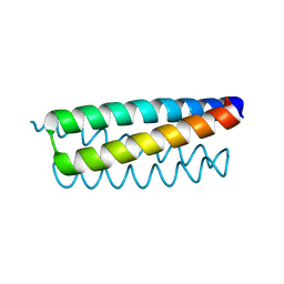
3K2R
 | | Crystal Structure of Spin Labeled T4 Lysozyme Mutant K65V1/R76V1 | | 分子名称: | CHLORIDE ION, HEXANE-1,6-DIOL, Lysozyme, ... | | 著者 | Toledo Warshaviak, D, Cascio, D, Khramtsov, V.V, Hubbell, W.L. | | 登録日 | 2009-09-30 | | 公開日 | 2010-10-13 | | 最終更新日 | 2023-09-06 | | 実験手法 | X-RAY DIFFRACTION (1.5 Å) | | 主引用文献 | Crystal Structure of Spin Labeled T4 Lysozyme Mutant K65V1/R76V1
To be Published
|
|
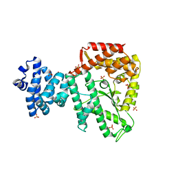
3K6V
 | | M. acetivorans Molybdate-Binding Protein (ModA) in Citrate-Bound Open Form | | 分子名称: | CITRIC ACID, Solute-binding protein MA_0280 | | 著者 | Chan, S, Giuroiu, I, Chernishof, I, Sawaya, M.R, Chiang, J, Gunsalus, R.P, Arbing, M.A, Perry, L.J. | | 登録日 | 2009-10-09 | | 公開日 | 2010-01-12 | | 最終更新日 | 2024-02-21 | | 実験手法 | X-RAY DIFFRACTION (1.69 Å) | | 主引用文献 | Apo and ligand-bound structures of ModA from the archaeon Methanosarcina acetivorans
Acta Crystallogr.,Sect.F, 66, 2010
|
|
3JUY
 | |
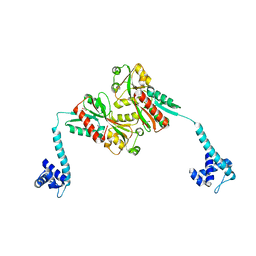
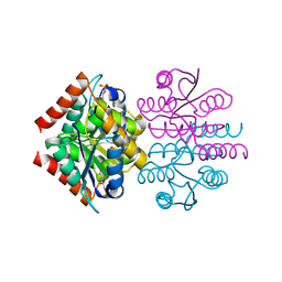
3K2L
 | | Crystal Structure of dual-specificity tyrosine phosphorylation regulated kinase 2 (DYRK2) | | 分子名称: | CHLORIDE ION, Dual specificity tyrosine-phosphorylation-regulated kinase 2, SODIUM ION, ... | | 著者 | Filippakopoulos, P, Myrianthopoulos, V, Soundararajan, M, Krojer, T, Hapka, E, Fedorov, O, Berridge, G, Wang, J, Shrestha, L, Pike, A.C.W, Ugochukwu, E, von Delft, F, Arrowsmith, C.H, Edwards, A, Weigelt, J, Bountra, C, Mikros, E, Knapp, S, Structural Genomics Consortium (SGC) | | 登録日 | 2009-09-30 | | 公開日 | 2009-10-13 | | 最終更新日 | 2023-11-22 | | 実験手法 | X-RAY DIFFRACTION (2.36 Å) | | 主引用文献 | Structures of Down Syndrome Kinases, DYRKs, Reveal Mechanisms of Kinase Activation and Substrate Recognition.
Structure, 21, 2013
|
|
3K5U
 | | Identification, SAR Studies and X-ray Cocrystal Analysis of a Novel Furano-pyrimidine Aurora Kinase A Inhibitor | | 分子名称: | 2-[(5,6-DIPHENYLFURO[2,3-D]PYRIMIDIN-4-YL)AMINO]ETHANOL, Serine/threonine-protein kinase 6 | | 著者 | Wu, J.S, Leou, J.S, Coumar, M.S, Hsieh, H.P, Wu, S.Y. | | 登録日 | 2009-10-08 | | 公開日 | 2010-10-13 | | 最終更新日 | 2023-11-01 | | 実験手法 | X-RAY DIFFRACTION (2.35 Å) | | 主引用文献 | Identification, SAR studies, and X-ray co-crystallographic analysis of a novel furanopyrimidine aurora kinase A inhibitor
Chemmedchem, 5, 2010
|
|
3JQ7
 | | Crystal structure of pteridine reductase 1 (PTR1) from Trypanosoma brucei in ternary complex with cofactor (NADP+) and inhibitor 6-phenylpteridine-2,4,7-triamine (DX2) | | 分子名称: | 2,3-DIHYDROXY-1,4-DITHIOBUTANE, 6-phenylpteridine-2,4,7-triamine, ACETATE ION, ... | | 著者 | Tulloch, L.B, Hunter, W.N. | | 登録日 | 2009-09-06 | | 公開日 | 2009-12-08 | | 最終更新日 | 2023-09-06 | | 実験手法 | X-RAY DIFFRACTION (1.8 Å) | | 主引用文献 | Structure-based design of pteridine reductase inhibitors targeting african sleeping sickness and the leishmaniases.
J.Med.Chem., 53, 2010
|
|
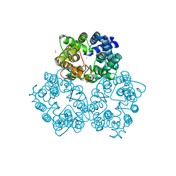
3K92
 | | Crystal structure of a E93K mutant of the majour Bacillus subtilis glutamate dehydrogenase RocG | | 分子名称: | DI(HYDROXYETHYL)ETHER, NAD-specific glutamate dehydrogenase | | 著者 | Gunka, K, Newman, J.A, Commichau, F.M, Herzberg, C, Rodrigues, C, Hewitt, L, Lewis, R.J, Stulke, J. | | 登録日 | 2009-10-15 | | 公開日 | 2010-06-02 | | 最終更新日 | 2024-03-20 | | 実験手法 | X-RAY DIFFRACTION (2.3 Å) | | 主引用文献 | Functional dissection of a trigger enzyme: mutations of the bacillus subtilis glutamate dehydrogenase RocG that affect differentially its catalytic activity and regulatory properties
J.Mol.Biol., 400, 2010
|
|
3JRV
 | |
3JUI
 | | Crystal Structure of the C-terminal Domain of Human Translation Initiation Factor eIF2B epsilon Subunit | | 分子名称: | GLYCEROL, Translation initiation factor eIF-2B subunit epsilon | | 著者 | Wei, J, Xu, H, Zhang, C, Wang, M, Gao, F, Gong, W. | | 登録日 | 2009-09-15 | | 公開日 | 2009-10-06 | | 最終更新日 | 2021-11-10 | | 実験手法 | X-RAY DIFFRACTION (2 Å) | | 主引用文献 | Crystal Structure of the C-terminal Domain of Human Translation Initiation Factor eIF2B epsilon Subunit
To be published
|
|
3JV3
 | |
3K1M
 | | Crystal Structure of full-length BenM, R156H mutant | | 分子名称: | CHLORIDE ION, GLYCEROL, HTH-type transcriptional regulator benM, ... | | 著者 | Ruangprasert, A, Momany, C, Neidle, E.L, Craven, S.H. | | 登録日 | 2009-09-28 | | 公開日 | 2010-09-29 | | 最終更新日 | 2023-09-06 | | 実験手法 | X-RAY DIFFRACTION (2.29 Å) | | 主引用文献 | Structure of Full-length BenM
To be Published
|
|
3K3F
 | |
3JQ6
 | | Crystal structure of pteridine reductase 1 (PTR1) from Trypanosoma brucei in ternary complex with cofactor (NADP+) and inhibitor 6,7-bis(1-methylethyl)pteridine-2,4-diamine (DX1) | | 分子名称: | 2,3-DIHYDROXY-1,4-DITHIOBUTANE, 6,7-bis(1-methylethyl)pteridine-2,4-diamine, NADP NICOTINAMIDE-ADENINE-DINUCLEOTIDE PHOSPHATE, ... | | 著者 | Tulloch, L.B, Hunter, W.N. | | 登録日 | 2009-09-06 | | 公開日 | 2009-12-08 | | 最終更新日 | 2023-09-06 | | 実験手法 | X-RAY DIFFRACTION (1.8 Å) | | 主引用文献 | Structure-based design of pteridine reductase inhibitors targeting african sleeping sickness and the leishmaniases.
J.Med.Chem., 53, 2010
|
|
3K6K
 | |
3K79
 | |
3K7D
 | | C-terminal (adenylylation) domain of E.coli Glutamine Synthetase Adenylyltransferase | | 分子名称: | Glutamate-ammonia-ligase adenylyltransferase, SULFATE ION | | 著者 | Xu, Y, Carr, P.D, Vasudevan, S.G, Ollis, D.L. | | 登録日 | 2009-10-12 | | 公開日 | 2009-12-15 | | 最終更新日 | 2023-09-06 | | 実験手法 | X-RAY DIFFRACTION (2.4 Å) | | 主引用文献 | Structure of the Adenylylation Domain of E. coli Glutamine Synthetase Adenylyl Transferase: Evidence for Gene Duplication and Evolution of a New Active Site.
J.Mol.Biol., 396, 2010
|
|
3K8C
 | |
3JSF
 | |
3JSS
 | |
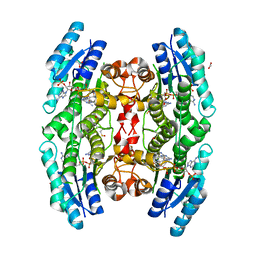
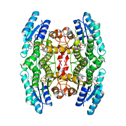
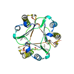
3KA6
 | | Frog M-ferritin, EED mutant, with cobalt | | 分子名称: | CHLORIDE ION, COBALT (II) ION, Ferritin, ... | | 著者 | Tosha, T, Ng, H.L, Theil, E, Alber, T, Bhattasali, O. | | 登録日 | 2009-10-18 | | 公開日 | 2010-10-06 | | 最終更新日 | 2023-09-06 | | 実験手法 | X-RAY DIFFRACTION (1.4 Å) | | 主引用文献 | Moving Metal Ions through Ferritin-Protein Nanocages from Three-Fold Pores to Catalytic Sites.
J.Am.Chem.Soc., 132, 2010
|
|
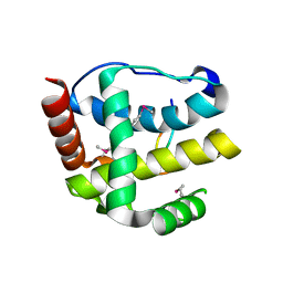
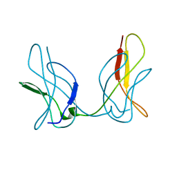
3JXF
 | | CA-like domain of human PTPRZ | | 分子名称: | Receptor-type tyrosine-protein phosphatase zeta | | 著者 | Bouyain, S. | | 登録日 | 2009-09-19 | | 公開日 | 2009-12-22 | | 最終更新日 | 2017-11-01 | | 実験手法 | X-RAY DIFFRACTION (2.004 Å) | | 主引用文献 | The protein tyrosine phosphatases PTPRZ and PTPRG bind to distinct members of the contactin family of neural recognition molecules.
Proc.Natl.Acad.Sci.USA, 107, 2010
|
|
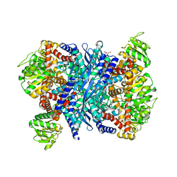
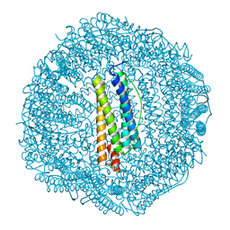
3K00
 | | Crystal structures of the GacH receptor of Streptomyces glaucescens GLA.O in the unliganded form and in complex with acarbose and an acarbose homolog. Comparison with acarbose-loaded maltose binding protein of Salmonella typhimurium. | | 分子名称: | Acarbose/maltose binding protein GacH, alpha-D-glucopyranose-(1-4)-alpha-D-glucopyranose-(1-4)-alpha-D-glucopyranose-(1-4)-alpha-D-glucopyranose | | 著者 | Vahedi-Faridi, A, Licht, A, Bulut, H, Schneider, E. | | 登録日 | 2009-09-24 | | 公開日 | 2010-02-16 | | 最終更新日 | 2024-02-21 | | 実験手法 | X-RAY DIFFRACTION (1.55 Å) | | 主引用文献 | Crystal Structures of the Solute Receptor GacH of Streptomyces glaucescens in Complex with Acarbose and an Acarbose Homolog: Comparison with the Acarbose-Loaded Maltose-Binding Protein of Salmonella typhimurium.
J.Mol.Biol., 397, 2010
|
|
3K2M
 | |
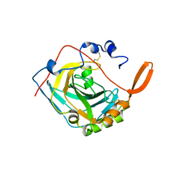
3K4D
 | | Crystal structure of E. coli beta-glucuronidase with the glucaro-d-lactam inhibitor bound | | 分子名称: | (2S,3R,4S,5R)-3,4,5-trihydroxy-6-oxopiperidine-2-carboxylic acid, Beta-glucuronidase | | 著者 | Wallace, B.D, Redinbo, M.R. | | 登録日 | 2009-10-05 | | 公開日 | 2010-11-17 | | 最終更新日 | 2023-09-06 | | 実験手法 | X-RAY DIFFRACTION (2.393 Å) | | 主引用文献 | Alleviating cancer drug toxicity by inhibiting a bacterial enzyme.
Science, 330, 2010
|
|


