2IAF
 
 | |
2IAG
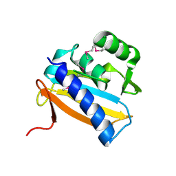
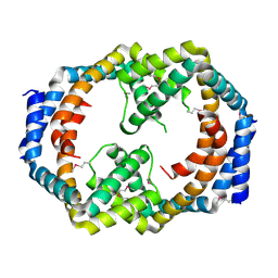
 | | Crystal structure of human prostacyclin synthase | | 分子名称: | PROTOPORPHYRIN IX CONTAINING FE, Prostacyclin synthase, SODIUM ION | | 著者 | Chiang, C.-W, Yeh, H.-C, Wang, L.-H, Chan, N.-L. | | 登録日 | 2006-09-08 | | 公開日 | 2006-10-10 | | 最終更新日 | 2024-03-13 | | 実験手法 | X-RAY DIFFRACTION (2.15 Å) | | 主引用文献 | Crystal Structure of the Human Prostacyclin Synthase
J.Mol.Biol., 364, 2006
|
|
2IAH
 | | Crystal structure of the ferripyoverdine receptor of the outer membrane of Pseudomonas aeruginosa bound to ferripyoverdine. | | 分子名称: | (1S)-1-CARBOXY-5-[(3-CARBOXYPROPANOYL)AMINO]-8,9-DIHYDROXY-1,2,3,4-TETRAHYDROPYRIMIDO[1,2-A]QUINOLIN-11-IUM, FE (III) ION, Ferripyoverdine receptor, ... | | 著者 | Wirth, C, Pattus, F, Cobessi, D. | | 登録日 | 2006-09-08 | | 公開日 | 2007-09-11 | | 最終更新日 | 2023-11-15 | | 実験手法 | X-RAY DIFFRACTION (2.73 Å) | | 主引用文献 | From the periplasmic signaling domain to the extracellular face of an outer membrane signal transducer of Pseudomonas aeruginosa: crystal structure of the ferric pyoverdine outer membrane receptor.
J.Mol.Biol., 368, 2007
|
|
2IAI
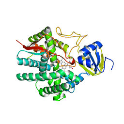
 | | Crystal structure of SCO3833, a member of the TetR transcriptional regulator family from Streptomyces coelicolor A3 | | 分子名称: | Putative transcriptional regulator SCO3833 | | 著者 | Zimmerman, M.D, Xu, X, Wang, S, Gu, J, Chruszcz, M, Cymborowski, M, Savchenko, A, Edwards, A, Joachimiak, A, Minor, W, Midwest Center for Structural Genomics (MCSG) | | 登録日 | 2006-09-08 | | 公開日 | 2006-09-26 | | 最終更新日 | 2022-04-13 | | 実験手法 | X-RAY DIFFRACTION (1.65 Å) | | 主引用文献 | Crystal structure of SCO3833, a member of the TetR transcriptional regulator family from Streptomyces coelicolor A3
To be Published
|
|
2IAJ
 | |
2IAK
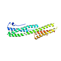 | |
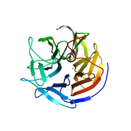
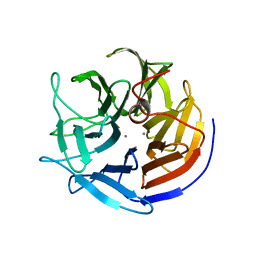
2IAL
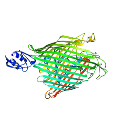
 | | Structural basis for recognition of mutant self by a tumor-specific, MHC class II-restricted TCR | | 分子名称: | CD4+ T cell receptor E8 alpha chain, CD4+ T cell receptor E8 beta chain | | 著者 | Deng, L, Langley, R.J, Mariuzza, R.A. | | 登録日 | 2006-09-08 | | 公開日 | 2007-04-03 | | 最終更新日 | 2023-10-25 | | 実験手法 | X-RAY DIFFRACTION (1.92 Å) | | 主引用文献 | Structural basis for the recognition of mutant self by a tumor-specific, MHC class II-restricted T cell receptor
Nat.Immunol., 8, 2007
|
|
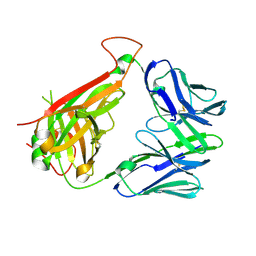
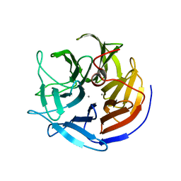
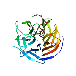
2IAM
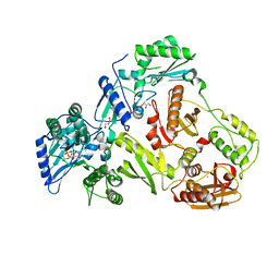
 | | Structural basis for recognition of mutant self by a tumor-specific, MHC class II-restricted TCR | | 分子名称: | 15-mer peptide from Triosephosphate isomerase, CD4+ T cell receptor E8 alpha chain, CD4+ T cell receptor E8 beta chain, ... | | 著者 | Deng, L, Langley, R.J, Mariuzza, R.A. | | 登録日 | 2006-09-08 | | 公開日 | 2007-04-03 | | 最終更新日 | 2023-10-25 | | 実験手法 | X-RAY DIFFRACTION (2.8 Å) | | 主引用文献 | Structural basis for the recognition of mutant self by a tumor-specific, MHC class II-restricted T cell receptor
Nat.Immunol., 8, 2007
|
|
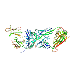
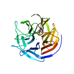
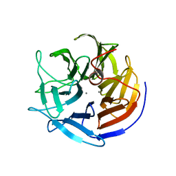
2IAN
 | | Structural basis for recognition of mutant self by a tumor-specific, MHC class II-restricted TCR | | 分子名称: | 15-mer peptide from Triosephosphate isomerase, CD4+ T cell receptor E8 alpha chain, CD4+ T cell receptor E8 beta chain, ... | | 著者 | Deng, L, Langley, R.J, Mariuzza, R.A. | | 登録日 | 2006-09-08 | | 公開日 | 2007-04-03 | | 最終更新日 | 2023-10-25 | | 実験手法 | X-RAY DIFFRACTION (2.8 Å) | | 主引用文献 | Structural basis for the recognition of mutant self by a tumor-specific, MHC class II-restricted T cell receptor
Nat.Immunol., 8, 2007
|
|
2IAO
 | | Crystal structure of squid ganglion DFPase E37Q mutant | | 分子名称: | CALCIUM ION, DIISOPROPYLFLUOROPHOSPHATASE | | 著者 | Scharff, E.I, Koepke, J, Fritzsch, G, Luecke, C, Rueterjans, H. | | 登録日 | 2006-09-08 | | 公開日 | 2006-09-26 | | 最終更新日 | 2023-08-30 | | 実験手法 | X-RAY DIFFRACTION (2 Å) | | 主引用文献 | Crystal structure of diisopropylfluorophosphatase from Loligo vulgaris
Structure, 9, 2001
|
|
2IAP
 | | Crystal structure of squid ganglion DFPase E21Q mutant | | 分子名称: | CALCIUM ION, Diisopropylfluorophosphatase | | 著者 | Scharff, E.I, Koepke, J, Fritzsch, G, Luecke, C, Rueterjans, H. | | 登録日 | 2006-09-08 | | 公開日 | 2006-09-26 | | 最終更新日 | 2023-08-30 | | 実験手法 | X-RAY DIFFRACTION (1.9 Å) | | 主引用文献 | Crystal structure of diisopropylfluorophosphatase from Loligo vulgaris
Structure, 9, 2001
|
|
2IAQ
 | | Crystal structure of squid ganglion DFPase S271A mutant | | 分子名称: | CALCIUM ION, Diisopropylfluorophosphatase | | 著者 | Scharff, E.I, Koepke, J, Fritzsch, G, Luecke, C, Rueterjans, H. | | 登録日 | 2006-09-08 | | 公開日 | 2006-09-26 | | 最終更新日 | 2023-08-30 | | 実験手法 | X-RAY DIFFRACTION (2.1 Å) | | 主引用文献 | Crystal structure of diisopropylfluorophosphatase from Loligo vulgaris
Structure, 9, 2001
|
|
2IAR
 | | Crystal structure of squid ganglion DFPase W244H mutant | | 分子名称: | CALCIUM ION, Diisopropylfluorophosphatase | | 著者 | Scharff, E.I, Koepke, J, Fritzsch, G, Luecke, C, Rueterjans, H. | | 登録日 | 2006-09-08 | | 公開日 | 2006-09-26 | | 最終更新日 | 2023-08-30 | | 実験手法 | X-RAY DIFFRACTION (1.9 Å) | | 主引用文献 | Crystal structure of diisopropylfluorophosphatase from Loligo vulgaris
Structure, 9, 2001
|
|
2IAS
 | | Crystal structure of squid ganglion DFPase W244F mutant | | 分子名称: | CALCIUM ION, Diisopropylfluorophosphatase | | 著者 | Scharff, E.I, Koepke, J, Fritzsch, G, Luecke, C, Rueterjans, H. | | 登録日 | 2006-09-08 | | 公開日 | 2006-09-26 | | 最終更新日 | 2023-08-30 | | 実験手法 | X-RAY DIFFRACTION (2 Å) | | 主引用文献 | Crystal structure of diisopropylfluorophosphatase from Loligo vulgaris
Structure, 9, 2001
|
|
2IAT
 | | Crystal structure of squid ganglion DFPase W244L mutant | | 分子名称: | CALCIUM ION, Diisopropylfluorophosphatase | | 著者 | Scharff, E.I, Koepke, J, Fritzsch, G, Luecke, C, Rueterjans, H. | | 登録日 | 2006-09-08 | | 公開日 | 2006-09-26 | | 最終更新日 | 2023-08-30 | | 実験手法 | X-RAY DIFFRACTION (1.9 Å) | | 主引用文献 | Crystal structure of diisopropylfluorophosphatase from Loligo vulgaris
Structure, 9, 2001
|
|
2IAU
 | | Crystal structure of squid ganglion DFPase W244Y mutant | | 分子名称: | CALCIUM ION, Diisopropylfluorophosphatase | | 著者 | Scharff, E.I, Koepke, J, Fritzsch, G, Luecke, C, Rueterjans, H. | | 登録日 | 2006-09-08 | | 公開日 | 2006-09-26 | | 最終更新日 | 2023-08-30 | | 実験手法 | X-RAY DIFFRACTION (2 Å) | | 主引用文献 | Crystal structure of diisopropylfluorophosphatase from Loligo vulgaris
Structure, 9, 2001
|
|
2IAV
 | | Crystal structure of squid ganglion DFPase H287A mutant | | 分子名称: | CALCIUM ION, Diisopropylfluorophosphatase | | 著者 | Katsemi, V, Luecke, C, Koepke, J, Loehr, F, Maurer, S, Fritzsch, G, Rueterjans, H. | | 登録日 | 2006-09-08 | | 公開日 | 2006-09-26 | | 最終更新日 | 2023-08-30 | | 実験手法 | X-RAY DIFFRACTION (1.07 Å) | | 主引用文献 | Mutational and structural studies of the diisopropylfluorophosphatase from Loligo vulgaris shed new light on the catalytic mechanism of the enzyme
Biochemistry, 44, 2005
|
|
2IAW
 | | Crystal structure of squid ganglion DFPase N175D mutant | | 分子名称: | CALCIUM ION, Diisopropylfluorophosphatase | | 著者 | Katsemi, V, Luecke, C, Koepke, J, Loehr, F, Maurer, S, Fritzsch, G, Rueterjans, H. | | 登録日 | 2006-09-08 | | 公開日 | 2006-09-26 | | 最終更新日 | 2023-08-30 | | 実験手法 | X-RAY DIFFRACTION (1.74 Å) | | 主引用文献 | Mutational and structural studies of the diisopropylfluorophosphatase from Loligo vulgaris shed new light on the catalytic mechanism of the enzyme
Biochemistry, 44, 2005
|
|
2IAX
 | | Crystal structure of squid ganglion DFPase D232S mutant | | 分子名称: | CALCIUM ION, Diisopropylfluorophosphatase | | 著者 | Katsemi, V, Luecke, C, Koepke, J, Loehr, F, Maurer, S, Fritzsch, G, Rueterjans, H. | | 登録日 | 2006-09-08 | | 公開日 | 2006-09-26 | | 最終更新日 | 2023-08-30 | | 実験手法 | X-RAY DIFFRACTION (1.1 Å) | | 主引用文献 | Mutational and structural studies of the diisopropylfluorophosphatase from Loligo vulgaris shed new light on the catalytic mechanism of the enzyme
Biochemistry, 44, 2005
|
|
2IAY
 | |
2IAZ
 | |
2IB0
 | | Crystal structure of a conserved hypothetical protein, rv2844, from Mycobacterium tuberculosis | | 分子名称: | CONSERVED HYPOTHETICAL ALANINE RICH PROTEIN | | 著者 | Yu, M, Bursey, E.H, Radhakannan, T, Kim, C.Y, Kaviratne, T, Woodruff, T, Segelke, B.W, Lekin, T, Toppani, D, Terwilliger, T.C, Hung, L.W, TB Structural Genomics Consortium (TBSGC), Integrated Center for Structure and Function Innovation (ISFI) | | 登録日 | 2006-09-08 | | 公開日 | 2006-09-26 | | 最終更新日 | 2024-02-21 | | 実験手法 | X-RAY DIFFRACTION (2 Å) | | 主引用文献 | Crystal structure of a conserved hypothetical protein, rv2844, from Mycobacterium tuberculosis
To be Published
|
|
2IB1
 | |
2IB5
 | |
2IB6
 | | Structural characterization of a blue chromoprotein and its yellow mutant from the sea anemone cnidopus japonicus | | 分子名称: | PHOSPHATE ION, Yellow mutant chromo protein | | 著者 | Chan, M.C.Y, Bosanac, I, Ho, D, Prive, G, Ikura, M. | | 登録日 | 2006-09-10 | | 公開日 | 2006-10-10 | | 最終更新日 | 2023-11-15 | | 実験手法 | X-RAY DIFFRACTION (2 Å) | | 主引用文献 | Structural Characterization of a Blue Chromoprotein and Its Yellow Mutant from the Sea Anemone Cnidopus Japonicus
J.Biol.Chem., 281, 2006
|
|


