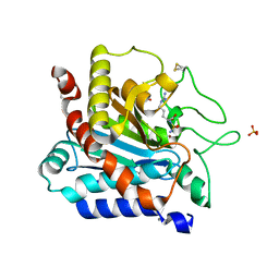
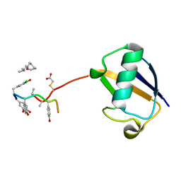
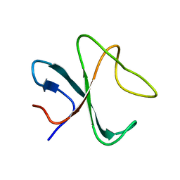
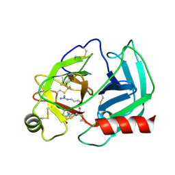
7V7X
 
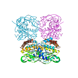
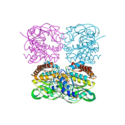
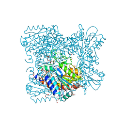
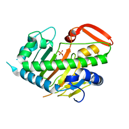 | | Structure of H194A AdaV | | 分子名称: | 2'-DEOXYADENOSINE-5'-MONOPHOSPHATE, AdaV | | 著者 | Zhang, Z.Y, Chen, W.Q, Zhai, G.Q, Zhang, M. | | 登録日 | 2021-08-22 | | 公開日 | 2022-08-31 | | 最終更新日 | 2024-05-29 | | 実験手法 | X-RAY DIFFRACTION (2.7 Å) | | 主引用文献 | Structural Insight into the Catalytic Mechanism of Non-Heme Iron Halogenase AdaV in 2'-Chloropentostatin Biosynthesis
Acs Catalysis, 12, 2022
|
|
6F79
 | |
6F75
 | |
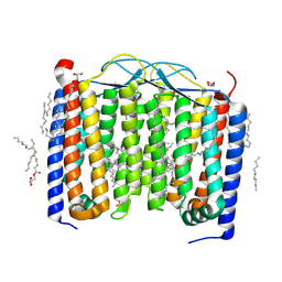
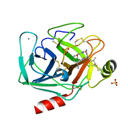
6SU4
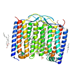 | | Crystal structure of the 48C12 heliorhodopsin in the blue form at pH 4.3 | | 分子名称: | (2R)-2,3-dihydroxypropyl (9Z)-octadec-9-enoate, 48C12 heliorhodopsin, ACETATE ION, ... | | 著者 | Kovalev, K, Volkov, D, Astashkin, R, Alekseev, A, Gushchin, I, Gordeliy, V. | | 登録日 | 2019-09-12 | | 公開日 | 2019-12-11 | | 最終更新日 | 2024-01-24 | | 実験手法 | X-RAY DIFFRACTION (1.5 Å) | | 主引用文献 | High-resolution structural insights into the heliorhodopsin family.
Proc.Natl.Acad.Sci.USA, 117, 2020
|
|
6F6Q
 | |
5AWV
 | |
4OIN
 | | Crystal structure of Thermus thermophilus transcription initiation complex soaked with GE23077 | | 分子名称: | (2Z)-2-methylbut-2-enoic acid, 5'-D(*CP*CP*T*GP*CP*AP*TP*CP*CP*GP*TP*GP*AP*GP*TP*CP*GP*AP*G)-3', 5'-D(*TP*AP*TP*AP*AP*TP*GP*GP*GP*AP*GP*CP*TP*GP*TP*CP*AP*CP*GP*GP*AP*TP*GP*CP*AP*GP*G)-3', ... | | 著者 | Zhang, Y, Ebright, R.H, Arnold, E. | | 登録日 | 2014-01-20 | | 公開日 | 2014-05-07 | | 最終更新日 | 2023-09-20 | | 実験手法 | X-RAY DIFFRACTION (2.8 Å) | | 主引用文献 | GE23077 binds to the RNA polymerase 'i' and 'i+1' sites and prevents the binding of initiating nucleotides.
Elife, 3, 2014
|
|
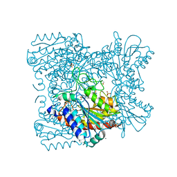
7XPR
 | | Crystal structrue of SeMet-MtdL:GDP | | 分子名称: | GUANOSINE-5'-DIPHOSPHATE, Transglycosylse | | 著者 | Li, F.D, He, C. | | 登録日 | 2022-05-05 | | 公開日 | 2023-03-29 | | 実験手法 | X-RAY DIFFRACTION (2.1 Å) | | 主引用文献 | Structures of the NDP-pyranose mutase belonging to glycosyltransferase family 75 reveal residues important for Mn 2+ coordination and substrate binding.
J.Biol.Chem., 299, 2023
|
|
7XPT
 | | Crystal structrue of MtdL:GDP:Mn soaked with GDP-Glc | | 分子名称: | GDP-ALPHA-D-GLUCOSE, MANGANESE (II) ION, Transglycosylse | | 著者 | Li, F.D, He, C. | | 登録日 | 2022-05-05 | | 公開日 | 2023-03-29 | | 最終更新日 | 2023-11-29 | | 実験手法 | X-RAY DIFFRACTION (2 Å) | | 主引用文献 | Structures of the NDP-pyranose mutase belonging to glycosyltransferase family 75 reveal residues important for Mn 2+ coordination and substrate binding.
J.Biol.Chem., 299, 2023
|
|
7XPS
 | | Crystal structrue of MtdL:GDP:Mn | | 分子名称: | GUANOSINE-5'-DIPHOSPHATE, MANGANESE (II) ION, Transglycosylse | | 著者 | Li, F.D, He, C. | | 登録日 | 2022-05-05 | | 公開日 | 2023-03-29 | | 最終更新日 | 2023-11-29 | | 実験手法 | X-RAY DIFFRACTION (2.1 Å) | | 主引用文献 | Structures of the NDP-pyranose mutase belonging to glycosyltransferase family 75 reveal residues important for Mn 2+ coordination and substrate binding.
J.Biol.Chem., 299, 2023
|
|
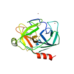
6SU3
 | | Crystal structure of the 48C12 heliorhodopsin in the violet form at pH 8.8 | | 分子名称: | (2R)-2,3-dihydroxypropyl (9Z)-octadec-9-enoate, 48C12 heliorhodopsin, EICOSANE, ... | | 著者 | Kovalev, K, Volkov, D, Astashkin, R, Alekseev, A, Gushchin, I, Gordeliy, V. | | 登録日 | 2019-09-12 | | 公開日 | 2019-12-11 | | 最終更新日 | 2024-01-24 | | 実験手法 | X-RAY DIFFRACTION (1.5 Å) | | 主引用文献 | High-resolution structural insights into the heliorhodopsin family.
Proc.Natl.Acad.Sci.USA, 117, 2020
|
|
7XPU
 | | crystal structure of MtdL-S228A-His soaked GDP-Fucp and Mn | | 分子名称: | GLYCEROL, GUANOSINE-5'-DIPHOSPHATE-BETA-L-FUCOPYRANOSE, MANGANESE (II) ION, ... | | 著者 | Li, F.D, He, C. | | 登録日 | 2022-05-05 | | 公開日 | 2023-03-29 | | 最終更新日 | 2023-11-29 | | 実験手法 | X-RAY DIFFRACTION (2.2 Å) | | 主引用文献 | Structures of the NDP-pyranose mutase belonging to glycosyltransferase family 75 reveal residues important for Mn 2+ coordination and substrate binding.
J.Biol.Chem., 299, 2023
|
|
4MQ9
 | | Crystal structure of Thermus thermophilus RNA polymerase holoenzyme in complex with GE23077 | | 分子名称: | (2Z)-2-methylbut-2-enoic acid, DNA-directed RNA polymerase subunit alpha, DNA-directed RNA polymerase subunit beta, ... | | 著者 | Ho, M.X, Arnold, E, Ebright, R.H, Zhang, Y, Tuske, S. | | 登録日 | 2013-09-16 | | 公開日 | 2014-05-07 | | 最終更新日 | 2023-09-20 | | 実験手法 | X-RAY DIFFRACTION (3.35 Å) | | 主引用文献 | GE23077 binds to the RNA polymerase 'i' and 'i+1' sites and prevents the binding of initiating nucleotides.
Elife, 3, 2014
|
|
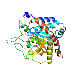
3V38
 | | Carboxypeptidase T mutant L254N | | 分子名称: | Carboxypeptidase T, GLYCEROL, SULFATE ION, ... | | 著者 | Timofeev, V.I, Kuznetsov, S.A, Akparov, V.K, Kuranova, I.P. | | 登録日 | 2011-12-13 | | 公開日 | 2012-12-19 | | 実験手法 | X-RAY DIFFRACTION (1.5 Å) | | 主引用文献 | Carboxypeptidase T mutant L254N
TO BE PUBLISHED
|
|
3VFK
 | | The structure of monodechloro-teicoplanin in complex with its ligand, using ubiquitin as a ligand carrier | | 分子名称: | 2-acetamido-2-deoxy-beta-D-glucopyranose, 2-amino-2-deoxy-beta-D-glucopyranose, 8-METHYLNONANOIC ACID, ... | | 著者 | Economou, N.J, Weeks, S.D, Grasty, K.C, Loll, P.J. | | 登録日 | 2012-01-09 | | 公開日 | 2013-01-09 | | 最終更新日 | 2023-12-06 | | 実験手法 | X-RAY DIFFRACTION (2.8 Å) | | 主引用文献 | Structure of the complex between teicoplanin and a bacterial cell-wall peptide: use of a carrier-protein approach.
Acta Crystallogr.,Sect.D, 69, 2013
|
|
3VFJ
 | | The structure of monodechloro-teicoplanin in complex with its ligand, using MBP as a ligand carrier | | 分子名称: | 2-acetamido-2-deoxy-beta-D-glucopyranose, 2-amino-2-deoxy-beta-D-glucopyranose, 8-METHYLNONANOIC ACID, ... | | 著者 | Economou, N.J, Weeks, S.D, Grasty, K.C, Loll, P.J. | | 登録日 | 2012-01-09 | | 公開日 | 2013-01-09 | | 最終更新日 | 2020-07-29 | | 実験手法 | X-RAY DIFFRACTION (2.05 Å) | | 主引用文献 | Structure of the complex between teicoplanin and a bacterial cell-wall peptide: use of a carrier-protein approach.
Acta Crystallogr.,Sect.D, 69, 2013
|
|
2MR8
 | |
2MR7
 | |
2MPX
 | |
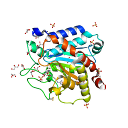
2AGI
 | | The leupeptin-trypsin covalent complex at 1.14 A resolution | | 分子名称: | CALCIUM ION, SULFATE ION, beta-trypsin, ... | | 著者 | Radisky, E.S, Lee, J.M, Lu, C.J, Koshland Jr, D.E. | | 登録日 | 2005-07-26 | | 公開日 | 2006-05-16 | | 最終更新日 | 2023-08-23 | | 実験手法 | X-RAY DIFFRACTION (1.14 Å) | | 主引用文献 | Insights into the serine protease mechanism from atomic resolution structures of trypsin reaction intermediates
Proc.Natl.Acad.Sci.USA, 103, 2006
|
|
2PSY
 | | Crystal Structure of Human Kallikrein 5 in complex with Leupeptin and Zinc | | 分子名称: | 2-acetamido-2-deoxy-beta-D-glucopyranose-(1-4)-2-acetamido-2-deoxy-beta-D-glucopyranose, Kallikrein-5, LEUPEPTIN, ... | | 著者 | Debela, M, Bode, W, Goettig, P. | | 登録日 | 2007-05-07 | | 公開日 | 2007-09-11 | | 最終更新日 | 2023-08-30 | | 実験手法 | X-RAY DIFFRACTION (2.3 Å) | | 主引用文献 | Structural basis of the zinc inhibition of human tissue kallikrein 5.
J.Mol.Biol., 373, 2007
|
|
4GM5
 | | Carboxypeptidase T with Sulphamoil Arginine | | 分子名称: | CALCIUM ION, Carboxypeptidase T, GLYCEROL, ... | | 著者 | Kuznetsov, S.A, Timofeev, V.I, Akparov, V.K, Kuranova, I.P. | | 登録日 | 2012-08-15 | | 公開日 | 2013-08-21 | | 実験手法 | X-RAY DIFFRACTION (1.39 Å) | | 主引用文献 | Carboxypeptidase T with Sulphamoil Arginine
To be Published
|
|
2PSX
 | |
2P0R
 | | Structure of Human Calpain 9 in complex with Leupeptin | | 分子名称: | CALCIUM ION, Calpain-9, leupeptin | | 著者 | Davis, T.L, Paramanathan, R, Walker, J.R, Butler-Cole, C, Finerty Jr, P.J, Weigelt, J, Sundstrom, M, Arrowsmith, C.H, Edwards, A.M, Bochkarev, A, Dhe-Paganon, S, Structural Genomics Consortium (SGC) | | 登録日 | 2007-03-01 | | 公開日 | 2007-03-20 | | 最終更新日 | 2023-08-30 | | 実験手法 | X-RAY DIFFRACTION (2.5 Å) | | 主引用文献 | Structures of Human Minicalpains bound to Inhibitors
To be Published
|
|
2OM5
 | |


