2HLF
 
 | | Structure of the Escherichis coli ClC chloride channel Y445E mutant and Fab complex | | 分子名称: | BROMIDE ION, Fab Fragment, Heavy chain, ... | | 著者 | Accardi, A, Lobet, S, Williams, C, Miller, C, Dutzler, R. | | 登録日 | 2006-07-07 | | 公開日 | 2006-09-19 | | 最終更新日 | 2023-08-30 | | 実験手法 | X-RAY DIFFRACTION (3.3 Å) | | 主引用文献 | Synergism Between Halide Binding and Proton Transport in a CLC-type Exchanger
J.Mol.Biol., 362, 2006
|
|
2HLG
 | |
2HLH
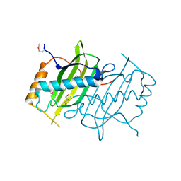
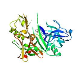
 | | Crystal structure of fucosyltransferase NodZ from Bradyrhizobium | | 分子名称: | Nodulation fucosyltransferase, PHOSPHATE ION | | 著者 | Brzezinski, K, Stepkowski, T, Panjikar, S, Bujacz, G, Jaskolski, M. | | 登録日 | 2006-07-07 | | 公開日 | 2007-07-17 | | 最終更新日 | 2023-08-30 | | 実験手法 | X-RAY DIFFRACTION (1.95 Å) | | 主引用文献 | High-resolution structure of NodZ fucosyltransferase involved in the biosynthesis of the nodulation factor.
Acta Biochim.Pol., 54, 2007
|
|
2HLI
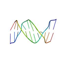 | | Solution structure of Crotonaldehyde-Derived N2-[3-Oxo-1(S)-methyl-propyl]-dG DNA Adduct in the 5'-CpG-3' Sequence | | 分子名称: | DNA dodecamer, DNA dodecamer with S-crotonaldehyde adduct | | 著者 | Cho, Y.-J, Wang, H, Kozekov, I.D, Kurtz, A.J, Jacob, J, Voehler, M, Smith, J, Harris, T.M, Rizzo, C.J, Stone, M.P. | | 登録日 | 2006-07-07 | | 公開日 | 2006-09-19 | | 最終更新日 | 2024-05-01 | | 実験手法 | SOLUTION NMR | | 主引用文献 | Orientation of the Crotonaldehyde-Derived N(2)-[3-Oxo-1(S)-methyl-propyl]-dG DNA Adduct Hinders Interstrand Cross-Link Formation in the 5'-CpG-3' Sequence
Chem.Res.Toxicol., 19, 2006
|
|
2HLJ
 | |
2HLN
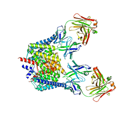
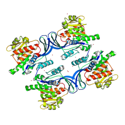
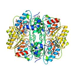 | | L-asparaginase from Erwinia carotovora in complex with glutamic acid | | 分子名称: | DI(HYDROXYETHYL)ETHER, GLUTAMIC ACID, L-asparaginase | | 著者 | Kravchenko, O.V, Kislitsin, Y.A, Popov, A.N, Nikonov, S.V, Kuranova, I.P. | | 登録日 | 2006-07-08 | | 公開日 | 2007-07-17 | | 最終更新日 | 2023-08-30 | | 実験手法 | X-RAY DIFFRACTION (2.2 Å) | | 主引用文献 | Three-dimensional structures of L-asparaginase from Erwinia carotovora complexed with aspartate and glutamate.
Acta Crystallogr.,Sect.D, 64, 2008
|
|
2HLO
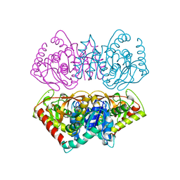
 | | Crystal Structure of Fragment D-dimer from Human Fibrin Complexed with Gly-hydroxyPro-Arg-Pro-amide | | 分子名称: | 2-acetamido-2-deoxy-alpha-D-glucopyranose-(1-4)-2-acetamido-2-deoxy-beta-D-glucopyranose, 2-acetamido-2-deoxy-beta-D-glucopyranose-(1-4)-2-acetamido-2-deoxy-beta-D-glucopyranose, CALCIUM ION, ... | | 著者 | Doolittle, R.F, Kollman, J.M, Chen, A, Pandi, L. | | 登録日 | 2006-07-08 | | 公開日 | 2007-07-17 | | 最終更新日 | 2023-08-30 | | 実験手法 | X-RAY DIFFRACTION (2.6 Å) | | 主引用文献 |
|
|
2HLP
 | | CRYSTAL STRUCTURE OF THE E267R MUTANT OF A HALOPHILIC MALATE DEHYDROGENASE IN THE APO FORM | | 分子名称: | CHLORIDE ION, MALATE DEHYDROGENASE, SODIUM ION | | 著者 | Richard, S.B, Madern, D, Garcin, E, Zaccai, G. | | 登録日 | 1999-04-23 | | 公開日 | 2000-02-04 | | 最終更新日 | 2023-08-30 | | 実験手法 | X-RAY DIFFRACTION (2.59 Å) | | 主引用文献 | Halophilic adaptation: novel solvent protein interactions observed in the 2.9 and 2.6 A resolution structures of the wild type and a mutant of malate dehydrogenase from Haloarcula marismortui.
Biochemistry, 39, 2000
|
|
2HLQ
 | |
2HLR
 | |
2HLS
 | |
2HLT
 | |
2HLU
 | |
2HLV
 | | Bovine Odorant Binding Protein deswapped triple mutant | | 分子名称: | 3,6-BIS(METHYLENE)DECANOIC ACID, GLYCEROL, Odorant-binding protein | | 著者 | Spinelli, S, Tegoni, M, Cambillau, C. | | 登録日 | 2006-07-10 | | 公開日 | 2008-03-25 | | 最終更新日 | 2023-08-30 | | 実験手法 | X-RAY DIFFRACTION (1.65 Å) | | 主引用文献 | Deswapping bovine odorant binding protein.
Biochim.Biophys.Acta, 1784, 2008
|
|
2HLW
 | | Solution Structure of the Human Ubiquitin-conjugating Enzyme Variant Uev1a | | 分子名称: | Ubiquitin-conjugating enzyme E2 variant 1 | | 著者 | Hau, D.D, Lewis, M.J, Saltibus, L.F, Pastushok, L, Xiao, W, Spyracopoulos, L. | | 登録日 | 2006-07-10 | | 公開日 | 2006-09-05 | | 最終更新日 | 2024-05-29 | | 実験手法 | SOLUTION NMR | | 主引用文献 | Structure and interactions of the ubiquitin-conjugating enzyme variant human uev1a: implications for enzymatic synthesis of polyubiquitin chains(,).
Biochemistry, 45, 2006
|
|
2HLY
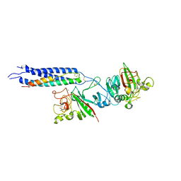
 | | The crystal structure of genomics APC5867 | | 分子名称: | Hypothetical protein Atu2299 | | 著者 | Dong, A, Xu, X, Zheng, H, Kim, Y, Edwards, A.M, Joachimiak, A, Savchenko, A, Midwest Center for Structural Genomics (MCSG) | | 登録日 | 2006-07-10 | | 公開日 | 2006-07-18 | | 最終更新日 | 2024-02-14 | | 実験手法 | X-RAY DIFFRACTION (1.6 Å) | | 主引用文献 | The crystal structure of genomics APC5867
TO BE PUBLISHED
|
|
2HLZ
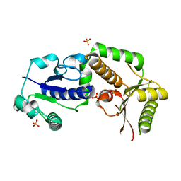
 | | Crystal Structure of human ketohexokinase | | 分子名称: | Ketohexokinase, UNKNOWN ATOM OR ION | | 著者 | Rabeh, W.M, Tempel, W, Nedyalkova, L, Landry, R, Arrowsmith, C.H, Edwards, A.M, Sundstrom, M, Weigelt, J, Bochkarev, A, Park, H, Structural Genomics Consortium (SGC) | | 登録日 | 2006-07-10 | | 公開日 | 2006-08-08 | | 最終更新日 | 2017-10-18 | | 実験手法 | X-RAY DIFFRACTION (1.85 Å) | | 主引用文献 | Crystal Structure of human ketohexokinase (CASP Target)
To be Published
|
|
2HM1
 | | Crystal Structure of human beta-secretase (BACE) in the presence of an inhibitor (2) | | 分子名称: | Beta-secretase 1, N-{(1S)-2-({(1S,2R)-1-(3,5-DIFLUOROBENZYL)-3-[(3-ETHYLBENZYL)AMINO]-2-HYDROXYPROPYL}AMINO)-2-OXO-1-[(PENTYLSULFONYL)METHYL]ETHYL}NICOTINAMIDE | | 著者 | Benson, T.E, Prince, D.B, Tomasselli, A.G, Emmons, T.L, Paddock, D.J. | | 登録日 | 2006-07-10 | | 公開日 | 2007-01-23 | | 最終更新日 | 2017-10-18 | | 実験手法 | X-RAY DIFFRACTION (2.2 Å) | | 主引用文献 | Design of potent inhibitors of human beta-secretase. Part 2.
Bioorg.Med.Chem.Lett., 17, 2007
|
|
2HM2
 | |
2HM3
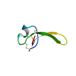
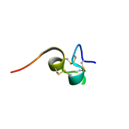 | | Nematocyst outer wall antigen, cysteine rich domain NW1 | | 分子名称: | Nematocyst outer wall antigen | | 著者 | Meier, S, Jensen, P.R, Grzesiek, S, Oezbek, S. | | 登録日 | 2006-07-11 | | 公開日 | 2007-02-06 | | 最終更新日 | 2022-03-09 | | 実験手法 | SOLUTION NMR | | 主引用文献 | Sequence-Structure and Structure-Function Analysis in Cysteine-rich Domains Forming the Ultrastable Nematocyst Wall.
J.Mol.Biol., 368, 2007
|
|
2HM4
 | | Nematocyst Outer Wall Antigen, NW1 K21P | | 分子名称: | Nematocyst outer wall antigen | | 著者 | Meier, S, Jensen, P.R, Grzesiek, S, Oezbek, S. | | 登録日 | 2006-07-11 | | 公開日 | 2007-02-06 | | 最終更新日 | 2021-10-20 | | 実験手法 | SOLUTION NMR | | 主引用文献 | Continuous molecular evolution of protein-domain structures by single amino Acid changes.
Curr.Biol., 17, 2007
|
|
2HM5
 | | NW1, K21P, Structural Species II | | 分子名称: | Nematocyst outer wall antigen | | 著者 | Meier, S, Jensen, P.R, Grzesiek, S, Oezbek, S. | | 登録日 | 2006-07-11 | | 公開日 | 2007-02-06 | | 最終更新日 | 2021-10-20 | | 実験手法 | SOLUTION NMR | | 主引用文献 | Continuous molecular evolution of protein-domain structures by single amino Acid changes.
Curr.Biol., 17, 2007
|
|
2HM6
 | | Nematocyst outer wall antigen, NW1 G11V K21P | | 分子名称: | Nematocyst outer wall antigen | | 著者 | Meier, S, Jensen, P.R, Grzesiek, S, Oezbek, S. | | 登録日 | 2006-07-11 | | 公開日 | 2007-02-06 | | 最終更新日 | 2021-10-20 | | 実験手法 | SOLUTION NMR | | 主引用文献 | Continuous molecular evolution of protein-domain structures by single amino Acid changes.
Curr.Biol., 17, 2007
|
|
2HM7
 | |
2HM8
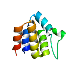
 | | Solution Structure of the C-terminal MA-3 domain of Pdcd4 | | 分子名称: | Pdcd4 C-terminal MA-3 domain | | 著者 | Waters, L.C, Veverka, V, Bohm, M, Muskett, F.W, Choong, P.T, Klempnauer, K.H, Carr, M.D. | | 登録日 | 2006-07-11 | | 公開日 | 2007-02-20 | | 最終更新日 | 2024-05-29 | | 実験手法 | SOLUTION NMR | | 主引用文献 | Structure of the C-terminal MA-3 domain of the tumour suppressor protein Pdcd4 and characterization of its interaction with eIF4A
Oncogene, 26, 2007
|
|


