6ML3
 
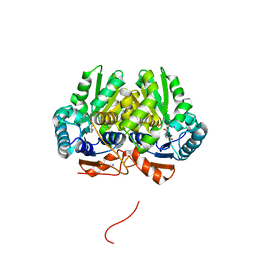
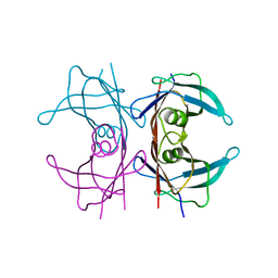
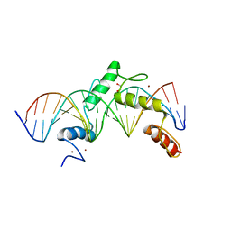 | | ZBTB24 Zinc Fingers 4-8 with 19+1mer DNA Oligonucleotide (Sequence 2) | | 分子名称: | 1,2-ETHANEDIOL, DNA (5'-D(*AP*CP*GP*CP*AP*GP*GP*TP*CP*CP*TP*GP*GP*AP*AP*GP*CP*TP*AP*A)-3'), DNA (5'-D(*TP*TP*TP*AP*GP*CP*TP*TP*CP*CP*AP*GP*GP*AP*CP*CP*TP*GP*CP*G)-3'), ... | | 著者 | Horton, J.R, Cheng, X, Ren, R. | | 登録日 | 2018-09-26 | | 公開日 | 2019-07-03 | | 最終更新日 | 2023-10-11 | | 実験手法 | X-RAY DIFFRACTION (1.683 Å) | | 主引用文献 | Structural basis of specific DNA binding by the transcription factor ZBTB24.
Nucleic Acids Res., 47, 2019
|
|
7YWV
 | | Eugenol oxidase from rhodococcus jostii: mutant S81H, D151E, A423M, H434Y, S394V, Q425S, I445D, S518P | | 分子名称: | 2-methoxy-4-(prop-2-en-1-yl)phenol, CALCIUM ION, FLAVIN-ADENINE DINUCLEOTIDE, ... | | 著者 | Alvigini, L, Mattevi, A. | | 登録日 | 2022-02-14 | | 公開日 | 2022-11-16 | | 最終更新日 | 2024-11-06 | | 実験手法 | X-RAY DIFFRACTION (2.4 Å) | | 主引用文献 | Structure- and computational-aided engineering of an oxidase to produce isoeugenol from a lignin-derived compound.
Nat Commun, 13, 2022
|
|
5EMT
 | |
6CPM
 | | Structure of the USP15 deubiquitinase domain in complex with a third-generation inhibitory Ubv | | 分子名称: | 1,2-ETHANEDIOL, CALCIUM ION, GLYCEROL, ... | | 著者 | Singer, A.U, Teyra, J, Boehmelt, G, Lenter, M, Sicheri, F, Sidhu, S.S. | | 登録日 | 2018-03-13 | | 公開日 | 2019-01-23 | | 最終更新日 | 2024-04-03 | | 実験手法 | X-RAY DIFFRACTION (2.011 Å) | | 主引用文献 | Structural and Functional Characterization of Ubiquitin Variant Inhibitors of USP15.
Structure, 27, 2019
|
|
5UN9
 | | The crystal structure of human O-GlcNAcase in complex with Thiamet-G | | 分子名称: | (3AR,5R,6S,7R,7AR)-2-(ETHYLAMINO)-5-(HYDROXYMETHYL)-5,6,7,7A-TETRAHYDRO-3AH-PYRANO[3,2-D][1,3]THIAZOLE-6,7-DIOL, Protein O-GlcNAcase | | 著者 | Li, B, Jiang, J. | | 登録日 | 2017-01-30 | | 公開日 | 2017-03-15 | | 最終更新日 | 2023-10-04 | | 実験手法 | X-RAY DIFFRACTION (2.5 Å) | | 主引用文献 | Structures of human O-GlcNAcase and its complexes reveal a new substrate recognition mode.
Nat. Struct. Mol. Biol., 24, 2017
|
|
5Z6W
 | | Crystal structure of paFAN1 bound to 2nt 5'flap DNA with gap with Manganese | | 分子名称: | DNA (5'-D(P*AP*TP*TP*CP*AP*A)-3'), DNA (5'-D(P*GP*AP*AP*TP*GP*TP*GP*TP*CP*TP*CP*AP*AP*TP*CP*CP*CP*AP*AP*CP*TP*T)-3'), DNA (5'-D(P*GP*TP*TP*GP*GP*GP*AP*TP*TP*G)-3'), ... | | 著者 | Jin, H, Cho, Y. | | 登録日 | 2018-01-25 | | 公開日 | 2018-03-14 | | 最終更新日 | 2023-11-22 | | 実験手法 | X-RAY DIFFRACTION (3.2 Å) | | 主引用文献 | Structural mechanism of DNA interstrand cross-link unhooking by the bacterial FAN1 nuclease.
J. Biol. Chem., 293, 2018
|
|
6MVA
 | | LDHA structure in complex with inhibitor 14 | | 分子名称: | (6R)-6-(3-aminophenyl)-3-[(2-chlorophenyl)sulfanyl]-4-hydroxy-6-(thiophen-3-yl)-5,6-dihydro-2H-pyran-2-one, 1,4-DIHYDRONICOTINAMIDE ADENINE DINUCLEOTIDE, 4-(2-HYDROXYETHYL)-1-PIPERAZINE ETHANESULFONIC ACID, ... | | 著者 | Eigenbrot, C.E, Ultsch, M, Wei, B. | | 登録日 | 2018-10-24 | | 公開日 | 2019-10-30 | | 最終更新日 | 2024-03-13 | | 実験手法 | X-RAY DIFFRACTION (2.02 Å) | | 主引用文献 | Structure-based Optimization of Potent, Cell-Active
Hydroxylactam Inhibitors of Lactate Dehydrogenase
To Be Published
|
|
1GCU
 | |
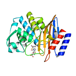
5IJW
 | | Glutamate Racemase (MurI) from Mycobacterium smegmatis with bound D-glutamate, 1.8 Angstrom resolution, X-ray diffraction | | 分子名称: | D-GLUTAMIC ACID, Glutamate racemase, IODIDE ION | | 著者 | Poen, S, Nakatani, Y, Krause, K. | | 登録日 | 2016-03-02 | | 公開日 | 2016-05-25 | | 最終更新日 | 2023-09-27 | | 実験手法 | X-RAY DIFFRACTION (1.76 Å) | | 主引用文献 | Exploring the structure of glutamate racemase from Mycobacterium tuberculosis as a template for anti-mycobacterial drug discovery.
Biochem. J., 473, 2016
|
|
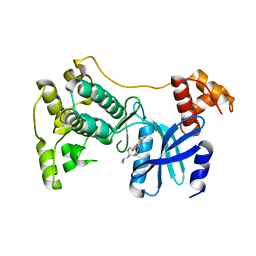
6N53
 | | Crystal structure of human uridine-cytidine kinase 2 complexed with 2'-azidouridine monophosphate | | 分子名称: | 2'-deoxy-2'-triaza-1,2-dien-2-ium-1-yl-uridine-5'-monophosphate, GLYCEROL, MAGNESIUM ION, ... | | 著者 | Cuthbert, B.J, Nainar, S, Spitale, R.C, Goulding, C.W. | | 登録日 | 2018-11-21 | | 公開日 | 2019-11-27 | | 最終更新日 | 2023-10-11 | | 実験手法 | X-RAY DIFFRACTION (2.7 Å) | | 主引用文献 | An optimized chemical-genetic method for cell-specific metabolic labeling of RNA.
Nat.Methods, 17, 2020
|
|
5TVT
 | | Structure of Maternal Embryonic Leucine Zipper Kinase | | 分子名称: | 9-(3,5-dichloro-4-hydroxyphenyl)-1-{trans-4-[(dimethylamino)methyl]cyclohexyl}-3-methyl-3,4-dihydropyrimido[5,4-c][1,5]naphthyridin-2(1H)-one, Maternal embryonic leucine zipper kinase | | 著者 | Seo, H.-Y, Huang, H, Gray, N.S, Dhe-Paganon, S. | | 登録日 | 2016-11-10 | | 公開日 | 2017-11-15 | | 最終更新日 | 2024-03-06 | | 実験手法 | X-RAY DIFFRACTION (2.28 Å) | | 主引用文献 | Structure of Maternal Embryonic Leucine Zipper Kinase
To Be Published
|
|
5TW6
 | | CTX-M-14 P167S:E166A mutant with acylated ceftazidime molecule | | 分子名称: | 1,2-ETHANEDIOL, ACYLATED CEFTAZIDIME, Beta-lactamase | | 著者 | Patel, M, Stojanoski, V, Sankaran, B, Prasad, B.V.V, Palzkill, T. | | 登録日 | 2016-11-11 | | 公開日 | 2017-06-28 | | 最終更新日 | 2023-10-04 | | 実験手法 | X-RAY DIFFRACTION (1.7 Å) | | 主引用文献 | The Drug-Resistant Variant P167S Expands the Substrate Profile of CTX-M beta-Lactamases for Oxyimino-Cephalosporin Antibiotics by Enlarging the Active Site upon Acylation.
Biochemistry, 56, 2017
|
|
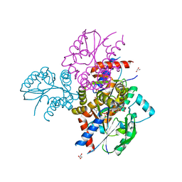
5TW1
 | | Crystal structure of a Mycobacterium smegmatis transcription initiation complex with RbpA | | 分子名称: | 1,2-ETHANEDIOL, DNA (26-MER), DNA (31-MER), ... | | 著者 | Hubin, E.A, Darst, S.A, Campbell, E.A. | | 登録日 | 2016-11-10 | | 公開日 | 2017-01-18 | | 最終更新日 | 2024-12-25 | | 実験手法 | X-RAY DIFFRACTION (2.76 Å) | | 主引用文献 | Structure and function of the mycobacterial transcription initiation complex with the essential regulator RbpA.
Elife, 6, 2017
|
|
5UUO
 | | Crystal structure of SARO_2595 from Novosphingobium aromaticivorans | | 分子名称: | 1,2-ETHANEDIOL, GLUTATHIONE, Glutathione S-transferase-like protein, ... | | 著者 | Bingman, C.A, Kontur, W.S, Olmsted, C.N, Fox, B.G, Donohue, T.J. | | 登録日 | 2017-02-17 | | 公開日 | 2018-02-28 | | 最終更新日 | 2024-05-22 | | 実験手法 | X-RAY DIFFRACTION (1.25 Å) | | 主引用文献 | Novosphingobium aromaticivoransuses a Nu-class glutathioneS-transferase as a glutathione lyase in breaking the beta-aryl ether bond of lignin.
J. Biol. Chem., 293, 2018
|
|
4HSX
 | | Structure of the L100F mutant of dehaloperoxidase-hemoglobin A from Amphitrite ornata with 4-bromophenol | | 分子名称: | 4-BROMOPHENOL, Dehaloperoxidase A, GLYCEROL, ... | | 著者 | Thompson, M.K, Plummer, A, Franzen, S. | | 登録日 | 2012-10-31 | | 公開日 | 2013-05-01 | | 最終更新日 | 2024-02-28 | | 実験手法 | X-RAY DIFFRACTION (1.12 Å) | | 主引用文献 | Role of polarity of the distal pocket in the control of inhibitor binding in dehaloperoxidase-hemoglobin.
Biochemistry, 52, 2013
|
|
5UTK
 | |
4PVN
 | | Neutron structure of human transthyretin (TTR) at room temperature to 2.3A resolution (monochromatic) | | 分子名称: | Transthyretin | | 著者 | Fisher, S.J, Blakeley, M.P, Haupt, M, Mason, S.A, Cooper, J.B, Mitchell, E.P, Forsyth, V.T. | | 登録日 | 2014-03-18 | | 公開日 | 2014-11-12 | | 最終更新日 | 2024-03-20 | | 実験手法 | NEUTRON DIFFRACTION (2.3 Å), X-RAY DIFFRACTION | | 主引用文献 | Binding site asymmetry in human transthyretin: insights from a joint neutron and X-ray crystallographic analysis using perdeuterated protein
IUCrJ, 1, 2014
|
|
4HSW
 | | Structure of the L100F mutant of dehaloperoxidase-hemoglobin A from Amphitrite ornata | | 分子名称: | Dehaloperoxidase A, GLYCEROL, PROTOPORPHYRIN IX CONTAINING FE, ... | | 著者 | Thompson, M.K, Plummer, A, Franzen, S. | | 登録日 | 2012-10-31 | | 公開日 | 2013-05-01 | | 最終更新日 | 2024-02-28 | | 実験手法 | X-RAY DIFFRACTION (1.22 Å) | | 主引用文献 | Role of polarity of the distal pocket in the control of inhibitor binding in dehaloperoxidase-hemoglobin.
Biochemistry, 52, 2013
|
|
1JDA
 | | MALTOTETRAOSE-FORMING EXO-AMYLASE | | 分子名称: | 1,4-ALPHA MALTOTETRAHYDROLASE, CALCIUM ION | | 著者 | Yoshioka, Y, Hasegawa, K, Matsuura, Y, Katsube, Y, Kubota, M. | | 登録日 | 1997-06-16 | | 公開日 | 1997-10-15 | | 最終更新日 | 2024-10-30 | | 実験手法 | X-RAY DIFFRACTION (2.2 Å) | | 主引用文献 | Crystal structures of a mutant maltotetraose-forming exo-amylase cocrystallized with maltopentaose.
J.Mol.Biol., 271, 1997
|
|
9FKK
 | | The structure of glycosynthase IXT6 (E241G mutant), the intracellular xylanase of G.proteiniphilus T-6 in complex with xylobiose-F and xylotetraose-F molecules | | 分子名称: | Beta-xylanase, SODIUM ION, beta-D-xylopyranose-(1-4)-1-fluoro-D-xylopyranose, ... | | 著者 | Hadad, N, Chmelnik, O, Dessau, M, Shoham, Y, Shoham, G. | | 登録日 | 2024-06-03 | | 公開日 | 2025-06-18 | | 実験手法 | X-RAY DIFFRACTION (2.4 Å) | | 主引用文献 | The structure of glycosynthase IXT6 (E241G mutant), the intracellular xylanase of G.proteiniphilus T-6 in complex with xylobiose-F and xylotetraose-F molecules
To Be Published
|
|
4DZ0
 | |
5B6Z
 | | A three dimensional movie of structural changes in bacteriorhodopsin: structure obtained 1.725 ms us after photoexcitation | | 分子名称: | 2,3-DI-PHYTANYL-GLYCEROL, Bacteriorhodopsin, DECANE, ... | | 著者 | Royant, A, Nango, E, Nakane, T, Tanaka, T, Arima, T, Neutze, R, Iwata, S. | | 登録日 | 2016-06-02 | | 公開日 | 2016-12-21 | | 最終更新日 | 2024-11-13 | | 実験手法 | X-RAY DIFFRACTION (2.1 Å) | | 主引用文献 | A three-dimensional movie of structural changes in bacteriorhodopsin
Science, 354, 2016
|
|
5X3G
 | |
5EWV
 | | Crystal structure of the human BRPF1 bromodomain in complex with SEED20 | | 分子名称: | 1,5-dimethyl-[1,2,4]triazolo[4,3-a]quinoline, NITRATE ION, Peregrin | | 著者 | Zhu, J, Wiedmer, L, Caflisch, A. | | 登録日 | 2015-11-21 | | 公開日 | 2016-11-02 | | 最終更新日 | 2024-01-10 | | 実験手法 | X-RAY DIFFRACTION (1.67 Å) | | 主引用文献 | Crystal structure of the human BRPF1 bromodomain in complex with SEED20
To Be Published
|
|
5EWK
 | | Scabin toxin from Streptomyces Scabies in complex with inhibitor PJ34 | | 分子名称: | N~2~,N~2~-DIMETHYL-N~1~-(6-OXO-5,6-DIHYDROPHENANTHRIDIN-2-YL)GLYCINAMIDE, Putative ADP-Ribosyltransferase Scabin | | 著者 | Ravulapalli, R, Lyons, B, Merrill, A.R. | | 登録日 | 2015-11-20 | | 公開日 | 2016-03-23 | | 最終更新日 | 2024-10-23 | | 実験手法 | X-RAY DIFFRACTION (1.6 Å) | | 主引用文献 | Scabin, a Novel DNA-acting ADP-ribosyltransferase from Streptomyces scabies.
J.Biol.Chem., 291, 2016
|
|


