2G67
 
 | |
2G69
 | |
2G6B
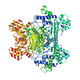
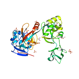
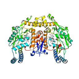
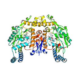
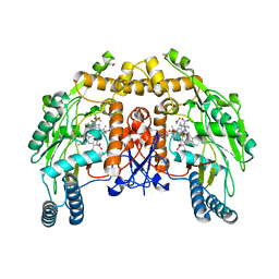
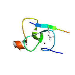
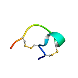
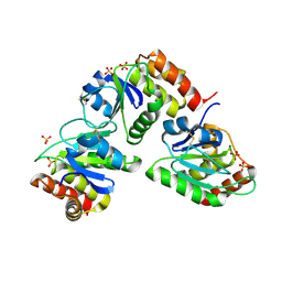
 | | Crystal structure of human RAB26 in complex with a GTP analogue | | 分子名称: | MAGNESIUM ION, PHOSPHOAMINOPHOSPHONIC ACID-GUANYLATE ESTER, Ras-related protein Rab-26, ... | | 著者 | Wang, J, Tempel, W, Shen, Y, Shen, L, Yaniw, D, Arrowsmith, C, Edwards, A, Sundstrom, M, Weigelt, J, Bochkarev, A, Park, H, Structural Genomics Consortium (SGC) | | 登録日 | 2006-02-24 | | 公開日 | 2006-03-14 | | 最終更新日 | 2024-04-03 | | 実験手法 | X-RAY DIFFRACTION (2 Å) | | 主引用文献 | Crystal structure of human RAB26 in complex with a GTP analogue
To be Published
|
|
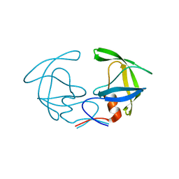
2G6D
 | | Human cathepsin S mutant with vinyl sulfone inhibitor CRA-14009 | | 分子名称: | N-[(1S)-1-[({(1S)-3-PHENYL-1-[2-(PHENYLSULFONYL)ETHYL]PROPYL}AMINO)CARBONYL]-3-(PHENYLSULFONYL)PROPYL]MORPHOLINE-4-CARBOXAMIDE, cathepsin S | | 著者 | Somoza, J.R. | | 登録日 | 2006-02-24 | | 公開日 | 2006-04-04 | | 最終更新日 | 2021-10-20 | | 実験手法 | X-RAY DIFFRACTION (2.5 Å) | | 主引用文献 | Human cathepsin S mutant with vinyl sulfone inhibitor CRA-14009
To be Published
|
|
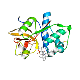
2G6E
 | | Structure of cyclized F64L S65A Y66S GFP variant | | 分子名称: | Green fluorescent protein, MAGNESIUM ION | | 著者 | Barondeau, D.P. | | 登録日 | 2006-02-24 | | 公開日 | 2006-04-18 | | 最終更新日 | 2023-11-15 | | 実験手法 | X-RAY DIFFRACTION (1.3 Å) | | 主引用文献 | Understanding GFP Posttranslational Chemistry: Structures of Designed Variants that Achieve Backbone Fragmentation, Hydrolysis, and Decarboxylation.
J.Am.Chem.Soc., 128, 2006
|
|
2G6F
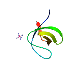 | |
2G6G
 | | Crystal structure of MltA from Neisseria gonorrhoeae | | 分子名称: | GLYCEROL, GNA33, SULFATE ION | | 著者 | Powell, A.J, Liu, Z.J, Nicholas, R.A, Davies, C. | | 登録日 | 2006-02-24 | | 公開日 | 2006-05-02 | | 最終更新日 | 2017-10-18 | | 実験手法 | X-RAY DIFFRACTION (2.2 Å) | | 主引用文献 | Crystal Structures of the Lytic Transglycosylase MltA from N.gonorrhoeae and E.coli: Insights into Interdomain Movements and Substrate Binding.
J.Mol.Biol., 359, 2006
|
|
2G6H
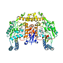
 | | Structure of rat nNOS heme domain (BH4 bound) in the reduced form | | 分子名称: | 5,6,7,8-TETRAHYDROBIOPTERIN, ACETATE ION, ARGININE, ... | | 著者 | Li, H, Igarashi, J, Jamal, J, Yang, W, Poulos, T.L. | | 登録日 | 2006-02-24 | | 公開日 | 2006-08-08 | | 最終更新日 | 2023-08-30 | | 実験手法 | X-RAY DIFFRACTION (2 Å) | | 主引用文献 | Structural studies of constitutive nitric oxide synthases with diatomic ligands bound.
J.Biol.Inorg.Chem., 11, 2006
|
|
2G6I
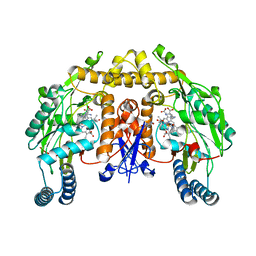
 | | Structure of rat nNOS heme domain (BH2-bound) in the reduced form | | 分子名称: | 7,8-DIHYDROBIOPTERIN, ACETATE ION, ARGININE, ... | | 著者 | Li, H, Igarashi, J, Jamal, J, Yang, W, Poulos, T.L. | | 登録日 | 2006-02-24 | | 公開日 | 2006-08-08 | | 最終更新日 | 2023-08-30 | | 実験手法 | X-RAY DIFFRACTION (1.9 Å) | | 主引用文献 | Structural studies of constitutive nitric oxide synthases with diatomic ligands bound.
J.Biol.Inorg.Chem., 11, 2006
|
|
2G6J
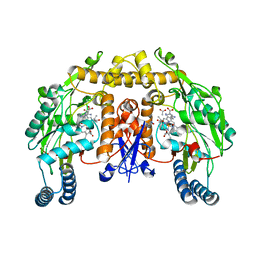
 | | Structure of rat nNOS (L337N) heme domain (4-aminobiopterin bound) complexed with NO | | 分子名称: | (1S,2S)-1-(2,4-DIAMINOPTERIDIN-6-YL)PROPANE-1,2-DIOL, ACETATE ION, ARGININE, ... | | 著者 | Li, H, Igarashi, J, Jamal, J, Yang, W, Poulos, T.L. | | 登録日 | 2006-02-24 | | 公開日 | 2006-08-08 | | 最終更新日 | 2023-08-30 | | 実験手法 | X-RAY DIFFRACTION (2.3 Å) | | 主引用文献 | Structural studies of constitutive nitric oxide synthases with diatomic ligands bound.
J.Biol.Inorg.Chem., 11, 2006
|
|
2G6K
 | | Structure of rat nNOS heme domain (BH4 bound) complexed with NO | | 分子名称: | 5,6,7,8-TETRAHYDROBIOPTERIN, ACETATE ION, ARGININE, ... | | 著者 | Li, H, Igarashi, J, Jamal, J, Yang, W, Poulos, T.L. | | 登録日 | 2006-02-24 | | 公開日 | 2006-08-08 | | 最終更新日 | 2023-08-30 | | 実験手法 | X-RAY DIFFRACTION (2 Å) | | 主引用文献 | Structural studies of constitutive nitric oxide synthases with diatomic ligands bound.
J.Biol.Inorg.Chem., 11, 2006
|
|
2G6L
 | | Structure of rat nNOS heme domain (BH2 bound) complexed with NO | | 分子名称: | 7,8-DIHYDROBIOPTERIN, ACETATE ION, ARGININE, ... | | 著者 | Li, H, Igarashi, J, Jamal, J, Yang, W, Poulos, T.L. | | 登録日 | 2006-02-24 | | 公開日 | 2006-08-08 | | 最終更新日 | 2023-08-30 | | 実験手法 | X-RAY DIFFRACTION (2.05 Å) | | 主引用文献 | Structural studies of constitutive nitric oxide synthases with diatomic ligands bound.
J.Biol.Inorg.Chem., 11, 2006
|
|
2G6M
 | | Structure of rat nNOS heme domain (BH4 bound) complexed with CO | | 分子名称: | 5,6,7,8-TETRAHYDROBIOPTERIN, ACETATE ION, ARGININE, ... | | 著者 | Li, H, Igarashi, J, Jamal, J, Yang, W, Poulos, T.L. | | 登録日 | 2006-02-24 | | 公開日 | 2006-08-08 | | 最終更新日 | 2023-08-30 | | 実験手法 | X-RAY DIFFRACTION (1.85 Å) | | 主引用文献 | Structural studies of constitutive nitric oxide synthases with diatomic ligands bound.
J.Biol.Inorg.Chem., 11, 2006
|
|
2G6N
 | | Strcture of rat nNOS heme domain (BH2 bound) complexed with CO | | 分子名称: | 7,8-DIHYDROBIOPTERIN, ACETATE ION, ARGININE, ... | | 著者 | Li, H, Igarashi, J, Jamal, J, Yang, W, Poulos, T.L. | | 登録日 | 2006-02-24 | | 公開日 | 2006-08-08 | | 最終更新日 | 2023-08-30 | | 実験手法 | X-RAY DIFFRACTION (1.9 Å) | | 主引用文献 | Structural studies of constitutive nitric oxide synthases with diatomic ligands bound.
J.Biol.Inorg.Chem., 11, 2006
|
|
2G6O
 | | Structure of bovine eNOS heme domain (BH4-free) complexed with CO | | 分子名称: | ACETATE ION, ARGININE, CACODYLATE ION, ... | | 著者 | Li, H, Igarashi, J, Jamal, J, Yang, W, Poulos, T.L. | | 登録日 | 2006-02-24 | | 公開日 | 2006-08-08 | | 最終更新日 | 2024-02-14 | | 実験手法 | X-RAY DIFFRACTION (1.9 Å) | | 主引用文献 | Structural studies of constitutive nitric oxide synthases with diatomic ligands bound.
J.Biol.Inorg.Chem., 11, 2006
|
|
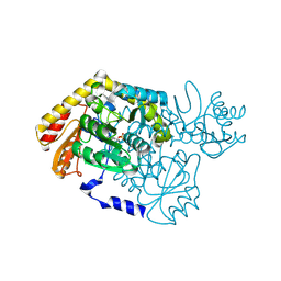
2G6P
 | | Crystal structure of truncated (delta 1-89) human methionine aminopeptidase Type 1 in complex with Pyridyl pyrimidine derivative | | 分子名称: | 4-(2-HYDROXYETHYL)-1-PIPERAZINE ETHANESULFONIC ACID, 5-CHLORO-6-METHYL-N-(2-PHENYLETHYL)-2-PYRIDIN-2-YLPYRIMIDIN-4-AMINE, COBALT (II) ION, ... | | 著者 | Addlagatta, A, Hu, X, Liu, J.O, Matthews, B.W. | | 登録日 | 2006-02-24 | | 公開日 | 2006-06-20 | | 最終更新日 | 2024-02-14 | | 実験手法 | X-RAY DIFFRACTION (1.9 Å) | | 主引用文献 | Identification of Pyridinylpyrimidines as Inhibitors of Human Methionine Aminopeptidases.
Angew.Chem.Int.Ed.Engl., 45, 2006
|
|
2G6Q
 | |
2G6T
 | |
2G6U
 | |
2G6V
 | | The crystal structure of ribD from Escherichia coli | | 分子名称: | Riboflavin biosynthesis protein ribD | | 著者 | Stenmark, P, Moche, M, Gurmu, D, Nordlund, P, Structural Proteomics in Europe (SPINE) | | 登録日 | 2006-02-25 | | 公開日 | 2007-02-06 | | 最終更新日 | 2011-07-13 | | 実験手法 | X-RAY DIFFRACTION (2.6 Å) | | 主引用文献 | The Crystal Structure of the Bifunctional Deaminase/Reductase RibD of the Riboflavin Biosynthetic Pathway in Escherichia coli: Implications for the Reductive Mechanism.
J.Mol.Biol., 373, 2007
|
|
2G6W
 | |
2G6X
 | |
2G6Y
 | |
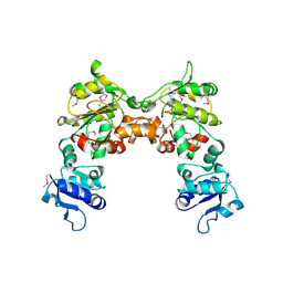
2G6Z
 | | Crystal structure of human DUSP5 | | 分子名称: | Dual specificity protein phosphatase 5, SULFATE ION | | 著者 | Kim, S.J, Ryu, S.E. | | 登録日 | 2006-02-26 | | 公開日 | 2007-01-16 | | 最終更新日 | 2011-07-13 | | 実験手法 | X-RAY DIFFRACTION (2.7 Å) | | 主引用文献 | Crystal structure of the catalytic domain of human DUSP5, a dual specificity MAP kinase protein phosphatase
Proteins, 66, 2007
|
|
2G70
 | | Structure of human PNMT in complex with inhibitor 3-hydroxymethyl-7-nitro-THIQ and AdoMet (SAM) | | 分子名称: | PHOSPHATE ION, Phenylethanolamine N-methyltransferase, S-ADENOSYLMETHIONINE, ... | | 著者 | Tyndall, J.D.A, Gee, C.L, Martin, J.L. | | 登録日 | 2006-02-27 | | 公開日 | 2007-02-13 | | 最終更新日 | 2023-10-25 | | 実験手法 | X-RAY DIFFRACTION (2.4 Å) | | 主引用文献 | Enzyme Adaptation to Inhibitor Binding: A Cryptic Binding Site in Phenylethanolamine N-Methyltransferase
J.Med.Chem., 50, 2007
|
|


