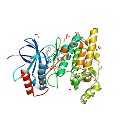
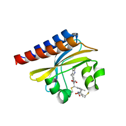
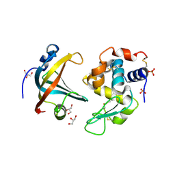
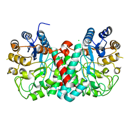
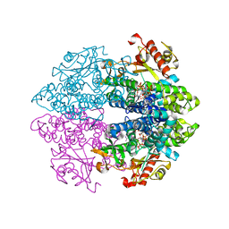
8IFD
 
 | | Dibekacin-added human 80S ribosome | | 分子名称: | 18S ribosomal RNA, 28S ribosomal RNA, 40S ribosomal protein S10, ... | | 著者 | Tomono, J, Asano, K, Chiashi, T, Tanaka, Y, Yokoyama, T. | | 登録日 | 2023-02-17 | | 公開日 | 2024-02-14 | | 最終更新日 | 2024-06-19 | | 実験手法 | ELECTRON MICROSCOPY (2.59 Å) | | 主引用文献 | Direct visualization of ribosomes in the cell-free system revealed the functional evolution of aminoglycoside.
J.Biochem., 175, 2024
|
|
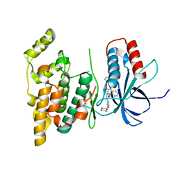
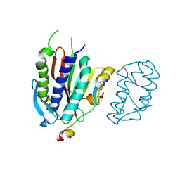
4QTD
 | | Structure of human JNK1 in complex with SCH772984 and the AMPPNP-hydrolysed triphosphate revealing the second type-I binding mode | | 分子名称: | (3R)-1-(2-oxo-2-{4-[4-(pyrimidin-2-yl)phenyl]piperazin-1-yl}ethyl)-N-[3-(pyridin-4-yl)-2H-indazol-5-yl]pyrrolidine-3-carboxamide, 1,2-ETHANEDIOL, 4-(2-HYDROXYETHYL)-1-PIPERAZINE ETHANESULFONIC ACID, ... | | 著者 | Chaikuad, A, Keates, T, von Delft, F, Arrowsmith, C.H, Edwards, A.M, Bountra, C, Knapp, S, Structural Genomics Consortium (SGC) | | 登録日 | 2014-07-07 | | 公開日 | 2014-07-23 | | 最終更新日 | 2023-09-20 | | 実験手法 | X-RAY DIFFRACTION (1.5 Å) | | 主引用文献 | A unique inhibitor binding site in ERK1/2 is associated with slow binding kinetics.
Nat.Chem.Biol., 10, 2014
|
|
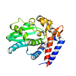
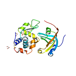
4U79
 | | Crystal structure of human JNK3 in complex with a benzenesulfonamide inhibitor. | | 分子名称: | Mitogen-activated protein kinase 10, N-{4-[(3-{2-[(trans-4-aminocyclohexyl)amino]pyrimidin-4-yl}pyridin-2-yl)oxy]naphthalen-1-yl}benzenesulfonamide | | 著者 | Mohr, C. | | 登録日 | 2014-07-30 | | 公開日 | 2014-10-08 | | 最終更新日 | 2023-09-27 | | 実験手法 | X-RAY DIFFRACTION (2.23 Å) | | 主引用文献 | Unfolded Protein Response in Cancer: IRE1 alpha Inhibition by Selective Kinase Ligands Does Not Impair Tumor Cell Viability.
Acs Med.Chem.Lett., 6, 2015
|
|
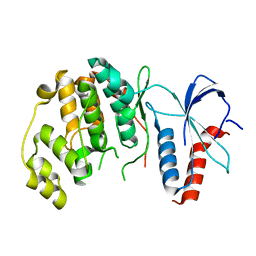
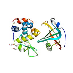
6BE0
 | | AvrA delL154 with IP6, CoA | | 分子名称: | AvrA, COENZYME A, INOSITOL HEXAKISPHOSPHATE | | 著者 | Labriola, J.M, Nagar, B. | | 登録日 | 2017-10-24 | | 公開日 | 2018-08-01 | | 最終更新日 | 2023-10-04 | | 実験手法 | X-RAY DIFFRACTION (2.438 Å) | | 主引用文献 | Structural Analysis of the Bacterial Effector AvrA Identifies a Critical Helix Involved in Substrate Recognition.
Biochemistry, 57, 2018
|
|
6RFO
 | | ERK2 MAP kinase with the activation loop of p38alpha | | 分子名称: | Mitogen-activated protein kinase 1,Mitogen-activated protein kinase 14,Mitogen-activated protein kinase 1 | | 著者 | Livnah, O, Eitan-Wexler, M. | | 登録日 | 2019-04-15 | | 公開日 | 2020-05-27 | | 最終更新日 | 2024-01-24 | | 実験手法 | X-RAY DIFFRACTION (1.7 Å) | | 主引用文献 | The bacterial metalloprotease NleD selectively cleaves mitogen-activated protein kinases that have high flexibility in their activation loop.
J.Biol.Chem., 295, 2020
|
|
4GLQ
 | | Crystal Structure of the blue-light absorbing form of the Thermosynechococcus elongatus PixJ GAF-domain | | 分子名称: | Methyl-accepting chemotaxis protein, Phycoviolobilin, blue light-absorbing form | | 著者 | Burgie, E.S, Walker, J.M, Phillips Jr, G.N, Vierstra, R.D. | | 登録日 | 2012-08-14 | | 公開日 | 2013-01-16 | | 最終更新日 | 2024-04-03 | | 実験手法 | X-RAY DIFFRACTION (1.772 Å) | | 主引用文献 | A Photo-Labile Thioether Linkage to Phycoviolobilin Provides the Foundation for the Blue/Green Photocycles in DXCF-Cyanobacteriochromes.
Structure, 21, 2013
|
|
4GK0
 | | Crystal structure of human Rev3-Rev7-Rev1 complex | | 分子名称: | DNA polymerase zeta catalytic subunit, DNA repair protein REV1, Mitotic spindle assembly checkpoint protein MAD2B | | 著者 | Tao, J, Min, X, Wei, X. | | 登録日 | 2012-08-10 | | 公開日 | 2013-03-13 | | 最終更新日 | 2023-11-08 | | 実験手法 | X-RAY DIFFRACTION (2.7 Å) | | 主引用文献 | Structural insights into the assembly of human translesion polymerase complexes
Protein Cell, 3, 2012
|
|
4GMI
 | | BACE-1 in complex with HEA-type macrocyclic inhibitor, MV078571 | | 分子名称: | (4S,8E)-4-[(1R)-2-{[2-(5-tert-butyl-1,3-oxazol-2-yl)propan-2-yl]amino}-1-hydroxyethyl]-16-methyl-6-oxa-3-azabicyclo[12.3.1]octadeca-1(18),8,14,16-tetraene-2,13-dione, ACETATE ION, Beta-secretase 1, ... | | 著者 | Lindberg, J.D, Derbyshire, D. | | 登録日 | 2012-08-16 | | 公開日 | 2013-09-04 | | 実験手法 | X-RAY DIFFRACTION (1.8 Å) | | 主引用文献 | Design and synthesis of novel BACE-1 inhibitors
To be Published
|
|
4G8J
 | | X-ray Structure of Uridine Phosphorylease from Vibrio cholerae Complexed with Thymidine at 2.12 A Resolution | | 分子名称: | 1,2-ETHANEDIOL, CHLORIDE ION, MAGNESIUM ION, ... | | 著者 | Lashkov, A.A, Gabdoulkhakov, A.G, Prokofev, I.I, Sotnichenko, S.E, Betzel, C, Mikhailov, A.M. | | 登録日 | 2012-07-23 | | 公開日 | 2013-07-24 | | 最終更新日 | 2023-09-13 | | 実験手法 | X-RAY DIFFRACTION (2.119 Å) | | 主引用文献 | X-ray structure of uridine phosphorylease from Vibrio cholerae complexed with thymidine
To be Published
|
|
4GLV
 | | OBody AM3L09 bound to hen egg-white lysozyme | | 分子名称: | 4-(2-HYDROXYETHYL)-1-PIPERAZINE ETHANESULFONIC ACID, GLYCEROL, Lysozyme C, ... | | 著者 | Steemson, J.D. | | 登録日 | 2012-08-15 | | 公開日 | 2013-08-21 | | 最終更新日 | 2014-02-12 | | 実験手法 | X-RAY DIFFRACTION (2.574 Å) | | 主引用文献 | Tracking Molecular Recognition at the Atomic Level with a New Protein Scaffold Based on the OB-Fold.
Plos One, 9, 2014
|
|
4GK5
 | | Crystal structure of human Rev3-Rev7-Rev1-Polkappa complex | | 分子名称: | DNA polymerase kappa, DNA polymerase zeta catalytic subunit, DNA repair protein REV1, ... | | 著者 | Tao, J, Min, X, Wei, X. | | 登録日 | 2012-08-10 | | 公開日 | 2013-03-13 | | 最終更新日 | 2023-11-08 | | 実験手法 | X-RAY DIFFRACTION (3.21 Å) | | 主引用文献 | Structural insights into the assembly of human translesion polymerase complexes
Protein Cell, 3, 2012
|
|
4GN5
 | |
4GN4
 | |
4GNV
 | |
6T3H
 | |
6T46
 | | Structure of the Rap conjugation gene regulator of the plasmid pLS20 in complex with the Phr* peptide | | 分子名称: | CHLORIDE ION, Quorum-sensing secretion protein (processed), Response regulator aspartate phosphatase, ... | | 著者 | Crespo, I, Bernardo, N, Meijer, W.J.J, Boer, D.R. | | 登録日 | 2019-10-12 | | 公開日 | 2020-06-24 | | 最終更新日 | 2024-01-24 | | 実験手法 | X-RAY DIFFRACTION (2.45 Å) | | 主引用文献 | Inactivation of the dimeric RappLS20 anti-repressor of the conjugation operon is mediated by peptide-induced tetramerization.
Nucleic Acids Res., 48, 2020
|
|
2WF4
 | | Human BACE-1 in complex with 6-ethyl-1-methyl-N-((1S)-2-oxo-1-(phenylmethyl)-3-(tetrahydro-2H-pyran-4-ylamino)propyl)-1,3,4,6- tetrahydro(1,2)thiazepino(5,4,3-cd)indole-8-carboxamide 2,2-dioxide | | 分子名称: | BETA-SECRETASE 1, N-[(1S)-1-BENZYL-2,2-DIHYDROXY-3-(TETRAHYDRO-2H-PYRAN-4-YLAMINO)PROPYL]-6-ETHYL-1-METHYL-1,3,4,6-TETRAHYDRO[1,2]THIAZEPINO[5,4,3-CD]INDOLE-8-CARBOXAMIDE 2,2-DIOXIDE | | 著者 | Charrier, N, Clarke, B, Cutler, L, Demont, E, Dingwall, C, Dunsdon, R, Hawkins, J, Howes, C, Hubbard, J, Hussain, I, Maile, G, Matico, R, Mosley, J, Naylor, A, O'Brien, A, Redshaw, S, Rowland, P, Soleil, V, Smith, K.J, Sweitzer, S, Theobald, P, Vesey, D, Walter, D.S, Wayne, G. | | 登録日 | 2009-04-02 | | 公開日 | 2009-05-12 | | 最終更新日 | 2019-05-15 | | 実験手法 | X-RAY DIFFRACTION (1.8 Å) | | 主引用文献 | Second Generation of Bace-1 Inhibitors Part 3: Towards Non Hydroxyethylamine Transition State Mimetics.
Bioorg.Med.Chem.Lett., 19, 2009
|
|
6TXF
 | | Crystal structure of tetrameric human D137N-SAMHD1 (residues 109-626) with XTP, dAMPNPP and Mn | | 分子名称: | 2'-deoxy-5'-O-[(R)-hydroxy{[(R)-hydroxy(phosphonooxy)phosphoryl]amino}phosphoryl]adenosine, Deoxynucleoside triphosphate triphosphohydrolase SAMHD1, FE (III) ION, ... | | 著者 | Morris, E.R, Kunzelmann, S, Caswell, S.J, Arnold, L.H, Purkiss, A.G, Kelly, G, Taylor, I.A. | | 登録日 | 2020-01-14 | | 公開日 | 2020-06-24 | | 最終更新日 | 2024-01-24 | | 実験手法 | X-RAY DIFFRACTION (2.25 Å) | | 主引用文献 | Crystal structures of SAMHD1 inhibitor complexes reveal the mechanism of water-mediated dNTP hydrolysis.
Nat Commun, 11, 2020
|
|
4ZK0
 | | Psoriasis pathogenesis - Pso p27 constitute a compact structure forming large aggregates. High pH structure | | 分子名称: | Serpin B4, ZINC ION | | 著者 | Helland, R, Lysvand, H, Slupphaug, G, Iversen, O.J. | | 登録日 | 2015-04-29 | | 公開日 | 2015-07-01 | | 最終更新日 | 2024-01-10 | | 実験手法 | X-RAY DIFFRACTION (2.15 Å) | | 主引用文献 | Psoriasis pathogenesis - Pso p27 constitutes a compact structure forming large aggregates.
Biochem Biophys Rep, 2, 2015
|
|
4ZK3
 | |
3VVB
 | | Crystal Structure of Capsular Polysaccharide Synthesizing Enzyme CapE from Staphylococcus aureus in apo form | | 分子名称: | CapE, NADP NICOTINAMIDE-ADENINE-DINUCLEOTIDE PHOSPHATE | | 著者 | Miyafusa, T, Caaveiro, J.M, Tanaka, Y, Tsumoto, K. | | 登録日 | 2012-07-18 | | 公開日 | 2013-06-12 | | 最終更新日 | 2023-11-08 | | 実験手法 | X-RAY DIFFRACTION (2.8 Å) | | 主引用文献 | Crystal structure of the capsular polysaccharide synthesizing protein CapE of Staphylococcus aureus.
Biosci.Rep., 33, 2013
|
|
7SFT
 | | Filamin complex-2 | | 分子名称: | Filamin-A, Integrin alpha-IIb light chain, form 2 | | 著者 | Liu, J, Qin, J. | | 登録日 | 2021-10-04 | | 公開日 | 2023-04-12 | | 最終更新日 | 2024-05-15 | | 実験手法 | SOLUTION NMR | | 主引用文献 | A mechanism of platelet integrin alpha IIb beta 3 outside-in signaling through a novel integrin alpha IIb subunit-filamin-actin linkage.
Blood, 141, 2023
|
|
7SC4
 | | filamin complex-1 | | 分子名称: | Filamin-A/Integrin alpha-IIb light chain, form 2 chimera, SULFATE ION | | 著者 | Liu, J, Qin, J. | | 登録日 | 2021-09-27 | | 公開日 | 2023-04-12 | | 最終更新日 | 2023-10-25 | | 実験手法 | X-RAY DIFFRACTION (1.85 Å) | | 主引用文献 | A mechanism of platelet integrin alpha IIb beta 3 outside-in signaling through a novel integrin alpha IIb subunit-filamin-actin linkage.
Blood, 141, 2023
|
|
3VTM
 | | Structure of heme transport protein IsdH-NEAT3 from S. aureus in complex with Indium-porphyrin | | 分子名称: | GLYCEROL, Iron-regulated surface determinant protein H, PROTOPORPHYRIN IX CONTAINING INDIUM | | 著者 | Vu, N.T, Caaveiro, J.M.M, Moriwaki, Y, Tsumoto, K. | | 登録日 | 2012-05-31 | | 公開日 | 2013-05-15 | | 最終更新日 | 2023-11-08 | | 実験手法 | X-RAY DIFFRACTION (2.8 Å) | | 主引用文献 | Selective binding of antimicrobial porphyrins to the heme-receptor IsdH-NEAT3 of Staphylococcus aureus
Protein Sci., 22, 2013
|
|
3W4T
 | | Crystal structure of MATE P26A mutant | | 分子名称: | (2R)-2,3-dihydroxypropyl (9Z)-octadec-9-enoate, Putative uncharacterized protein | | 著者 | Tanaka, Y, Ishitani, R, Nureki, O. | | 登録日 | 2013-01-16 | | 公開日 | 2013-04-03 | | 最終更新日 | 2024-03-20 | | 実験手法 | X-RAY DIFFRACTION (2.096 Å) | | 主引用文献 | Structural basis for the drug extrusion mechanism by a MATE multidrug transporter.
Nature, 496, 2013
|
|


