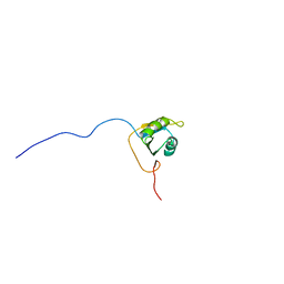
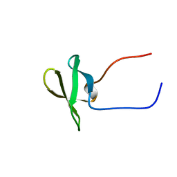
2E5E
 
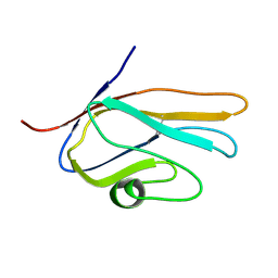 | | Solution Structure of Variable-type Domain of Human Receptor for Advanced Glycation Endproducts | | 分子名称: | Advanced glycosylation end product-specific receptor | | 著者 | Matsumoto, S, Yoshida, T, Yasumatsu, I, Yamamoto, H, Kobayashi, Y, Ohkubo, T. | | 登録日 | 2006-12-20 | | 公開日 | 2007-12-25 | | 最終更新日 | 2022-03-09 | | 実験手法 | SOLUTION NMR | | 主引用文献 | Solution Structure of Variable-type Domain of Human Receptor for Advanced Glycation Endproducts
to be published
|
|
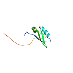
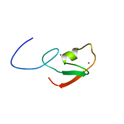
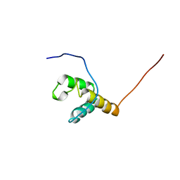
2E5F
 | |
2E5G
 | | Solution structure of RNA binding domain in RNA binding motif protein 21 | | 分子名称: | U6 snRNA-specific terminal uridylyltransferase 1 | | 著者 | Tsuda, K, Muto, Y, Inoue, M, Kigawa, T, Terada, T, Shirouzu, M, Yokoyama, S, RIKEN Structural Genomics/Proteomics Initiative (RSGI) | | 登録日 | 2006-12-21 | | 公開日 | 2007-06-26 | | 最終更新日 | 2024-05-29 | | 実験手法 | SOLUTION NMR | | 主引用文献 | Solution structure of RNA binding domain in RNA binding motif protein 21
To be Published
|
|
2E5H
 | | Solution structure of RNA binding domain in Zinc finger CCHC-type and RNA binding motif 1 | | 分子名称: | Zinc finger CCHC-type and RNA-binding motif-containing protein 1 | | 著者 | Iibuchi, H, Tsuda, K, Muto, Y, Inoue, M, Kigawa, T, Terada, T, Shirouzu, M, Yokoyama, S, RIKEN Structural Genomics/Proteomics Initiative (RSGI) | | 登録日 | 2006-12-21 | | 公開日 | 2007-06-26 | | 最終更新日 | 2024-05-29 | | 実験手法 | SOLUTION NMR | | 主引用文献 | Solution structure of RNA binding domain in Zinc finger CCHC-type and RNA binding motif 1
To be Published
|
|
2E5I
 | | Solution structure of RNA binding domain 2 in Heterogeneous nuclear ribonucleoprotein L-like | | 分子名称: | Heterogeneous nuclear ribonucleoprotein L-like | | 著者 | Tsuda, K, Muto, Y, Inoue, M, Kigawa, T, Terada, T, Shirouzu, M, Yokoyama, S, RIKEN Structural Genomics/Proteomics Initiative (RSGI) | | 登録日 | 2006-12-21 | | 公開日 | 2007-06-26 | | 最終更新日 | 2024-05-29 | | 実験手法 | SOLUTION NMR | | 主引用文献 | Solution structure of RNA binding domain 2 in Heterogeneous nuclear ribonucleoprotein L-like
To be Published
|
|
2E5J
 | | Solution structure of RNA binding domain in Methenyltetrahydrofolate synthetase domain containing | | 分子名称: | Methenyltetrahydrofolate synthetase domain containing | | 著者 | Tsuda, K, Muto, Y, Inoue, M, Kigawa, T, Terada, T, Shirouzu, M, Yokoyama, S, RIKEN Structural Genomics/Proteomics Initiative (RSGI) | | 登録日 | 2006-12-21 | | 公開日 | 2007-06-26 | | 最終更新日 | 2024-05-29 | | 実験手法 | SOLUTION NMR | | 主引用文献 | Solution structure of RNA binding domain in Methenyltetrahydrofolate synthetase domain containing
To be Published
|
|
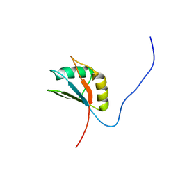
2E5K
 | | Solution structure of SH3 domain in Suppressor of T-cell receptor signaling 1 | | 分子名称: | Suppressor of T-cell receptor signaling 1 | | 著者 | Sukegawa, S, Tsuda, K, Muto, Y, Inoue, M, Kigawa, T, Terada, T, Shirouzu, M, Yokoyama, S, RIKEN Structural Genomics/Proteomics Initiative (RSGI) | | 登録日 | 2006-12-21 | | 公開日 | 2007-06-26 | | 最終更新日 | 2024-05-29 | | 実験手法 | SOLUTION NMR | | 主引用文献 | Solution structure of SH3 domain in Suppressor of T-cell receptor signaling 1
To be Published
|
|
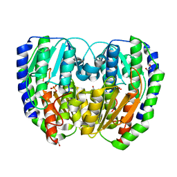
2E5L
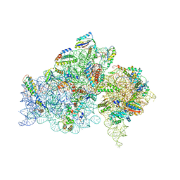 | | A snapshot of the 30S ribosomal subunit capturing mRNA via the Shine- Dalgarno interaction | | 分子名称: | 16S ribosomal RNA, 30S ribosomal protein S10, 30S ribosomal protein S11, ... | | 著者 | Kaminishi, T, Wilson, D.N, Takemoto, C, Harms, J.M, Kawazoe, M, Schluenzen, F, Hanawa-Suetsugu, K, Shirouzu, M, Fucini, P, Yokoyama, S, RIKEN Structural Genomics/Proteomics Initiative (RSGI) | | 登録日 | 2006-12-21 | | 公開日 | 2007-05-15 | | 最終更新日 | 2024-03-13 | | 実験手法 | X-RAY DIFFRACTION (3.3 Å) | | 主引用文献 | A snapshot of the 30S ribosomal subunit capturing mRNA via the Shine-Dalgarno interaction
Structure, 15, 2007
|
|
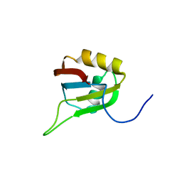
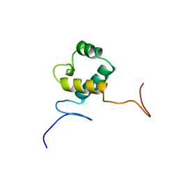
2E5M
 | |
2E5N
 | | Solution structure of the ELL_N2 domain of target of RNA polymerase II elongation factor ELL2 | | 分子名称: | RNA polymerase II elongation factor ELL2 | | 著者 | Dang, W, Muto, Y, Inoue, M, Kigawa, T, Shirouzu, M, Terada, T, Yokoyama, S, RIKEN Structural Genomics/Proteomics Initiative (RSGI) | | 登録日 | 2006-12-22 | | 公開日 | 2007-06-26 | | 最終更新日 | 2024-05-29 | | 実験手法 | SOLUTION NMR | | 主引用文献 | Solution structure of the ELL_N2 domain of target of RNA polymerase II elongation factor ELL2
To be Published
|
|
2E5O
 | | 'Solution structure of the TRIP_4C domain of target of activating signal cointegrator 1 | | 分子名称: | Activating signal cointegrator 1 | | 著者 | Dang, W, Muto, Y, Inoue, M, Kigawa, T, Shirouzu, M, Terada, T, Yokoyama, S, RIKEN Structural Genomics/Proteomics Initiative (RSGI) | | 登録日 | 2006-12-22 | | 公開日 | 2008-01-01 | | 最終更新日 | 2024-05-29 | | 実験手法 | SOLUTION NMR | | 主引用文献 | Solution structure of the TRIP_4C domain of target of activating signal cointegrator 1
To be Published
|
|
2E5P
 | | Solution structure of the TUDOR domain of PHD finger protein 1 (PHF1 protein) | | 分子名称: | PHD finger protein 1 | | 著者 | Dang, W, Muto, Y, Inoue, M, Kigawa, T, Shirouzu, M, Terada, T, Yokoyama, S, RIKEN Structural Genomics/Proteomics Initiative (RSGI) | | 登録日 | 2006-12-22 | | 公開日 | 2007-06-26 | | 最終更新日 | 2024-05-29 | | 実験手法 | SOLUTION NMR | | 主引用文献 | Solution structure of the TUDOR domain of PHD finger protein 1 (PHF1 protein)
To be Published
|
|
2E5Q
 | | Solution structure of the TUDOR domain of PHD finger protein 19, isoform b [Homo sapiens] | | 分子名称: | PHD finger protein 19 | | 著者 | Dang, W, Muto, Y, Inoue, M, Kigawa, T, Shirouzu, M, Terada, T, Yokoyama, S, RIKEN Structural Genomics/Proteomics Initiative (RSGI) | | 登録日 | 2006-12-22 | | 公開日 | 2007-06-26 | | 最終更新日 | 2024-05-29 | | 実験手法 | SOLUTION NMR | | 主引用文献 | Solution structure of the TUDOR domain of PHD finger protein 19,
isoform b [Homo sapiens]
To be Published
|
|
2E5R
 | | Solution structure of the ZZ domain of Dystrobrevin alpha (Dystrobrevin-alpha) | | 分子名称: | Dystrobrevin alpha, ZINC ION | | 著者 | Dang, W, Muto, Y, Inoue, M, Kigawa, T, Shirouzu, M, Terada, T, Yokoyama, S, RIKEN Structural Genomics/Proteomics Initiative (RSGI) | | 登録日 | 2006-12-22 | | 公開日 | 2007-06-26 | | 最終更新日 | 2024-05-29 | | 実験手法 | SOLUTION NMR | | 主引用文献 | Solution structure of the ZZ domain of Dystrobrevin alpha (Dystrobrevin-alpha)
To be Published
|
|
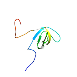
2E5S
 | | Solution structure of the zf-CCCHx2 domain of muscleblind-like 2, isoform 1 [Homo sapiens] | | 分子名称: | OTTHUMP00000018578, ZINC ION | | 著者 | Dang, W, Muto, Y, Inoue, M, Kigawa, T, Shirouzu, M, Terada, T, Yokoyama, S, RIKEN Structural Genomics/Proteomics Initiative (RSGI) | | 登録日 | 2006-12-22 | | 公開日 | 2007-06-26 | | 最終更新日 | 2024-05-29 | | 実験手法 | SOLUTION NMR | | 主引用文献 | Solution structure of the RNA binding domain in the human muscleblind-like protein 2
Protein Sci., 18, 2009
|
|
2E5T
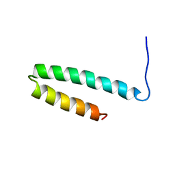
 | |
2E5U
 | |
2E5V
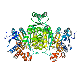
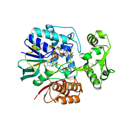 | | Crystal structure of L-Aspartate Oxidase from hyperthermophilic archaeon Sulfolobus tokodaii | | 分子名称: | CHLORIDE ION, FLAVIN-ADENINE DINUCLEOTIDE, L-aspartate oxidase | | 著者 | Yoneda, K, Sakuraba, H, Asai, I, Tsuge, H, Katunuma, N, Ohshima, T. | | 登録日 | 2006-12-25 | | 公開日 | 2008-01-01 | | 最終更新日 | 2024-03-13 | | 実験手法 | X-RAY DIFFRACTION (2.09 Å) | | 主引用文献 | Structure of l-aspartate oxidase from the hyperthermophilic archaeon Sulfolobus tokodaii
Biochim.Biophys.Acta, 1784, 2008
|
|
2E5W
 | |
2E5X
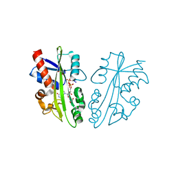 | | Structure of nucleotide triphosphate pyrophosphatase from pyrococcus horikoshii OT3 | | 分子名称: | 1,2-ETHANEDIOL, Hypothetical protein PH1917, INOSINE 5'-TRIPHOSPHATE, ... | | 著者 | Mizutani, H, Lokanath, N.K, Kunishima, N, RIKEN Structural Genomics/Proteomics Initiative (RSGI) | | 登録日 | 2006-12-25 | | 公開日 | 2007-06-26 | | 最終更新日 | 2023-10-25 | | 実験手法 | X-RAY DIFFRACTION (2 Å) | | 主引用文献 | Structure of nucleotide triphosphate pyrophosphatase from pyrococcus horikoshii OT3
To be Published
|
|
2E5Y
 | |
2E5Z
 | | Solution structure of the surp2 domain in splicing factor, arginine/serine-rich 8 | | 分子名称: | Splicing factor, arginine/serine-rich 8 | | 著者 | He, F, Muto, Y, Inoue, M, Kigawa, T, Shirouzu, M, Terada, T, Yokoyama, S, RIKEN Structural Genomics/Proteomics Initiative (RSGI) | | 登録日 | 2006-12-25 | | 公開日 | 2007-06-26 | | 最終更新日 | 2024-05-29 | | 実験手法 | SOLUTION NMR | | 主引用文献 | Solution structure of the surp2 domain in splicing factor, arginine/serine-rich 8
To be Published
|
|
2E60
 | | Solution structure of the surp1 domain in splicing factor, arginine/serine-rich 8 | | 分子名称: | Splicing factor, arginine/serine-rich 8 | | 著者 | He, F, Muto, Y, Inoue, M, Kigawa, T, Shirouzu, M, Terada, T, Yokoyama, S, RIKEN Structural Genomics/Proteomics Initiative (RSGI) | | 登録日 | 2006-12-25 | | 公開日 | 2007-06-26 | | 最終更新日 | 2024-05-29 | | 実験手法 | SOLUTION NMR | | 主引用文献 | Solution structure of the surp1 domain in splicing factor, arginine/serine-rich 8
To be Published
|
|
2E61
 | | Solution structure of the zf-CW domain in zinc finger CW-type PWWP domain protein 1 | | 分子名称: | ZINC ION, Zinc finger CW-type PWWP domain protein 1 | | 著者 | He, F, Muto, Y, Inoue, M, Kigawa, T, Shirouzu, M, Terada, T, Yokoyama, S, RIKEN Structural Genomics/Proteomics Initiative (RSGI) | | 登録日 | 2006-12-25 | | 公開日 | 2007-06-26 | | 最終更新日 | 2024-05-01 | | 実験手法 | SOLUTION NMR | | 主引用文献 | Structural insight into the zinc finger CW domain as a histone modification reader
Structure, 18, 2010
|
|
2E62
 | | Solution structure of the cwf21 domain in protein AAK25922 | | 分子名称: | protein At5g25060 | | 著者 | He, F, Muto, Y, Inoue, M, Kigawa, T, Shirouzu, M, Terada, T, Yokoyama, S, RIKEN Structural Genomics/Proteomics Initiative (RSGI) | | 登録日 | 2006-12-25 | | 公開日 | 2007-06-26 | | 最終更新日 | 2024-05-29 | | 実験手法 | SOLUTION NMR | | 主引用文献 | Solution structure of the cwf21 domain in protein AAK25922
To be Published
|
|


