8J50
 
 | |
8J4Z
 | |
8J4J
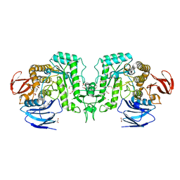
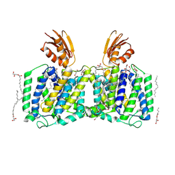
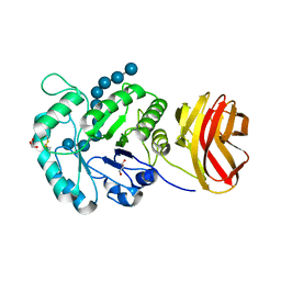
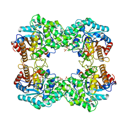
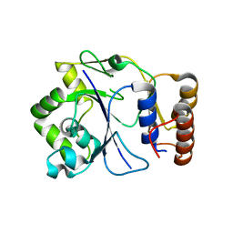
 | | X-ray structure of a ferric ion-binding protein A (FbpA) from Vibrio metschnikovii in complex with ferric ion | | 分子名称: | CARBONATE ION, FE (III) ION, Ferric iron ABC transporter iron-binding protein | | 著者 | Lu, P, Jiang, J, Nagata, K. | | 登録日 | 2023-04-20 | | 公開日 | 2024-01-10 | | 最終更新日 | 2024-02-07 | | 実験手法 | X-RAY DIFFRACTION (2.15 Å) | | 主引用文献 | Molecular mechanism of Fe 3+ binding inhibition to Vibrio metschnikovii ferric ion-binding protein, FbpA, by rosmarinic acid and its hydrolysate, danshensu.
Protein Sci., 33, 2024
|
|
8J4H
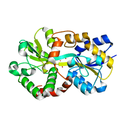
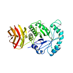
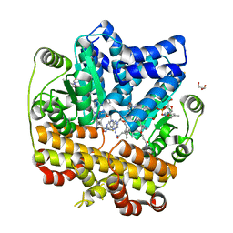
 | | X-ray structure of a ferric ion-binding protein A (FbpA) from Vibrio metschnikovii in complex with Danshensu (DSS) | | 分子名称: | (2~{R})-3-[3,4-bis(oxidanyl)phenyl]-2-oxidanyl-propanoic acid, Ferric iron ABC transporter iron-binding protein | | 著者 | Lu, P, Jiang, J, Nagata, K. | | 登録日 | 2023-04-20 | | 公開日 | 2024-01-10 | | 最終更新日 | 2024-02-07 | | 実験手法 | X-RAY DIFFRACTION (2.01 Å) | | 主引用文献 | Molecular mechanism of Fe 3+ binding inhibition to Vibrio metschnikovii ferric ion-binding protein, FbpA, by rosmarinic acid and its hydrolysate, danshensu.
Protein Sci., 33, 2024
|
|
8J4F
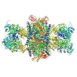
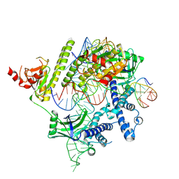
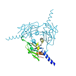
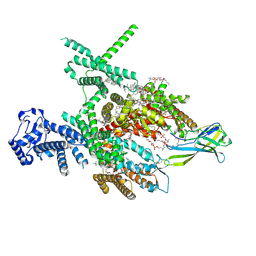 | | Structure of human Nav1.7 in complex with Hardwickii acid | | 分子名称: | (3beta,14beta,17beta,25R)-3-[4-methoxy-3-(methoxymethyl)butoxy]spirost-5-en, (4~{a}~{R},5~{S},6~{R},8~{a}~{R})-5-[2-(furan-3-yl)ethyl]-5,6,8~{a}-trimethyl-3,4,4~{a},6,7,8-hexahydronaphthalene-1-carboxylic acid, 1,2-DIOLEOYL-SN-GLYCERO-3-PHOSPHOCHOLINE, ... | | 著者 | Wu, Q.R, Yan, N. | | 登録日 | 2023-04-19 | | 公開日 | 2023-06-14 | | 実験手法 | ELECTRON MICROSCOPY (3 Å) | | 主引用文献 | Structural mapping of Na v 1.7 antagonists.
Nat Commun, 14, 2023
|
|
8J4C
 | | YeeE(TsuA)-YeeD(TsuB) complex for thiosulfate uptake | | 分子名称: | (2R)-2,3-dihydroxypropyl (9Z)-octadec-9-enoate, Spirochaeta thermophila YeeE(TsuA)-YeeD(TsuB),UPF0033 domain-containing protein, SirA-like domain-containing protein (chimera), ... | | 著者 | Ikei, M, Miyazaki, R, Monden, K, Naito, Y, Takeuchi, A, Takahashi, Y.S, Tanaka, Y, Ichikawa, M, Tsukazaki, T. | | 登録日 | 2023-04-19 | | 公開日 | 2024-03-27 | | 実験手法 | X-RAY DIFFRACTION (3.34 Å) | | 主引用文献 | Structure and function of YeeE-YeeD complex for sophisticated thiosulfate uptake
To Be Published
|
|
8J45
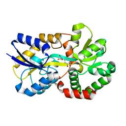
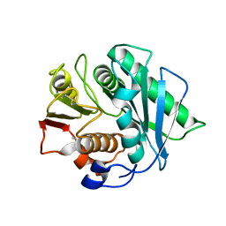 | | Crystal structure of a Pichia pastoris-expressed IsPETase variant | | 分子名称: | 2-acetamido-2-deoxy-beta-D-glucopyranose, Poly(ethylene terephthalate) hydrolase | | 著者 | Li, X, He, H.L, Long, X, Niu, D, Huang, J.-W, Chen, C.-C, Guo, R.-T. | | 登録日 | 2023-04-19 | | 公開日 | 2024-01-17 | | 実験手法 | X-RAY DIFFRACTION (1.49 Å) | | 主引用文献 | Complete decomposition of poly(ethylene terephthalate) by crude PET hydrolytic enzyme produced in Pichia pastoris
Chem Eng J, 2023
|
|
8J40
 | | Crystal Structure of CATB8 in complex with chloramphenicol | | 分子名称: | CHLORAMPHENICOL, CatB8, GLYCEROL, ... | | 著者 | Liao, J, Kuang, L, Jiang, Y. | | 登録日 | 2023-04-18 | | 公開日 | 2024-02-28 | | 最終更新日 | 2024-03-20 | | 実験手法 | X-RAY DIFFRACTION (1.57 Å) | | 主引用文献 | Chloramphenicol Binding Sites of Acinetobacter baumannii Chloramphenicol Acetyltransferase CatB8.
Acs Infect Dis., 10, 2024
|
|
8J3Y
 | |
8J3X
 | |
8J3R
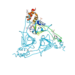
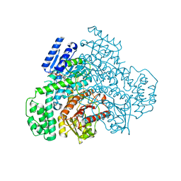
 | | Cryo-EM structure of the AsCas12f-HKRA-sgRNAS3-5v7-target DNA | | 分子名称: | DNA (37-MER), DNA (38-MER), MAGNESIUM ION, ... | | 著者 | Hino, T, Omura, N.S, Nakagawa, R, Togashi, T, Takeda, N.S, Hiramoto, T, Tasaka, S, Hirano, H, Tokuyama, T, Uosaki, H, Ishiguro, H, Yamano, H, Ozaki, Y, Motooka, D, Mori, H, Kirita, Y, Kise, Y, Itoh, Y, Matoba, S, Aburatani, H, Yachie, N, Siksnys, V, Ohmori, T, Hoshino, A, Nureki, O. | | 登録日 | 2023-04-18 | | 公開日 | 2023-09-27 | | 実験手法 | ELECTRON MICROSCOPY (2.95 Å) | | 主引用文献 | Minimal and most efficient genome editing Cas enzyme
To Be Published
|
|
8J3P
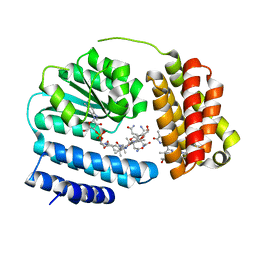
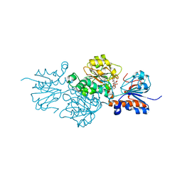 | | Formate dehydrogenase mutant from from Candida dubliniensis M4 complexed with NADP+ | | 分子名称: | Formate dehydrogenase, NADP NICOTINAMIDE-ADENINE-DINUCLEOTIDE PHOSPHATE | | 著者 | Ma, W, Zheng, Y.C, Geng, Q, Chen, C, Xu, J.H. | | 登録日 | 2023-04-17 | | 公開日 | 2023-09-20 | | 最終更新日 | 2023-11-01 | | 実験手法 | X-RAY DIFFRACTION (2.3 Å) | | 主引用文献 | Engineering a Formate Dehydrogenase for NADPH Regeneration.
Chembiochem, 24, 2023
|
|
8J3M
 | |
8J2Y
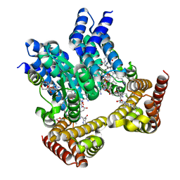
 | | Acidimicrobiaceae bacterium photocobilins protein, dark state | | 分子名称: | 5'-DEOXYADENOSINE, COBALAMIN, DI(HYDROXYETHYL)ETHER, ... | | 著者 | Zhang, S, Poddar, H, Levy, W.C, Leys, D. | | 登録日 | 2023-04-15 | | 公開日 | 2024-04-10 | | 実験手法 | X-RAY DIFFRACTION (2.3 Å) | | 主引用文献 | Photocobilins integrate B12 and bilin photochemistry for enzyme control.
Nat Commun, 15, 2024
|
|
8J2X
 | | Saccharothrix syringae photocobilins protein, light state | | 分子名称: | BILIVERDINE IX ALPHA, COBALAMIN, Cobalamin-binding protein, ... | | 著者 | Zhang, S, Poddar, H, Levy, C, Leys, D. | | 登録日 | 2023-04-15 | | 公開日 | 2024-04-10 | | 実験手法 | X-RAY DIFFRACTION (1.98 Å) | | 主引用文献 | Photocobilins integrate B12 and bilin photochemistry for enzyme control.
Nat Commun, 15, 2024
|
|
8J2W
 | | Saccharothrix syringae photocobilins protein, dark state | | 分子名称: | 1,4-DIETHYLENE DIOXIDE, 5'-DEOXYADENOSINE, BILIVERDINE IX ALPHA, ... | | 著者 | Zhang, S, Poddar, H, Levy, W.C, Leys, D. | | 登録日 | 2023-04-15 | | 公開日 | 2024-04-10 | | 実験手法 | X-RAY DIFFRACTION (1.7 Å) | | 主引用文献 | Photocobilins integrate B12 and bilin photochemistry for enzyme control.
Nat Commun, 15, 2024
|
|
8J2Q
 | | Crystal structure of Cypovirus Polyhedra mutant fused with c-Myc fragment | | 分子名称: | Polyhedrin,Myc proto-oncogene protein | | 著者 | Kojima, M, Ueno, T, Abe, S, Hirata, K. | | 登録日 | 2023-04-15 | | 公開日 | 2024-04-17 | | 最終更新日 | 2024-07-03 | | 実験手法 | X-RAY DIFFRACTION (1.92 Å) | | 主引用文献 | High-throughput structure determination of an intrinsically disordered protein using cell-free protein crystallization.
Proc.Natl.Acad.Sci.USA, 121, 2024
|
|
8J2N
 | |
8J2M
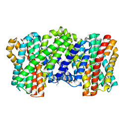 | | The truncated rice Na+/H+ antiporter SOS1 (1-976) in a constitutively active state | | 分子名称: | (2R)-3-{[(S)-(2-aminoethoxy)(hydroxy)phosphoryl]oxy}-2-(tetradecanoyloxy)propyl tetradecanoate, Na+/H+ antiporter | | 著者 | Zhang, X.Y, Tang, L.H, Zhang, C.R, Nie, J.W. | | 登録日 | 2023-04-14 | | 公開日 | 2023-11-22 | | 最終更新日 | 2023-11-29 | | 実験手法 | ELECTRON MICROSCOPY (3.4 Å) | | 主引用文献 | Structure and activation mechanism of the rice Salt Overly Sensitive 1 (SOS1) Na + /H + antiporter.
Nat.Plants, 9, 2023
|
|
8J2L
 | | Crystal structure of a bright green fluorescent protein (StayGold) with double mutations (N137A, Y187F) in jellyfish Cytaeis uchidae from Biortus | | 分子名称: | 4-(2-HYDROXYETHYL)-1-PIPERAZINE ETHANESULFONIC ACID, GLYCEROL, SODIUM ION, ... | | 著者 | Wu, J, Wang, F, Gui, W, Cheng, W, Yang, Y. | | 登録日 | 2023-04-14 | | 公開日 | 2023-12-13 | | 実験手法 | X-RAY DIFFRACTION (1.7 Å) | | 主引用文献 | Crystal structure of a bright green fluorescent protein (StayGold) in jellyfish Cytaeis uchidae from Biortus
To Be Published
|
|
8J2H
 | | Crystal structure of a bright green fluorescent protein (StayGold) with single mutation (N137A) in jellyfish Cytaeis uchidae from Biortus | | 分子名称: | GLYCEROL, SODIUM ION, StayGold(N137A) | | 著者 | Wu, J, Wang, F, Gui, W, Cheng, W, Yang, Y. | | 登録日 | 2023-04-14 | | 公開日 | 2023-12-13 | | 実験手法 | X-RAY DIFFRACTION (1.7 Å) | | 主引用文献 | Crystal structure of a bright green fluorescent protein (StayGold) in jellyfish Cytaeis uchidae from Biortus
To Be Published
|
|
8J2E
 | |
8J2D
 | |
8J2C
 | |
8J2B
 | |


