8CSQ
 
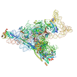
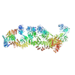
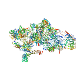 | | Human mitochondrial small subunit assembly intermediate (State B) | | 分子名称: | 12S mitochondrial rRNA, 28S ribosomal protein S10, mitochondrial, ... | | 著者 | Harper, N.J, Burnside, C, Klinge, S. | | 登録日 | 2022-05-13 | | 公開日 | 2022-12-14 | | 最終更新日 | 2024-06-12 | | 実験手法 | ELECTRON MICROSCOPY (2.54 Å) | | 主引用文献 | Principles of mitoribosomal small subunit assembly in eukaryotes.
Nature, 614, 2023
|
|
8CSU
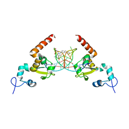
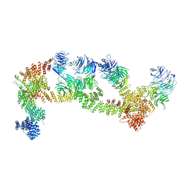
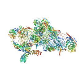 | | Human mitochondrial small subunit assembly intermediate (State C*) | | 分子名称: | 12S mitochondrial rRNA, 28S ribosomal protein S10, mitochondrial, ... | | 著者 | Harper, N.J, Burnside, C, Klinge, S. | | 登録日 | 2022-05-13 | | 公開日 | 2022-12-14 | | 最終更新日 | 2023-02-08 | | 実験手法 | ELECTRON MICROSCOPY (3.03 Å) | | 主引用文献 | Principles of mitoribosomal small subunit assembly in eukaryotes.
Nature, 614, 2023
|
|
8CST
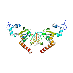
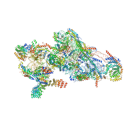 | | Human mitochondrial small subunit assembly intermediate (State E) | | 分子名称: | 12S mitochondrial rRNA, 28S ribosomal protein S10, mitochondrial, ... | | 著者 | Harper, N.J, Burnside, C, Klinge, S. | | 登録日 | 2022-05-13 | | 公開日 | 2022-12-14 | | 最終更新日 | 2023-02-08 | | 実験手法 | ELECTRON MICROSCOPY (2.85 Å) | | 主引用文献 | Principles of mitoribosomal small subunit assembly in eukaryotes.
Nature, 614, 2023
|
|
8D8K
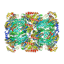
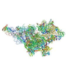 | | Yeast mitochondrial small subunit assembly intermediate (State 2) | | 分子名称: | 15S ribosomal RNA, 37S ribosomal protein MRP1, mitochondrial, ... | | 著者 | Burnside, C, Harper, N.J, Klinge, S. | | 登録日 | 2022-06-08 | | 公開日 | 2022-12-21 | | 最終更新日 | 2023-02-15 | | 実験手法 | ELECTRON MICROSCOPY (3.13 Å) | | 主引用文献 | Principles of mitoribosomal small subunit assembly in eukaryotes.
Nature, 614, 2023
|
|
8D8J
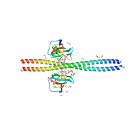
 | | Yeast mitochondrial small subunit assembly intermediate (State 1) | | 分子名称: | 15S ribosomal RNA, 37S ribosomal protein MRP13, mitochondrial, ... | | 著者 | Burnside, C, Harper, N, Klinge, S. | | 登録日 | 2022-06-08 | | 公開日 | 2022-12-21 | | 最終更新日 | 2024-06-12 | | 実験手法 | ELECTRON MICROSCOPY (3.8 Å) | | 主引用文献 | Principles of mitoribosomal small subunit assembly in eukaryotes.
Nature, 614, 2023
|
|
6WVX
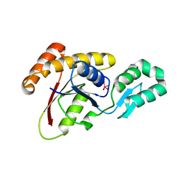
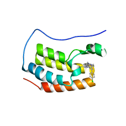 | | Crystal structure of the first bromodomain of human BRD4 with benzodiazepine inhibitor | | 分子名称: | 6'-(4-chlorophenyl)-1'-methyl-8'-(1-methyl-1H-pyrazol-4-yl)spiro[cyclopropane-1,4'-[1,2,4]triazolo[4,3-a][1,4]benzodiazepine], Bromodomain-containing protein 4 | | 著者 | Eron, S.J. | | 登録日 | 2020-05-07 | | 公開日 | 2021-09-01 | | 最終更新日 | 2023-10-18 | | 実験手法 | X-RAY DIFFRACTION (1.55 Å) | | 主引用文献 | Structural Characterization of Degrader-Induced Ternary Complexes Using Hydrogen-Deuterium Exchange Mass Spectrometry and Computational Modeling: Implications for Structure-Based Design.
Acs Chem.Biol., 16, 2021
|
|
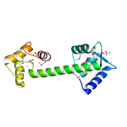
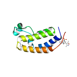
1H7V
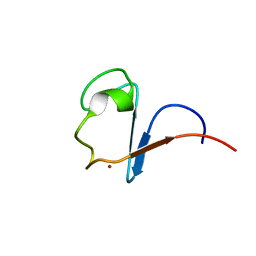 | | Rubredoxin from Guillardia Theta | | 分子名称: | RUBREDOXIN, ZINC ION | | 著者 | Schweimer, K, Hoffmann, S, Wastl, J, Maier, U.G, Roesch, P, Sticht, H. | | 登録日 | 2001-01-16 | | 公開日 | 2002-01-29 | | 最終更新日 | 2024-05-15 | | 実験手法 | SOLUTION NMR | | 主引用文献 | Solution structure of a zinc substituted eukaryotic rubredoxin from the cryptomonad alga Guillardia theta.
Protein Sci., 9, 2000
|
|
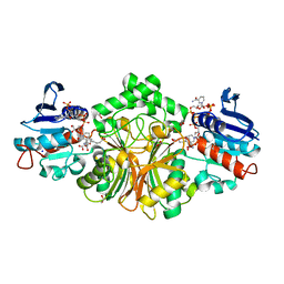
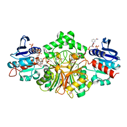
2HDJ
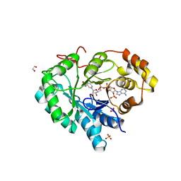 | | Crystal structure of human type 3 3alpha-hydroxysteroid dehydrogenase in complex with NADP(H) | | 分子名称: | 1,2-ETHANEDIOL, Aldo-keto reductase family 1 member C2, NADPH DIHYDRO-NICOTINAMIDE-ADENINE-DINUCLEOTIDE PHOSPHATE, ... | | 著者 | Faucher, F, Pereira de Jesus-Tran, K, Cantin, L, Luu-the, V, Labrie, F, Breton, R. | | 登録日 | 2006-06-20 | | 公開日 | 2006-12-05 | | 最終更新日 | 2023-08-30 | | 実験手法 | X-RAY DIFFRACTION (2 Å) | | 主引用文献 | Crystal Structures of Mouse 17alpha-Hydroxysteroid Dehydrogenase (Apoenzyme and Enzyme-NADP(H) Binary Complex): Identification of Molecular Determinants Responsible for the Unique 17alpha-reductive Activity of this Enzyme.
J.Mol.Biol., 364, 2006
|
|
7PXA
 | |
5LCL
 | | STRUCTURE OF the RAD14 DNA-binding domain IN COMPLEX WITH C8-aminofluorene- GUANINE CONTAINING DNA | | 分子名称: | DNA (5'-D(*GP*TP*GP*AP*TP*GP*AP*CP*GP*TP*AP*GP*AP*G)-3'), DNA repair protein RAD14, GCTCTAC(8AF)TCATCA, ... | | 著者 | Schneider, S, Carell, T, Ebert, C, Simon, N. | | 登録日 | 2016-06-22 | | 公開日 | 2017-05-10 | | 最終更新日 | 2024-01-10 | | 実験手法 | X-RAY DIFFRACTION (2.2 Å) | | 主引用文献 | Structural Insights into the Recognition of N(2) -Aryl- and C8-Aryl DNA Lesions by the Repair Protein XPA/Rad14.
Chembiochem, 18, 2017
|
|
5LCM
 | | STRUCTURE OF the RAD14 DNA-binding domain IN COMPLEX WITH N2-acetylaminonaphtyl- GUANINE CONTAINING DNA | | 分子名称: | DNA (5'-D(*GP*CP*TP*CP*TP*AP*CP*(AAN)P*TP*CP*AP*TP*CP*A)-3'), DNA (5'-D(*GP*TP*GP*AP*TP*GP*AP*CP*GP*TP*AP*GP*AP*G)-3'), DNA repair protein RAD14, ... | | 著者 | Schneider, S, Ebert, C, Simon, N, Carell, T. | | 登録日 | 2016-06-22 | | 公開日 | 2017-05-10 | | 最終更新日 | 2024-05-08 | | 実験手法 | X-RAY DIFFRACTION (1.9 Å) | | 主引用文献 | Structural Insights into the Recognition of N(2) -Aryl- and C8-Aryl DNA Lesions by the Repair Protein XPA/Rad14.
Chembiochem, 18, 2017
|
|
6OQA
 | | Crystal structure of CEP250 bound to FKBP12 in the presence of FK506-like novel natural product | | 分子名称: | (3R,4E,7E,10R,11S,12R,13S,16R,17R,24aS)-11,17-dihydroxy-10,12,16-trimethyl-3-[(2R)-1-phenylbutan-2-yl]-6,9,10,11,12,13,14,15,16,17,22,23,24,24a-tetradecahydro-3H-13,17-epoxypyrido[2,1-c][1,4]oxazacyclohenicosine-1,18,19(21H)-trione, 1,2-ETHANEDIOL, 3,6,9,12,15,18,21-HEPTAOXATRICOSANE-1,23-DIOL, ... | | 著者 | Lee, S.-J, Shigdel, U.K, Townson, S.A, Verdine, G.L. | | 登録日 | 2019-04-26 | | 公開日 | 2020-04-29 | | 最終更新日 | 2024-03-13 | | 実験手法 | X-RAY DIFFRACTION (2.2 Å) | | 主引用文献 | Genomic discovery of an evolutionarily programmed modality for small-molecule targeting of an intractable protein surface.
Proc.Natl.Acad.Sci.USA, 117, 2020
|
|
8F5P
 | | Structure of Leishmania tarentolae IFT-A (state 2) | | 分子名称: | Intraflagellar transport protein 122 homolog, Intraflagellar transport protein 122B, putative, ... | | 著者 | Zhou, H, Brown, A. | | 登録日 | 2022-11-14 | | 公開日 | 2022-12-21 | | 最終更新日 | 2024-05-22 | | 実験手法 | ELECTRON MICROSCOPY (3.4 Å) | | 主引用文献 | Mechanism of IFT-A polymerization into trains for ciliary transport.
Cell, 185, 2022
|
|
8F5O
 | | Structure of Leishmania tarentolae IFT-A (state 1) | | 分子名称: | Intraflagellar transport protein 122 homolog, Intraflagellar transport protein 122B, putative, ... | | 著者 | Zhou, H, Brown, A. | | 登録日 | 2022-11-14 | | 公開日 | 2022-12-21 | | 最終更新日 | 2024-05-22 | | 実験手法 | ELECTRON MICROSCOPY (3.5 Å) | | 主引用文献 | Mechanism of IFT-A polymerization into trains for ciliary transport.
Cell, 185, 2022
|
|
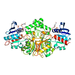
3RGD
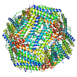 | | Iron loaded frog M ferritin. Short soaking time | | 分子名称: | FE (III) ION, Ferritin, middle subunit | | 著者 | Bertini, I, Lalli, D, Mangani, S, Pozzi, C, Rosa, C, Theil, E.C, Turano, P. | | 登録日 | 2011-04-08 | | 公開日 | 2012-04-11 | | 最終更新日 | 2023-09-13 | | 実験手法 | X-RAY DIFFRACTION (2.89 Å) | | 主引用文献 | Structural insights into the ferroxidase site of ferritins from higher eukaryotes.
J.Am.Chem.Soc., 134, 2012
|
|
2IY5
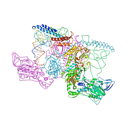 | | PHENYLALANYL-TRNA SYNTHETASE FROM THERMUS THERMOPHILUS complexed with tRNA and a phenylalanyl-adenylate analog | | 分子名称: | ADENOSINE-5'-[PHENYLALANINOL-PHOSPHATE], MAGNESIUM ION, PHENYLALANYL-TRNA SYNTHETASE ALPHA CHAIN, ... | | 著者 | Moor, N, Kotik-Kogan, O, Tworowski, D, Sukhanova, M, Safro, M. | | 登録日 | 2006-07-12 | | 公開日 | 2006-09-06 | | 最終更新日 | 2023-12-13 | | 実験手法 | X-RAY DIFFRACTION (3.1 Å) | | 主引用文献 | The crystal structure of the ternary complex of phenylalanyl-tRNA synthetase with tRNAPhe and a phenylalanyl-adenylate analogue reveals a conformational switch of the CCA end.
Biochemistry, 45, 2006
|
|
3OMX
 | | Crystal structure of Ssu72 with vanadate complex | | 分子名称: | CG14216, VANADATE ION | | 著者 | Zhang, Y, Zhang, M, Zhang, Y. | | 登録日 | 2010-08-27 | | 公開日 | 2011-01-19 | | 最終更新日 | 2023-09-06 | | 実験手法 | X-RAY DIFFRACTION (2.3366 Å) | | 主引用文献 | Crystal structure of Ssu72, an essential eukaryotic phosphatase specific for the C-terminal domain of RNA polymerase II, in complex with a transition state analogue.
Biochem.J., 434, 2011
|
|
1Y6W
 | | Trapped intermediate of calmodulin | | 分子名称: | (4S)-2-METHYL-2,4-PENTANEDIOL, CALCIUM ION, Calmodulin, ... | | 著者 | Grabarek, Z. | | 登録日 | 2004-12-07 | | 公開日 | 2005-03-01 | | 最終更新日 | 2021-10-20 | | 実験手法 | X-RAY DIFFRACTION (2.4 Å) | | 主引用文献 | Structure of a Trapped Intermediate of Calmodulin: Calcium Regulation of EF-hand Proteins from a New Perspective.
J.Mol.Biol., 346, 2005
|
|
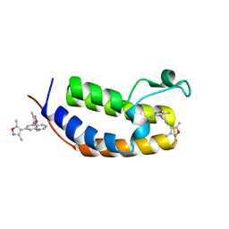
2JU4
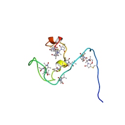 | | NMR structure of the gamma subunit of cGMP phosphodiesterase | | 分子名称: | (3'R)-1'-oxyl-2',2',5',5'-tetramethyl-1,3'-bipyrrolidine-2,5-dione, Retinal rod rhodopsin-sensitive cGMP 3',5'-cyclic phosphodiesterase subunit gamma | | 著者 | Song, J, Guo, L.W, Ruoho, A.E, Markley, J.L, Center for Eukaryotic Structural Genomics (CESG) | | 登録日 | 2007-08-14 | | 公開日 | 2007-10-16 | | 最終更新日 | 2020-02-19 | | 実験手法 | SOLUTION NMR | | 主引用文献 | Intrinsically disordered gamma-subunit of cGMP phosphodiesterase encodes functionally relevant transient secondary and tertiary structure.
Proc.Natl.Acad.Sci.USA, 105, 2008
|
|
2PI6
 | | Crystal structure of the sheep signalling glycoprotein (SPS-40) complex with 2-methyl-2-4-pentanediol at 1.65A resolution reveals specific binding characteristics of SPS-40 | | 分子名称: | (4S)-2-METHYL-2,4-PENTANEDIOL, Chitinase-3-like protein 1, ETHANOL, ... | | 著者 | Sharma, P, Singh, N, Sharma, S, Kaur, P, Betzel, C, Singh, T.P. | | 登録日 | 2007-04-13 | | 公開日 | 2007-05-01 | | 最終更新日 | 2023-10-25 | | 実験手法 | X-RAY DIFFRACTION (1.65 Å) | | 主引用文献 | Tryptophan as a three-way switch in regulating the function of the secretory signalling glycoprotein (SPS-40) from mammary glands: structure of SPS-40 complexed with 2-methylpentane-2,4-diol at 1.6 A resolution.
Acta Crystallogr.,Sect.D, 65, 2009
|
|
4R4J
 | | Crystal structure of complex sp_ASADH with 3-carboxypropyl-phthalic acid and Nicotinamide Adenine dinucleotide phosphate | | 分子名称: | 1,2-ETHANEDIOL, 3-(3-carboxypropyl)benzene-1,2-dicarboxylic acid, Aspartate-semialdehyde dehydrogenase, ... | | 著者 | Pavlovsky, A.G, Thangavelu, B, Bhansali, P, Viola, R.E. | | 登録日 | 2014-08-19 | | 公開日 | 2014-12-10 | | 最終更新日 | 2023-09-20 | | 実験手法 | X-RAY DIFFRACTION (1.51 Å) | | 主引用文献 | A cautionary tale of structure-guided inhibitor development against an essential enzyme in the aspartate-biosynthetic pathway.
Acta Crystallogr.,Sect.D, 70, 2014
|
|
4R51
 | | Crystal complex structure of sp-Aspartate-Semialdehyde-Dehydrogenase with Nicotinamide Adenine dinucleotide phosphate and phthalic acid | | 分子名称: | 1,2-ETHANEDIOL, ACETATE ION, Aspartate-semialdehyde dehydrogenase, ... | | 著者 | Pavlovsky, A.G, Viola, R.E. | | 登録日 | 2014-08-20 | | 公開日 | 2014-12-10 | | 最終更新日 | 2023-09-20 | | 実験手法 | X-RAY DIFFRACTION (1.81 Å) | | 主引用文献 | A cautionary tale of structure-guided inhibitor development against an essential enzyme in the aspartate-biosynthetic pathway.
Acta Crystallogr.,Sect.D, 70, 2014
|
|
4R41
 | | Complex Crystal structure of 4-nitro-2-phosphono-benzoic acid with sp-Aspartate-Semialdehyde Dehydrogenase and Nicotinamide-dinucleotide | | 分子名称: | 1,2-ETHANEDIOL, 4-nitro-2-phosphonobenzoic acid, ACETATE ION, ... | | 著者 | Pavlovsky, A.G, Viola, R.E. | | 登録日 | 2014-08-18 | | 公開日 | 2014-12-10 | | 最終更新日 | 2023-09-20 | | 実験手法 | X-RAY DIFFRACTION (1.61 Å) | | 主引用文献 | A cautionary tale of structure-guided inhibitor development against an essential enzyme in the aspartate-biosynthetic pathway.
Acta Crystallogr.,Sect.D, 70, 2014
|
|
6MO9
 | | N-terminal bromodomain of human BRD2 in complex with N-cyclopentyl-7-(3,5-dimethylisoxazol-4-yl)quinoline-5-sulfonamide inhibitor | | 分子名称: | Bromodomain-containing protein 2, N-cyclopentyl-7-(3,5-dimethyl-1,2-oxazol-4-yl)quinoline-5-sulfonamide | | 著者 | Lansdon, E.B, Newby, Z.E.R. | | 登録日 | 2018-10-04 | | 公開日 | 2019-01-23 | | 最終更新日 | 2024-03-13 | | 実験手法 | X-RAY DIFFRACTION (1.801 Å) | | 主引用文献 | Structure-guided discovery of a novel, potent, and orally bioavailable 3,5-dimethylisoxazole aryl-benzimidazole BET bromodomain inhibitor.
Bioorg. Med. Chem., 27, 2019
|
|
6MO8
 | | N-terminal bromodomain of human BRD2 in complex with 4,4'-(quinoline-5,7-diyl)bis(3,5-dimethylisoxazole) inhibitor | | 分子名称: | 5,7-bis(3,5-dimethyl-1,2-oxazol-4-yl)quinoline, Bromodomain-containing protein 2, SULFATE ION | | 著者 | Lansdon, E.B, Newby, Z.E.R. | | 登録日 | 2018-10-04 | | 公開日 | 2019-01-23 | | 最終更新日 | 2024-03-13 | | 実験手法 | X-RAY DIFFRACTION (1.8 Å) | | 主引用文献 | Structure-guided discovery of a novel, potent, and orally bioavailable 3,5-dimethylisoxazole aryl-benzimidazole BET bromodomain inhibitor.
Bioorg. Med. Chem., 27, 2019
|
|


