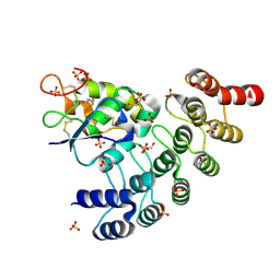
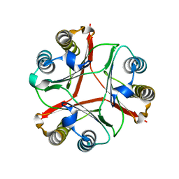
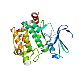
7DPW
 
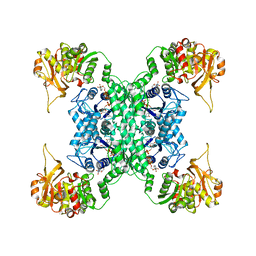 | | Structural basis for ligand binding modes of CTP synthase | | 分子名称: | CTP synthase, CYTIDINE-5'-TRIPHOSPHATE, MAGNESIUM ION | | 著者 | Liu, J.L, Zhou, X, Guo, C.J, Chang, C.C. | | 登録日 | 2020-12-21 | | 公開日 | 2021-09-15 | | 最終更新日 | 2024-06-05 | | 実験手法 | ELECTRON MICROSCOPY (2.65 Å) | | 主引用文献 | Structural basis for ligand binding modes of CTP synthase.
Proc.Natl.Acad.Sci.USA, 118, 2021
|
|
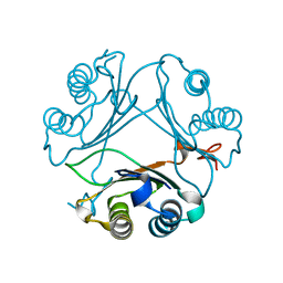
5AB7
 | |
6VC1
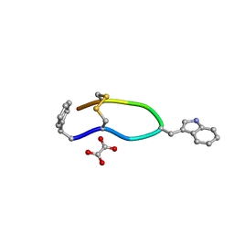 | | Octreotide oxalate | | 分子名称: | OXALATE ION, Octreotide | | 著者 | Spiliopoulou, M, Karavassili, F, Triandafillidis, D, Valmas, A, Kosinas, C, Fili, S, Barlos, K, Barlos, K.K, Morin, M, Reinle-Schmitt, M, Gozzo, F, Margiolaki, I. | | 登録日 | 2019-12-20 | | 公開日 | 2020-12-23 | | 最終更新日 | 2021-05-19 | | 実験手法 | POWDER DIFFRACTION | | 主引用文献 | New perspectives in macromolecular powder diffraction using single-photon-counting strip detectors: high-resolution structure of the pharmaceutical peptide octreotide.
Acta Crystallogr.,Sect.A, 77, 2021
|
|
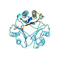
6VL5
 | |
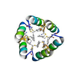
6NE2
 | | Designed repeat protein in complex with Fz7 | | 分子名称: | 1,2-ETHANEDIOL, 2-acetamido-2-deoxy-beta-D-glucopyranose, ACETATE ION, ... | | 著者 | Miao, Y, Jude, K.M, Garcia, K.C. | | 登録日 | 2018-12-15 | | 公開日 | 2019-05-15 | | 最終更新日 | 2023-10-11 | | 実験手法 | X-RAY DIFFRACTION (1.299 Å) | | 主引用文献 | Receptor subtype discrimination using extensive shape complementary designed interfaces.
Nat.Struct.Mol.Biol., 26, 2019
|
|
6NE4
 | | Designed repeat protein specifically in complex with Fz7CRD | | 分子名称: | 1,2-ETHANEDIOL, Designed repeat binding protein, Frizzled-7, ... | | 著者 | Miao, Y, Jude, K.M, Garcia, K.C. | | 登録日 | 2018-12-16 | | 公開日 | 2019-05-15 | | 最終更新日 | 2023-10-11 | | 実験手法 | X-RAY DIFFRACTION (1.648 Å) | | 主引用文献 | Receptor subtype discrimination using extensive shape complementary designed interfaces.
Nat.Struct.Mol.Biol., 26, 2019
|
|
6NDZ
 | | Designed repeat protein in complex with Fz8 | | 分子名称: | (4S)-2-METHYL-2,4-PENTANEDIOL, ACETATE ION, ACETIC ACID, ... | | 著者 | Miao, Y, Jude, K.M, Garcia, K.C. | | 登録日 | 2018-12-14 | | 公開日 | 2019-05-15 | | 最終更新日 | 2023-10-11 | | 実験手法 | X-RAY DIFFRACTION (2.264 Å) | | 主引用文献 | Receptor subtype discrimination using extensive shape complementary designed interfaces.
Nat.Struct.Mol.Biol., 26, 2019
|
|
6D02
 | | Cross-alpha Amyloid-like Structure alphaAmL, 2nd form | | 分子名称: | (4S)-2-METHYL-2,4-PENTANEDIOL, CARBONATE ION, PHOSPHATE ION, ... | | 著者 | Zhang, S.-Q, Liu, L, Degrado, W.F. | | 登録日 | 2018-04-10 | | 公開日 | 2019-02-20 | | 最終更新日 | 2023-10-04 | | 実験手法 | X-RAY DIFFRACTION (2.5 Å) | | 主引用文献 | Designed peptides that assemble into cross-alpha amyloid-like structures.
Nat. Chem. Biol., 14, 2018
|
|
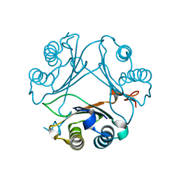
6ZLR
 | |
6WMK
 | | Crystal structure of beta sheet heterodimer LHD29 | | 分子名称: | Beta sheet heterodimer LHD29 - Chain A, Beta sheet heterodimer LHD29 - Chain B | | 著者 | Bera, A.K, Sahtoe, D.D, Kang, A, Sankaran, B, Baker, D. | | 登録日 | 2020-04-21 | | 公開日 | 2021-11-10 | | 最終更新日 | 2024-04-03 | | 実験手法 | X-RAY DIFFRACTION (2.2 Å) | | 主引用文献 | Reconfigurable asymmetric protein assemblies through implicit negative design.
Science, 375, 2022
|
|
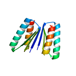
6B17
 | | Design of a short thermally stable alpha-helix embedded in a macrocycle | | 分子名称: | 3,3'-dimethyl-1,1'-biphenyl, Capped-strapped peptide | | 著者 | Wu, H, Acharyya, A, Wu, Y, Liu, L, Jo, H, Gai, F, DeGrado, W.F. | | 登録日 | 2017-09-17 | | 公開日 | 2018-02-21 | | 最終更新日 | 2024-04-03 | | 実験手法 | X-RAY DIFFRACTION (1.25 Å) | | 主引用文献 | Design of a Short Thermally Stable alpha-Helix Embedded in a Macrocycle.
Chembiochem, 19, 2018
|
|
1MM2
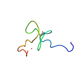 | | Solution structure of the 2nd PHD domain from Mi2b | | 分子名称: | Mi2-beta, ZINC ION | | 著者 | Kwan, A.H.Y, Gell, D.A, Verger, A, Crossley, M, Matthews, J.M, Mackay, J.P. | | 登録日 | 2002-09-02 | | 公開日 | 2003-07-22 | | 最終更新日 | 2024-05-29 | | 実験手法 | SOLUTION NMR | | 主引用文献 | Engineering a Protein Scaffold from a PHD Finger
structure, 11, 2003
|
|
8VG5
 | | Crystal Structure of V113N Variant of D-Dopachrome Tautomerase (D-DT) Bound with 4CPPC | | 分子名称: | 4-(3-carboxyphenyl)pyridine-2,5-dicarboxylic acid, CITRIC ACID, D-dopachrome decarboxylase | | 著者 | Parkins, A, Pilien, A, Thompson, M.C, Pantouris, G. | | 登録日 | 2023-12-22 | | 公開日 | 2024-05-01 | | 最終更新日 | 2024-05-22 | | 実験手法 | X-RAY DIFFRACTION (1.5 Å) | | 主引用文献 | The C-terminal Region of D-DT Regulates Molecular Recognition for Protein-Ligand Complexes.
J.Med.Chem., 67, 2024
|
|
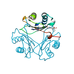
2R5W
 | | Crystal structure of a bifunctional NMN adenylyltransferase/ADP ribose pyrophosphatase from Francisella tularensis | | 分子名称: | CHLORIDE ION, MAGNESIUM ION, Nicotinamide-nucleotide adenylyltransferase | | 著者 | Huang, N, Sorci, L, Zhang, X, Brautigan, C, Li, X, Raffaelli, N, Grishin, N, Osterman, A, Zhang, H. | | 登録日 | 2007-09-04 | | 公開日 | 2008-03-04 | | 最終更新日 | 2011-07-13 | | 実験手法 | X-RAY DIFFRACTION (2.3 Å) | | 主引用文献 | Bifunctional NMN Adenylyltransferase/ADP-Ribose Pyrophosphatase: Structure and Function in Bacterial NAD Metabolism.
Structure, 16, 2008
|
|
8VFK
 | | Crystal Structure of Delta 109-117 D-Dopachrome Tautomerase (D-DT) | | 分子名称: | CITRATE ANION, D-dopachrome decarboxylase, SODIUM ION | | 著者 | Parkins, A, Pilien, A, Wolff, A, Thompson, M.C, Pantouris, G. | | 登録日 | 2023-12-21 | | 公開日 | 2024-05-01 | | 最終更新日 | 2024-05-22 | | 実験手法 | X-RAY DIFFRACTION (1.59 Å) | | 主引用文献 | The C-terminal Region of D-DT Regulates Molecular Recognition for Protein-Ligand Complexes.
J.Med.Chem., 67, 2024
|
|
8VDY
 | | Crystal Structure of Delta 114-117 D-Dopachrome Tautomerase (D-DT) | | 分子名称: | D-dopachrome decarboxylase | | 著者 | Parkins, A, Pilien, A, Wolff, A, Thompson, M.C, Pantouris, G. | | 登録日 | 2023-12-18 | | 公開日 | 2024-05-01 | | 最終更新日 | 2024-05-22 | | 実験手法 | X-RAY DIFFRACTION (2.44 Å) | | 主引用文献 | The C-terminal Region of D-DT Regulates Molecular Recognition for Protein-Ligand Complexes.
J.Med.Chem., 67, 2024
|
|
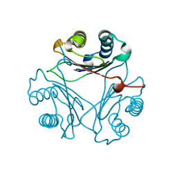
2RBL
 | |
8VG8
 | | Crystal Structure of T115A Variant of D-Dopachrome Tautomerase (D-DT) Bound to 4CPPC | | 分子名称: | 4-(3-carboxyphenyl)pyridine-2,5-dicarboxylic acid, D-dopachrome decarboxylase | | 著者 | Parkins, A, Pilien, A, Wolff, A, Thompson, M.C, Pantouris, G. | | 登録日 | 2023-12-23 | | 公開日 | 2024-05-01 | | 最終更新日 | 2024-05-22 | | 実験手法 | X-RAY DIFFRACTION (1.33 Å) | | 主引用文献 | The C-terminal Region of D-DT Regulates Molecular Recognition for Protein-Ligand Complexes.
J.Med.Chem., 67, 2024
|
|
8VFO
 | | Crystal Structure of L117G Variant of D-Dopachrome Tautomerase (D-DT) | | 分子名称: | CITRIC ACID, D-dopachrome decarboxylase | | 著者 | Parkins, A, Pilien, A, Thompson, M.C, Pantouris, G. | | 登録日 | 2023-12-21 | | 公開日 | 2024-05-01 | | 最終更新日 | 2024-05-22 | | 実験手法 | X-RAY DIFFRACTION (1.35 Å) | | 主引用文献 | The C-terminal Region of D-DT Regulates Molecular Recognition for Protein-Ligand Complexes.
J.Med.Chem., 67, 2024
|
|
8VFL
 | | Crystal Structure of WT D-Dopachrome Tautomerase (D-DT) at 290K | | 分子名称: | D-dopachrome decarboxylase | | 著者 | Parkins, A, Pilien, A, Wolff, A, Thompson, M.C, Pantouris, G. | | 登録日 | 2023-12-21 | | 公開日 | 2024-05-01 | | 最終更新日 | 2024-05-22 | | 実験手法 | X-RAY DIFFRACTION (1.23 Å) | | 主引用文献 | The C-terminal Region of D-DT Regulates Molecular Recognition for Protein-Ligand Complexes.
J.Med.Chem., 67, 2024
|
|
8VG7
 | | Crystal Structure of T115A Variant of D-Dopachrome Tautomerase (D-DT) | | 分子名称: | D-dopachrome decarboxylase | | 著者 | Parkins, A, Pilien, A, Wolff, A, Thompson, M.C, Pantouris, G. | | 登録日 | 2023-12-23 | | 公開日 | 2024-05-01 | | 最終更新日 | 2024-05-22 | | 実験手法 | X-RAY DIFFRACTION (1.24 Å) | | 主引用文献 | The C-terminal Region of D-DT Regulates Molecular Recognition for Protein-Ligand Complexes.
J.Med.Chem., 67, 2024
|
|
8VFN
 | | Crystal Structure of WT D-Dopachrome Tautomerase (D-DT) at 310K | | 分子名称: | D-dopachrome decarboxylase | | 著者 | Parkins, A, Pilien, A, Wolff, A, Thompson, M.C, Pantouris, G. | | 登録日 | 2023-12-21 | | 公開日 | 2024-05-01 | | 最終更新日 | 2024-05-22 | | 実験手法 | X-RAY DIFFRACTION (1.29 Å) | | 主引用文献 | The C-terminal Region of D-DT Regulates Molecular Recognition for Protein-Ligand Complexes.
J.Med.Chem., 67, 2024
|
|
8VFW
 | | Crystal Structure of V113N D-Dopachrome Tautomerase (D-DT) | | 分子名称: | CITRIC ACID, D-dopachrome decarboxylase | | 著者 | Parkins, A, Wolff, A, Thompson, M.C, Pantouris, G. | | 登録日 | 2023-12-22 | | 公開日 | 2024-05-01 | | 最終更新日 | 2024-05-22 | | 実験手法 | X-RAY DIFFRACTION (1.31 Å) | | 主引用文献 | The C-terminal Region of D-DT Regulates Molecular Recognition for Protein-Ligand Complexes.
J.Med.Chem., 67, 2024
|
|
3VBX
 | |
4UP1
 | |


