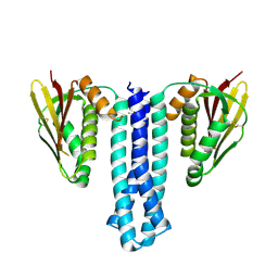
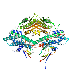
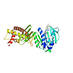
7EAS
 
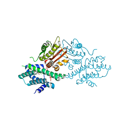 | | Crystal structure of human pyruvate dehydrogenase kinase 2 in complex with compound 2 | | 分子名称: | 1H-pyrrolo[2,3-b]pyridine-3-carbonitrile, ACETATE ION, CHLORIDE ION, ... | | 著者 | Orita, T, Doi, S, Iwanaga, T, Adachi, T. | | 登録日 | 2021-03-08 | | 公開日 | 2021-08-04 | | 最終更新日 | 2023-11-29 | | 実験手法 | X-RAY DIFFRACTION (1.97 Å) | | 主引用文献 | Fragment-based lead discovery to identify novel inhibitors that target the ATP binding site of pyruvate dehydrogenase kinases.
Bioorg.Med.Chem., 44, 2021
|
|
4H7Q
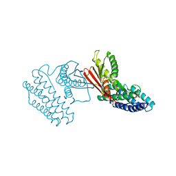 | | Crystal structure of branched-chain alpha-ketoacid dehydrogenase kinase in complex with alpha-ketoisocaproic acid and ADP | | 分子名称: | 2-OXO-4-METHYLPENTANOIC ACID, ADENOSINE-5'-DIPHOSPHATE, MAGNESIUM ION, ... | | 著者 | Tso, S.C, Chuang, J.L, Gui, W.J, Wynn, R.M, Li, J, Chuang, D.T. | | 登録日 | 2012-09-20 | | 公開日 | 2013-06-05 | | 最終更新日 | 2024-02-28 | | 実験手法 | X-RAY DIFFRACTION (2.1 Å) | | 主引用文献 | Structure-based design and mechanisms of allosteric inhibitors for mitochondrial branched-chain alpha-ketoacid dehydrogenase kinase.
Proc.Natl.Acad.Sci.USA, 110, 2013
|
|
4H85
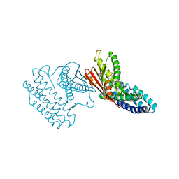 | | Crystal structure of branched-chain alpha-ketoacid dehydrogenase kinase/(R)-alpha-chloroisocaproate complex with ADP | | 分子名称: | ADENOSINE-5'-DIPHOSPHATE, ALPHA-CHLOROISOCAPROIC ACID, MAGNESIUM ION, ... | | 著者 | Tso, S.C, Chuang, J.L, Gui, W.J, Wynn, R.M, Li, J, Chuang, D.T. | | 登録日 | 2012-09-21 | | 公開日 | 2013-06-05 | | 最終更新日 | 2024-02-28 | | 実験手法 | X-RAY DIFFRACTION (2.1 Å) | | 主引用文献 | Structure-based design and mechanisms of allosteric inhibitors for mitochondrial branched-chain alpha-ketoacid dehydrogenase kinase.
Proc.Natl.Acad.Sci.USA, 110, 2013
|
|
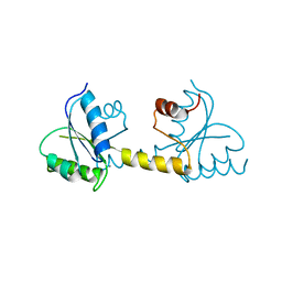
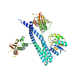
4CTI
 | | Escherichia coli EnvZ histidine kinase catalytic part fused to Archaeoglobus fulgidus Af1503 HAMP domain | | 分子名称: | OSMOLARITY SENSOR PROTEIN ENVZ, AF1503 | | 著者 | Ferris, H.U, Coles, M, Lupas, A.N, Hartmann, M.D. | | 登録日 | 2014-03-13 | | 公開日 | 2014-04-09 | | 最終更新日 | 2023-12-20 | | 実験手法 | X-RAY DIFFRACTION (2.847 Å) | | 主引用文献 | Crystallographic Snapshot of the Escherichia Coli Envz Histidine Kinase in an Active Conformation.
J.Struct.Biol., 186, 2014
|
|
6PAJ
 | |
7P8E
 | | Crystal structure of the Receiver domain of M. truncatula cytokinin receptor MtCRE1 | | 分子名称: | CALCIUM ION, Receiver domain of histidine kinase | | 著者 | Tran, L.H, Urbanowicz, A, Jasinski, M, Jaskolski, M, Ruszkowski, M. | | 登録日 | 2021-07-21 | | 公開日 | 2021-10-20 | | 最終更新日 | 2024-01-31 | | 実験手法 | X-RAY DIFFRACTION (2.5 Å) | | 主引用文献 | 3D Domain Swapping Dimerization of the Receiver Domain of Cytokinin Receptor CRE1 From Arabidopsis thaliana and Medicago truncatula .
Front Plant Sci, 12, 2021
|
|
7N0E
 | |
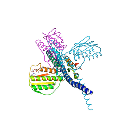
5UHT
 | | Structure of the Thermotoga maritima HK853-BeF3-RR468 complex at pH 5.0 | | 分子名称: | ADENOSINE-5'-DIPHOSPHATE, GLYCEROL, MAGNESIUM ION, ... | | 著者 | Liu, Y, Rose, J, Jiang, L, Zhou, P. | | 登録日 | 2017-01-12 | | 公開日 | 2017-12-27 | | 最終更新日 | 2024-11-06 | | 実験手法 | X-RAY DIFFRACTION (2.68 Å) | | 主引用文献 | A pH-gated conformational switch regulates the phosphatase activity of bifunctional HisKA-family histidine kinases.
Nat Commun, 8, 2017
|
|
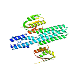
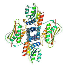
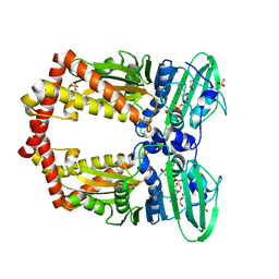
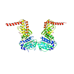
3CRK
 | | Crystal structure of the PDHK2-L2 complex. | | 分子名称: | Dihydrolipoyllysine-residue acetyltransferase component of pyruvate dehydrogenase complex, mitochondrial, POTASSIUM ION, ... | | 著者 | Green, T.J, Popov, K.M, Luo, M, Grigorian, A, Klyuyeva, A, Tuganova, A. | | 登録日 | 2008-04-07 | | 公開日 | 2008-04-29 | | 最終更新日 | 2025-03-26 | | 実験手法 | X-RAY DIFFRACTION (2.3 Å) | | 主引用文献 | Structural and functional insights into the molecular mechanisms responsible for the regulation of pyruvate dehydrogenase kinase 2.
J.Biol.Chem., 283, 2008
|
|
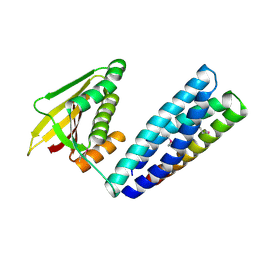
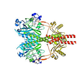
1EI1
 | | DIMERIZATION OF E. COLI DNA GYRASE B PROVIDES A STRUCTURAL MECHANISM FOR ACTIVATING THE ATPASE CATALYTIC CENTER | | 分子名称: | DNA GYRASE B, GLYCEROL, PHOSPHOAMINOPHOSPHONIC ACID-ADENYLATE ESTER, ... | | 著者 | Brino, L, Urzhumtsev, A, Oudet, P, Moras, D. | | 登録日 | 2000-02-23 | | 公開日 | 2000-03-31 | | 最終更新日 | 2024-02-07 | | 実験手法 | X-RAY DIFFRACTION (2.3 Å) | | 主引用文献 | Dimerization of Escherichia coli DNA-gyrase B provides a structural mechanism for activating the ATPase catalytic center.
J.Biol.Chem., 275, 2000
|
|
2IOQ
 | |
6J90
 | | Crystal Structure of GyraseB N-Terminal Domain complex with ATP from Salmonella Typhi at 2.2A Resolution | | 分子名称: | ADENOSINE-5'-TRIPHOSPHATE, CHLORIDE ION, DI(HYDROXYETHYL)ETHER, ... | | 著者 | Kaur, G, Sachdeva, E, Tiwari, P, Gupta, D, Ethayathulla, A.S, Kaur, P. | | 登録日 | 2019-01-21 | | 公開日 | 2020-01-22 | | 最終更新日 | 2023-11-22 | | 実験手法 | X-RAY DIFFRACTION (2.2 Å) | | 主引用文献 | Crystal Structure of GyraseB N-Terminal Domain complex with ATP from Salmonella Typhi at 2.2A Resolution
To Be Published
|
|
3LNU
 | |
3LPS
 | |
8C5V
 | |
6ENG
 | | Crystal structure of the 43K ATPase domain of Escherichia coli gyrase B in complex with an aminocoumarin | | 分子名称: | CHLORIDE ION, Coumermycin A1, DNA gyrase subunit B, ... | | 著者 | Vanden Broeck, A, McEwen, A.G, Lamour, V. | | 登録日 | 2017-10-04 | | 公開日 | 2019-04-10 | | 最終更新日 | 2024-01-17 | | 実験手法 | X-RAY DIFFRACTION (2.3 Å) | | 主引用文献 | Structural Basis for DNA Gyrase Interaction with Coumermycin A1.
J.Med.Chem., 62, 2019
|
|
7RZW
 | | CryoEM structure of Arabidopsis thaliana phytochrome B | | 分子名称: | 3-[5-[[(3~{R},4~{R})-3-ethyl-4-methyl-5-oxidanylidene-3,4-dihydropyrrol-2-yl]methyl]-2-[[5-[(4-ethyl-3-methyl-5-oxidanylidene-pyrrol-2-yl)methyl]-3-(3-hydroxy-3-oxopropyl)-4-methyl-1~{H}-pyrrol-2-yl]methyl]-4-methyl-1~{H}-pyrrol-3-yl]propanoic acid, Phytochrome B | | 著者 | Li, H, Burgie, E.S, Vierstra, R.D, Li, H. | | 登録日 | 2021-08-27 | | 公開日 | 2022-04-13 | | 最終更新日 | 2024-11-13 | | 実験手法 | ELECTRON MICROSCOPY (3.3 Å) | | 主引用文献 | Plant phytochrome B is an asymmetric dimer with unique signalling potential.
Nature, 604, 2022
|
|
7SSI
 | |
7SSJ
 | |
8SGK
 | | CryoEM structure of Deinococcus radiodurans BphP photosensory module in Pr state | | 分子名称: | 3-[2-[(Z)-[3-(2-carboxyethyl)-5-[(Z)-(4-ethenyl-3-methyl-5-oxidanylidene-pyrrol-2-ylidene)methyl]-4-methyl-pyrrol-1-ium -2-ylidene]methyl]-5-[(Z)-[(3E)-3-ethylidene-4-methyl-5-oxidanylidene-pyrrolidin-2-ylidene]methyl]-4-methyl-1H-pyrrol-3- yl]propanoic acid, Bacteriophytochrome | | 著者 | Li, H, Li, H. | | 登録日 | 2023-04-12 | | 公開日 | 2024-05-22 | | 最終更新日 | 2024-10-23 | | 実験手法 | ELECTRON MICROSCOPY (3.7 Å) | | 主引用文献 | CryoEM structure of Deinococcus radiodurans BphP photosensory module in Pr state
To Be Published
|
|
8S7O
 | | M. tuberculosis gyrase holocomplex with 150 bp DNA and BDM71403 | | 分子名称: | 6-[[2-[1-(6-methoxy-1,5-naphthyridin-4-yl)-1,2,3-triazol-4-yl]ethylamino]methyl]-4H-1,4-benzothiazin-3-one, DNA (5'-D(*CP*CP*GP*GP*AP*AP*GP*GP*GP*GP*TP*AP*AP*TP*AP*CP*T)-3'), DNA gyrase subunit A, ... | | 著者 | Gedeon, A, Yab, E, Dinut, A, Sadowski, E, Capton, E, Dreneau, A, Gioia, B, Piveteau, C, Djaout, K, Lecat, E, Wehenkel, A.M, Gubellini, F, Mechaly, A, Alzari, P.M, Deprez, B, Baulard, A, Aubry, A, Willand, N, Petrella, S. | | 登録日 | 2024-03-04 | | 公開日 | 2025-03-19 | | 実験手法 | ELECTRON MICROSCOPY (2.8 Å) | | 主引用文献 | M. tuberculosis gyrase holocomplex with 150 bp DNA and BDM71403
To Be Published
|
|
9JLB
 | | Cryo-EM structure of phyB-PIF6beta complex | | 分子名称: | 3-[5-[[(3~{R},4~{R})-3-ethyl-4-methyl-5-oxidanylidene-3,4-dihydropyrrol-2-yl]methyl]-2-[[5-[(4-ethyl-3-methyl-5-oxidanylidene-pyrrol-2-yl)methyl]-3-(3-hydroxy-3-oxopropyl)-4-methyl-1~{H}-pyrrol-2-yl]methyl]-4-methyl-1~{H}-pyrrol-3-yl]propanoic acid, Phytochrome B, Transcription factor PIF6 | | 著者 | Jia, H.L, Guan, Z.Y, Ding, J.Y, Wang, X.Y, Ma, L, Yin, P. | | 登録日 | 2024-09-18 | | 公開日 | 2025-06-04 | | 実験手法 | ELECTRON MICROSCOPY (3 Å) | | 主引用文献 | Structural insight into PIF6-mediated red light signal transduction of plant phytochrome B.
Cell Discov, 11, 2025
|
|
9ITF
 | | Cryo-EM structure of full-length phyB(Y276H)-PIF6beta complex | | 分子名称: | 3-[5-[[(3~{R},4~{R})-3-ethyl-4-methyl-5-oxidanylidene-3,4-dihydropyrrol-2-yl]methyl]-2-[[5-[(4-ethyl-3-methyl-5-oxidanylidene-pyrrol-2-yl)methyl]-3-(3-hydroxy-3-oxopropyl)-4-methyl-1~{H}-pyrrol-2-yl]methyl]-4-methyl-1~{H}-pyrrol-3-yl]propanoic acid, Phytochrome B, Transcription factor PIF6 | | 著者 | Jia, H.L, Guan, Z.Y, Ding, J.Y, Wang, X.Y, Ma, L, Yin, P. | | 登録日 | 2024-07-20 | | 公開日 | 2025-06-04 | | 実験手法 | ELECTRON MICROSCOPY (2.9 Å) | | 主引用文献 | Structural insight into PIF6-mediated red light signal transduction of plant phytochrome B.
Cell Discov, 11, 2025
|
|
9GBV
 | |
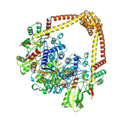
6GAV
 | | Extremely 'open' clamp structure of DNA gyrase: role of the Corynebacteriales GyrB specific insert | | 分子名称: | 2-(N-MORPHOLINO)-ETHANESULFONIC ACID, DNA gyrase subunit B,DNA gyrase subunit A | | 著者 | Petrella, S, Capton, E, Alzari, P.M, Aubry, A, MAyer, C. | | 登録日 | 2018-04-12 | | 公開日 | 2019-02-20 | | 最終更新日 | 2024-01-17 | | 実験手法 | X-RAY DIFFRACTION (2.6 Å) | | 主引用文献 | Overall Structures of Mycobacterium tuberculosis DNA Gyrase Reveal the Role of a Corynebacteriales GyrB-Specific Insert in ATPase Activity.
Structure, 27, 2019
|
|


