9BB4
 
 | |
9BB3
 | |
9BB2
 | |
9BB1
 | |
9B94
 | |
9B93
 | |
9B92
 | |
9B91
 | |
9B90
 | |
9B8Z
 | |
9B8Y
 | |
9B8X
 | |
9B8W
 | |
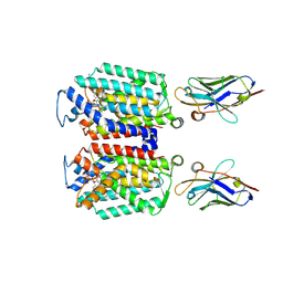
9B8Q
 | |
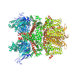
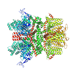
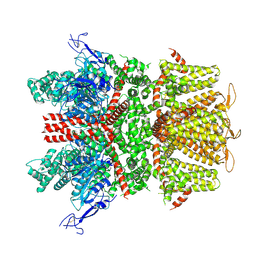
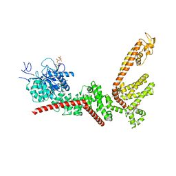
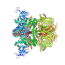
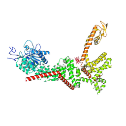
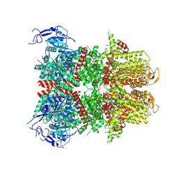
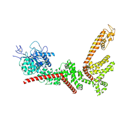
9B8O
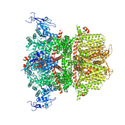
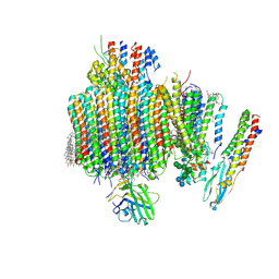 | | Synaptic Vesicle V-ATPase with synaptophysin and SidK, State 3, Vo | | 分子名称: | (7R)-4,7-DIHYDROXY-N,N,N-TRIMETHYL-10-OXO-3,5,9-TRIOXA-4-PHOSPHAHEPTACOSAN-1-AMINIUM 4-OXIDE, 1,2-DIACYL-SN-GLYCERO-3-PHOSPHOCHOLINE, 2-acetamido-2-deoxy-beta-D-glucopyranose, ... | | 著者 | Coupland, C.E, Rubinstein, J.L. | | 登録日 | 2024-03-31 | | 公開日 | 2024-06-26 | | 実験手法 | ELECTRON MICROSCOPY (3.2 Å) | | 主引用文献 | High-resolution electron cryomicroscopy of V-ATPase in native synaptic vesicles
Science, 2024
|
|
9B8E
 | | Structure of S-nitrosylated Legionella pneumophila Ceg10. | | 分子名称: | 1,2-ETHANEDIOL, ACETATE ION, CHLORIDE ION, ... | | 著者 | Tomchick, D.R, Heisler, D.B, Alto, N.M. | | 登録日 | 2024-03-29 | | 公開日 | 2024-04-10 | | 最終更新日 | 2024-05-29 | | 実験手法 | X-RAY DIFFRACTION (1.4 Å) | | 主引用文献 | Exploiting bacterial effector proteins to uncover evolutionarily conserved antiviral host machinery.
Plos Pathog., 20, 2024
|
|
9B8D
 | | Structure of Legionella pneumophila Ceg10 | | 分子名称: | 1,2-ETHANEDIOL, Ceg10, PHOSPHATE ION | | 著者 | Tomchick, D.R, Heisler, D.B, Alto, N.M. | | 登録日 | 2024-03-29 | | 公開日 | 2024-04-10 | | 最終更新日 | 2024-05-29 | | 実験手法 | X-RAY DIFFRACTION (1.72 Å) | | 主引用文献 | Exploiting bacterial effector proteins to uncover evolutionarily conserved antiviral host machinery.
Plos Pathog., 20, 2024
|
|
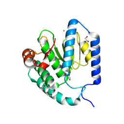
9B7F
 | | S_SAD structure of HEWL using lossless default compression | | 分子名称: | 1,2-ETHANEDIOL, CHLORIDE ION, Lysozyme C, ... | | 著者 | Jakoncic, J, Bernstein, H.J, Soares, A.S, Horvat, K. | | 登録日 | 2024-03-27 | | 公開日 | 2024-04-10 | | 実験手法 | X-RAY DIFFRACTION (1.65 Å) | | 主引用文献 | Investigation of fast and efficient lossless compression algorithms for macromolecular crystallography experiments
To Be Published
|
|
9B7E
 | | S_SAD structure of HEWL using lossy compression data with a compression ratio of 422 | | 分子名称: | 1,2-ETHANEDIOL, CHLORIDE ION, Lysozyme C, ... | | 著者 | Jakoncic, J, Bernstein, H.J, Soares, A.S, Horvat, K. | | 登録日 | 2024-03-27 | | 公開日 | 2024-04-10 | | 実験手法 | X-RAY DIFFRACTION (1.65 Å) | | 主引用文献 | Investigation of fast and efficient lossless compression algorithms for macromolecular crystallography experiments
To Be Published
|
|
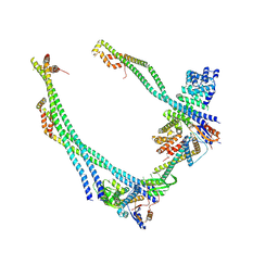
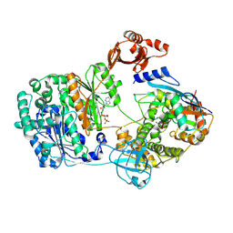
9B71
 | | Cryo-EM structure of MraY in complex with analogue 3 | | 分子名称: | (2~{S},3~{S})-3-[(2~{S},3~{R},4~{S},5~{R})-5-(aminomethyl)-3,4-bis(oxidanyl)oxolan-2-yl]oxy-3-[(2~{S},3~{S},4~{R},5~{R})-5-[2,4-bis(oxidanylidene)pyrimidin-1-yl]-3,4-bis(oxidanyl)oxolan-2-yl]-2-[[4-[[[(2~{S})-5-carbamimidamido-2-(hexadecanoylamino)pentanoyl]amino]methyl]phenyl]methylamino]propanoic acid, MraYAA Nanobody, Phospho-N-acetylmuramoyl-pentapeptide-transferase | | 著者 | Hao, A, Lee, S.-Y. | | 登録日 | 2024-03-26 | | 公開日 | 2024-06-26 | | 実験手法 | ELECTRON MICROSCOPY (2.7 Å) | | 主引用文献 | Development of a natural product optimization strategy for inhibitors against MraY, a promising antibacterial target.
Nat Commun, 15, 2024
|
|
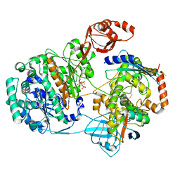
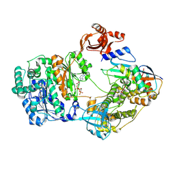
9B70
 | | Cryo-EM structure of MraY in complex with analogue 2 | | 分子名称: | (2~{S},3~{S})-3-[(2~{S},3~{R},4~{S},5~{R})-5-(aminomethyl)-3,4-bis(oxidanyl)oxolan-2-yl]oxy-2-[[3-[[[(2~{S})-6-azanyl-2-(hexadecanoylamino)hexanoyl]amino]methyl]phenyl]methylamino]-3-[(2~{S},3~{S},4~{R},5~{R})-5-[2,4-bis(oxidanylidene)pyrimidin-1-yl]-3,4-bis(oxidanyl)oxolan-2-yl]propanoic acid, MraYAA nanobody, Phospho-N-acetylmuramoyl-pentapeptide-transferase | | 著者 | Hao, A, Lee, S.-Y. | | 登録日 | 2024-03-26 | | 公開日 | 2024-06-26 | | 実験手法 | ELECTRON MICROSCOPY (2.88 Å) | | 主引用文献 | Development of a natural product optimization strategy for inhibitors against MraY, a promising antibacterial target.
Nat Commun, 15, 2024
|
|
9B5X
 | |
9B5W
 | |
9B5V
 | |
9B5U
 | |


