1HNV
 
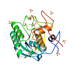
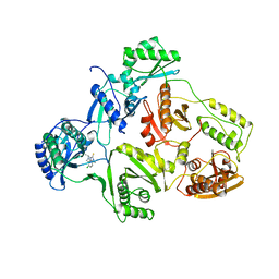 | | STRUCTURE OF HIV-1 RT(SLASH)TIBO R 86183 COMPLEX REVEALS SIMILARITY IN THE BINDING OF DIVERSE NONNUCLEOSIDE INHIBITORS | | 分子名称: | 5-CHLORO-8-METHYL-7-(3-METHYL-BUT-2-ENYL)-6,7,8,9-TETRAHYDRO-2H-2,7,9A-TRIAZA-BENZO[CD]AZULENE-1-THIONE, HIV-1 REVERSE TRANSCRIPTASE (SUBUNIT P51), HIV-1 REVERSE TRANSCRIPTASE (SUBUNIT P66) | | 著者 | Das, K, Ding, J, Arnold, E. | | 登録日 | 1995-03-30 | | 公開日 | 1995-07-10 | | 最終更新日 | 2024-02-07 | | 実験手法 | X-RAY DIFFRACTION (3 Å) | | 主引用文献 | Structure of HIV-1 RT/TIBO R 86183 complex reveals similarity in the binding of diverse nonnucleoside inhibitors.
Nat.Struct.Biol., 2, 1995
|
|
4R49
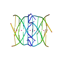
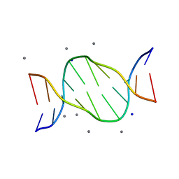 | | Racemic crystal structure of a calcium-bound B-DNA duplex | | 分子名称: | 5'-D(*CP*CP*GP*GP*TP*AP*CP*CP*GP*G)-3', CALCIUM ION, SODIUM ION | | 著者 | Mandal, P.K, Collie, G.W, Kauffmann, B, Huc, I. | | 登録日 | 2014-08-19 | | 公開日 | 2014-11-12 | | 最終更新日 | 2023-09-20 | | 実験手法 | X-RAY DIFFRACTION (1.28 Å) | | 主引用文献 | Racemic DNA crystallography.
Angew.Chem.Int.Ed.Engl., 53, 2014
|
|
4R4D
 | | Racemic crystal structure of a magnesium-bound B-DNA duplex | | 分子名称: | 5'-D(*CP*CP*GP*GP*TP*AP*CP*CP*GP*G)-3', MAGNESIUM ION, SODIUM ION | | 著者 | Mandal, P.K, Collie, G.W, Kauffmann, B, Huc, I. | | 登録日 | 2014-08-19 | | 公開日 | 2014-11-12 | | 最終更新日 | 2023-09-20 | | 実験手法 | X-RAY DIFFRACTION (1.29 Å) | | 主引用文献 | Racemic DNA crystallography.
Angew.Chem.Int.Ed.Engl., 53, 2014
|
|
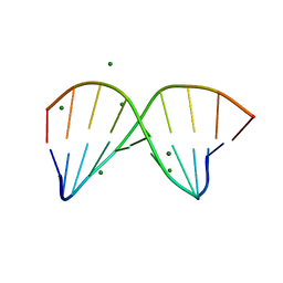
4R47
 | | Racemic crystal structure of a bimolecular DNA G-quadruplex (P21/n) | | 分子名称: | 5'-D(*GP*GP*GP*GP*TP*TP*TP*TP*GP*GP*GP*G)-3', MAGNESIUM ION, POTASSIUM ION | | 著者 | Mandal, P.K, Collie, G.W, Kauffmann, B, Huc, I. | | 登録日 | 2014-08-19 | | 公開日 | 2014-11-12 | | 最終更新日 | 2023-09-20 | | 実験手法 | X-RAY DIFFRACTION (1.85 Å) | | 主引用文献 | Racemic DNA crystallography.
Angew.Chem.Int.Ed.Engl., 53, 2014
|
|
8BVO
 | |
8BXR
 | |
8BW6
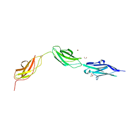
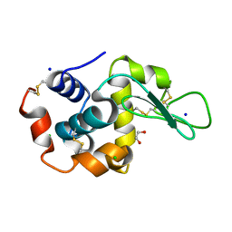
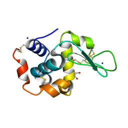
 | | Titin FnIII-domain I110 (I/A6) from the MIR region | | 分子名称: | 1,2-ETHANEDIOL, CALCIUM ION, DI(HYDROXYETHYL)ETHER, ... | | 著者 | Mayans, O, Fleming, J.R. | | 登録日 | 2022-12-06 | | 公開日 | 2023-02-15 | | 最終更新日 | 2023-03-08 | | 実験手法 | X-RAY DIFFRACTION (1.95 Å) | | 主引用文献 | Immunological and Structural Characterization of Titin Main Immunogenic Region; I110 Domain Is the Target of Titin Antibodies in Myasthenia Gravis.
Biomedicines, 11, 2023
|
|
4R44
 | | Racemic crystal structure of a tetramolecular DNA G-quadruplex | | 分子名称: | 5'-D(*TP*GP*GP*GP*GP*T)-3', POTASSIUM ION, SODIUM ION | | 著者 | Mandal, P.K, Collie, G.W, Kauffmann, B, Huc, I. | | 登録日 | 2014-08-19 | | 公開日 | 2014-11-12 | | 最終更新日 | 2023-09-20 | | 実験手法 | X-RAY DIFFRACTION (2.695 Å) | | 主引用文献 | Racemic DNA crystallography.
Angew.Chem.Int.Ed.Engl., 53, 2014
|
|
5H65
 | | Crystal structure of human POT1 and TPP1 | | 分子名称: | Adrenocortical dysplasia protein homolog, Protection of telomeres protein 1, ZINC ION | | 著者 | Chen, C, Wu, J, Lei, M. | | 登録日 | 2016-11-10 | | 公開日 | 2017-05-31 | | 最終更新日 | 2024-03-20 | | 実験手法 | X-RAY DIFFRACTION (2.1 Å) | | 主引用文献 | Structural insights into POT1-TPP1 interaction and POT1 C-terminal mutations in human cancer.
Nat Commun, 8, 2017
|
|
6QXW
 | | Pink beam serial crystallography: Lysozyme, 5 us exposure, 24344 patterns merged (3 chips) | | 分子名称: | 1,2-ETHANEDIOL, CHLORIDE ION, Lysozyme C, ... | | 著者 | Lieske, J, Tolstikova, A, Meents, A. | | 登録日 | 2019-03-08 | | 公開日 | 2019-09-25 | | 最終更新日 | 2024-01-24 | | 実験手法 | X-RAY DIFFRACTION (1.7 Å) | | 主引用文献 | 1 kHz fixed-target serial crystallography using a multilayer monochromator and an integrating pixel detector.
Iucrj, 6, 2019
|
|
6QXY
 | | Pink beam serial crystallography: Lysozyme, 5 us exposure, 8813 patterns merged (1 chip) | | 分子名称: | 1,2-ETHANEDIOL, CHLORIDE ION, Lysozyme C, ... | | 著者 | Lieske, J, Tolstikova, A, Meents, A. | | 登録日 | 2019-03-08 | | 公開日 | 2019-10-16 | | 最終更新日 | 2024-01-24 | | 実験手法 | X-RAY DIFFRACTION (1.7 Å) | | 主引用文献 | 1 kHz fixed-target serial crystallography using a multilayer monochromator and an integrating pixel detector.
Iucrj, 6, 2019
|
|
6QY2
 | | Pink beam serial crystallography: Lysozyme, 5 us exposure, 750 patterns merged | | 分子名称: | 1,2-ETHANEDIOL, CHLORIDE ION, Lysozyme C, ... | | 著者 | Lieske, J, Tolstikova, A, Meents, A. | | 登録日 | 2019-03-08 | | 公開日 | 2019-09-25 | | 最終更新日 | 2024-01-24 | | 実験手法 | X-RAY DIFFRACTION (1.7 Å) | | 主引用文献 | 1 kHz fixed-target serial crystallography using a multilayer monochromator and an integrating pixel detector.
Iucrj, 6, 2019
|
|
6QY5
 | | Pink beam serial crystallography: Lysozyme, 1 us exposure, 4448 patterns merged (1 chip) | | 分子名称: | 1,2-ETHANEDIOL, CHLORIDE ION, Lysozyme C, ... | | 著者 | Lieske, J, Tolstikova, A, Meents, A. | | 登録日 | 2019-03-08 | | 公開日 | 2019-10-16 | | 最終更新日 | 2024-01-24 | | 実験手法 | X-RAY DIFFRACTION (1.84 Å) | | 主引用文献 | 1 kHz fixed-target serial crystallography using a multilayer monochromator and an integrating pixel detector.
Iucrj, 6, 2019
|
|
6QXV
 | | Pink beam serial crystallography: Proteinase K, 1 us exposure, 1585 patterns merged (2 chips) | | 分子名称: | CALCIUM ION, CHLORIDE ION, Proteinase K, ... | | 著者 | Tolstikova, A, Oberthuer, D, Meents, A. | | 登録日 | 2019-03-08 | | 公開日 | 2019-09-25 | | 最終更新日 | 2024-01-24 | | 実験手法 | X-RAY DIFFRACTION (1.94 Å) | | 主引用文献 | 1 kHz fixed-target serial crystallography using a multilayer monochromator and an integrating pixel detector.
Iucrj, 6, 2019
|
|
6QY0
 | | Pink beam serial crystallography: Lysozyme, 5 us exposure, 3000 patterns merged | | 分子名称: | 1,2-ETHANEDIOL, CHLORIDE ION, Lysozyme C, ... | | 著者 | Lieske, J, Tolstikova, A, Meents, A. | | 登録日 | 2019-03-08 | | 公開日 | 2019-09-25 | | 最終更新日 | 2024-01-24 | | 実験手法 | X-RAY DIFFRACTION (1.7 Å) | | 主引用文献 | 1 kHz fixed-target serial crystallography using a multilayer monochromator and an integrating pixel detector.
Iucrj, 6, 2019
|
|
6QY4
 | | Pink beam serial crystallography: Lysozyme, 1 us exposure, 14793 patterns merged | | 分子名称: | 1,2-ETHANEDIOL, CHLORIDE ION, Lysozyme C, ... | | 著者 | Lieske, J, Tolstikova, A, Meents, A. | | 登録日 | 2019-03-08 | | 公開日 | 2019-09-25 | | 最終更新日 | 2024-01-24 | | 実験手法 | X-RAY DIFFRACTION (1.84 Å) | | 主引用文献 | 1 kHz fixed-target serial crystallography using a multilayer monochromator and an integrating pixel detector.
Iucrj, 6, 2019
|
|
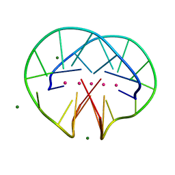
4RZD
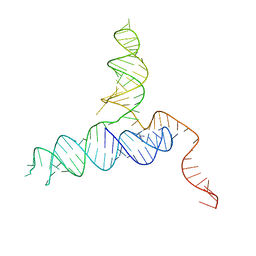 | | Crystal Structure of a PreQ1 Riboswitch | | 分子名称: | 7-DEAZA-7-AMINOMETHYL-GUANINE, PreQ1-III Riboswitch (Class 3) | | 著者 | Wedekind, J.E, Liberman, J.A, Salim, M. | | 登録日 | 2014-12-20 | | 公開日 | 2015-07-01 | | 最終更新日 | 2024-02-28 | | 実験手法 | X-RAY DIFFRACTION (2.75 Å) | | 主引用文献 | Structural analysis of a class III preQ1 riboswitch reveals an aptamer distant from a ribosome-binding site regulated by fast dynamics.
Proc.Natl.Acad.Sci.USA, 112, 2015
|
|
6QY1
 | | Pink beam serial crystallography: Lysozyme, 5 us exposure, 1500 patterns merged | | 分子名称: | 1,2-ETHANEDIOL, CHLORIDE ION, Lysozyme C, ... | | 著者 | Lieske, J, Tolstikova, A, Meents, A. | | 登録日 | 2019-03-08 | | 公開日 | 2019-09-25 | | 最終更新日 | 2024-01-24 | | 実験手法 | X-RAY DIFFRACTION (1.7 Å) | | 主引用文献 | 1 kHz fixed-target serial crystallography using a multilayer monochromator and an integrating pixel detector.
Iucrj, 6, 2019
|
|
6QXX
 | | Pink beam serial crystallography: Lysozyme, 5 us exposure, 14793 patterns merged | | 分子名称: | 1,2-ETHANEDIOL, CHLORIDE ION, Lysozyme C, ... | | 著者 | Lieske, J, Tolstikova, A, Meents, A. | | 登録日 | 2019-03-08 | | 公開日 | 2019-10-16 | | 最終更新日 | 2024-01-24 | | 実験手法 | X-RAY DIFFRACTION (1.7 Å) | | 主引用文献 | 1 kHz fixed-target serial crystallography using a multilayer monochromator and an integrating pixel detector.
Iucrj, 6, 2019
|
|
4UIF
 | | Cryo-EM structure of Dengue virus serotype 2 in complex with antigen-binding fragments of human antibody 2D22 | | 分子名称: | ANTIGEN-BINDING FRAGMENT OF HUMAN ANTIBODY 2D22 - HEAVY CHAIN, ANTIGEN-BINDING FRAGMENT OF HUMAN ANTIBODY 2D22 - LIGHT CHAIN, DENGUE VIRUS SEROTYPE 2 STRAIN PVP94 07 - ENVELOPE PROTEIN, ... | | 著者 | Fibriansah, G, Ibarra, K.D, Ng, T.-S, Smith, S.A, Tan, J.L, Lim, X.-N, Ooi, J.S.G, Kostyuchenko, V.A, Wang, J, de Silva, A.M, Harris, E, Crowe Junior, J.E, Lok, S.-M. | | 登録日 | 2015-03-30 | | 公開日 | 2015-07-15 | | 最終更新日 | 2024-05-08 | | 実験手法 | ELECTRON MICROSCOPY (6.5 Å) | | 主引用文献 | Cryo-EM structure of an antibody that neutralizes dengue virus type 2 by locking E protein dimers.
Science, 349, 2015
|
|
4UT1
 | |
1ULP
 | |
1ULO
 | |
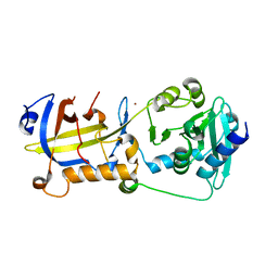
4PQF
 | | Mycobacterium tuberculosis RecA glycerol bound low temperature structure IIB-CR | | 分子名称: | 1,2-ETHANEDIOL, GLYCEROL, Protein RecA, ... | | 著者 | Chandran, A.V, Prabu, J.R, Patil, N.K, Muniyappa, K, Vijayan, M. | | 登録日 | 2014-03-03 | | 公開日 | 2015-03-18 | | 最終更新日 | 2023-11-08 | | 実験手法 | X-RAY DIFFRACTION (2.8 Å) | | 主引用文献 | Structural studies on Mycobacterium tuberculosis RecA: Molecular plasticity and interspecies variability
J.Biosci., 40, 2015
|
|
4PQR
 | | Mycobacterium tuberculosis RecA glycerol bound low temperature structure IIB-BN | | 分子名称: | 1,2-ETHANEDIOL, GLYCEROL, Protein RecA, ... | | 著者 | Chandran, A.V, Prabu, J.R, Patil, N.K, Muniyappa, K, Vijayan, M. | | 登録日 | 2014-03-04 | | 公開日 | 2015-03-18 | | 最終更新日 | 2023-11-08 | | 実験手法 | X-RAY DIFFRACTION (2.8 Å) | | 主引用文献 | Structural studies on Mycobacterium tuberculosis RecA: Molecular plasticity and interspecies variability
J.Biosci., 40, 2015
|
|


