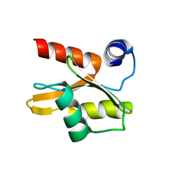
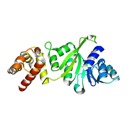
2GF4
 
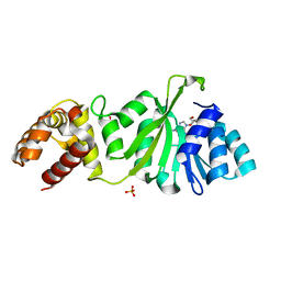
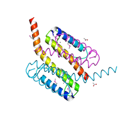 | | Crystal structure of Vng1086c from Halobacterium salinarium (Halobacterium halobium). Northeast Structural Genomics Target HsR14 | | 分子名称: | ACETATE ION, CALCIUM ION, Protein Vng1086c | | 著者 | Benach, J, Zhou, W, Jayaraman, S, Forouhar, F.F, Janjua, H, Xiao, R, Ma, L.-C, Cunningham, K, Wang, D, Acton, T.B, Montelione, G.T, Tong, L, Hunt, J.F, Northeast Structural Genomics Consortium (NESG) | | 登録日 | 2006-03-21 | | 公開日 | 2006-04-18 | | 最終更新日 | 2017-10-18 | | 実験手法 | X-RAY DIFFRACTION (2.07 Å) | | 主引用文献 | Crystal structure of Vng1086c from Halobacterium salinarium (Halobacterium halobium). Northeast Structural Genomics Target HsR14
To be Published
|
|
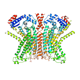
7A0G
 | |
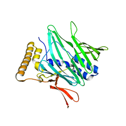
8OQ9
 | | Crystal structure of the titin domain Fn3-56 | | 分子名称: | CHLORIDE ION, Titin, ZINC ION | | 著者 | Rees, M, Gautel, M. | | 登録日 | 2023-04-11 | | 公開日 | 2023-08-23 | | 最終更新日 | 2023-08-30 | | 実験手法 | X-RAY DIFFRACTION (1.65 Å) | | 主引用文献 | Structure determination and analysis of titin A-band fibronectin type III domains provides insights for disease-linked variants and protein oligomerisation.
J.Struct.Biol., 215, 2023
|
|
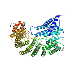
7RX2
 | |
2L9W
 | |
7A26
 | |
8OMW
 | | Crystal structure of the titin domain Fn3-20 | | 分子名称: | CHLORIDE ION, Titin | | 著者 | Rees, M, Gautel, M. | | 登録日 | 2023-03-31 | | 公開日 | 2023-08-23 | | 最終更新日 | 2023-08-30 | | 実験手法 | X-RAY DIFFRACTION (1.05 Å) | | 主引用文献 | Structure determination and analysis of titin A-band fibronectin type III domains provides insights for disease-linked variants and protein oligomerisation.
J.Struct.Biol., 215, 2023
|
|
8ORL
 | |
7P59
 | | Variant Surface Glycoprotein 3 (VSG3, MiTat1.3, VSG224) with two O-linked post-translational modifications | | 分子名称: | Variant surface glycoprotein, alpha-D-glucopyranose, alpha-D-mannopyranose-(1-2)-alpha-D-mannopyranose-(1-3)-[alpha-D-mannopyranose-(1-6)]beta-D-mannopyranose-(1-4)-2-acetamido-2-deoxy-beta-D-glucopyranose-(1-4)-2-acetamido-2-deoxy-beta-D-glucopyranose | | 著者 | Gkeka, A, Arest-Branco, F, Stebbins, C.E, Papavasiliou, F.N. | | 登録日 | 2021-07-14 | | 公開日 | 2022-07-27 | | 最終更新日 | 2024-02-07 | | 実験手法 | X-RAY DIFFRACTION (1.27 Å) | | 主引用文献 | Immunodominant surface epitopes power immune evasion in the African trypanosome.
Cell Rep, 42, 2023
|
|
3GRR
 | |
8OS3
 | |
8OTY
 | |
8OT5
 | | Crystal structure of the titin domain Fn3-85 | | 分子名称: | CHLORIDE ION, SODIUM ION, Titin | | 著者 | Nikoopour, R, Rees, M, Gautel, M. | | 登録日 | 2023-04-20 | | 公開日 | 2023-08-23 | | 最終更新日 | 2023-08-30 | | 実験手法 | X-RAY DIFFRACTION (1.56 Å) | | 主引用文献 | Structure determination and analysis of titin A-band fibronectin type III domains provides insights for disease-linked variants and protein oligomerisation.
J.Struct.Biol., 215, 2023
|
|
7P5B
 | | Variant Surface Glycoprotein 3 (VSG3, MiTat1.3, VSG224) mutant (serine 319 to alanine), single O-linked glycosylated at Ser317 | | 分子名称: | Variant surface glycoprotein, alpha-D-glucopyranose, alpha-D-mannopyranose-(1-2)-alpha-D-mannopyranose-(1-3)-[alpha-D-mannopyranose-(1-6)]beta-D-mannopyranose-(1-4)-2-acetamido-2-deoxy-beta-D-glucopyranose-(1-4)-2-acetamido-2-deoxy-beta-D-glucopyranose | | 著者 | Gkeka, A, Aresta-Branco, F, Stebbins, C.E, Papavasiliou, F.N. | | 登録日 | 2021-07-14 | | 公開日 | 2022-07-27 | | 最終更新日 | 2024-02-07 | | 実験手法 | X-RAY DIFFRACTION (1.13 Å) | | 主引用文献 | Immunodominant surface epitopes power immune evasion in the African trypanosome.
Cell Rep, 42, 2023
|
|
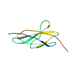
2LKQ
 | | NMR structure of the lambda 5 22-45 peptide | | 分子名称: | Immunoglobulin lambda-like polypeptide 1 | | 著者 | Elantak, L, Espeli, M, Boned, A, Bornet, O, Breton, C, Feracci, M, Roche, P, Guerlesquin, F, Schiff, C. | | 登録日 | 2011-10-19 | | 公開日 | 2012-10-24 | | 最終更新日 | 2024-05-15 | | 実験手法 | SOLUTION NMR | | 主引用文献 | Structural Basis for Galectin-1-dependent Pre-B Cell Receptor (Pre-BCR) Activation.
J.Biol.Chem., 287, 2012
|
|
7RX3
 | |
3GRV
 | |
8P7A
 | |
7P5A
 | | Variant Surface Glycoprotein 3 (VSG3, MiTat1.3, VSG224) mutant (serine 317 to alanine), single O-linked glycosylated at Ser319 | | 分子名称: | Variant surface glycoprotein, alpha-D-glucopyranose, alpha-D-mannopyranose-(1-2)-alpha-D-mannopyranose-(1-3)-[alpha-D-mannopyranose-(1-6)]beta-D-mannopyranose-(1-4)-2-acetamido-2-deoxy-beta-D-glucopyranose-(1-4)-2-acetamido-2-deoxy-beta-D-glucopyranose | | 著者 | Gkeka, A, Aresta-Branco, F, Stebbins, C.E, Papavasiliou, F.N. | | 登録日 | 2021-07-14 | | 公開日 | 2022-07-27 | | 最終更新日 | 2024-02-07 | | 実験手法 | X-RAY DIFFRACTION (1.95 Å) | | 主引用文献 | Immunodominant surface epitopes power immune evasion in the African trypanosome.
Cell Rep, 42, 2023
|
|
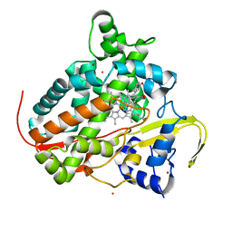
5K09
 | | Crystal Structure of COMT in complex with a thiazole ligand | | 分子名称: | 5-{3-[(4-methoxyphenyl)methyl]-1H-pyrazol-5-yl}-2,4-dimethyl-1,3-thiazole, Catechol O-methyltransferase, PHOSPHATE ION, ... | | 著者 | Ehler, A, Rodriguez-Sarmiento, R.M, Rudolph, M.G. | | 登録日 | 2016-05-17 | | 公開日 | 2016-09-07 | | 最終更新日 | 2024-05-08 | | 実験手法 | X-RAY DIFFRACTION (2.7 Å) | | 主引用文献 | Design of Potent and Druglike Nonphenolic Inhibitors for Catechol O-Methyltransferase Derived from a Fragment Screening Approach Targeting the S-Adenosyl-l-methionine Pocket.
J. Med. Chem., 59, 2016
|
|
7RXG
 | |
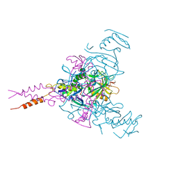
7A9W
 | | Structure of yeast Rmd9p in complex with 20nt target RNA | | 分子名称: | CHLORIDE ION, Protein RMD9, mitochondrial, ... | | 著者 | Hillen, H.S, Markov, D.A, Ireneusz, W.D, Hofmann, K.B, Cowan, A.T, Jones, J.L, Temiakov, D, Cramer, P, Anikin, M. | | 登録日 | 2020-09-02 | | 公開日 | 2021-04-07 | | 最終更新日 | 2021-05-05 | | 実験手法 | X-RAY DIFFRACTION (2.55 Å) | | 主引用文献 | The pentatricopeptide repeat protein Rmd9 recognizes the dodecameric element in the 3'-UTRs of yeast mitochondrial mRNAs.
Proc.Natl.Acad.Sci.USA, 118, 2021
|
|
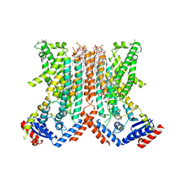
5JQ7
 | | Crystal structure of Ebola glycoprotein in complex with toremifene | | 分子名称: | 2-acetamido-2-deoxy-beta-D-glucopyranose, DIMETHYL SULFOXIDE, Envelope glycoprotein 1,Envelope glycoprotein 1,Envelope glycoprotein 1, ... | | 著者 | Zhao, Y, Ren, J, Stuart, D.I. | | 登録日 | 2016-05-04 | | 公開日 | 2016-06-29 | | 最終更新日 | 2024-01-10 | | 実験手法 | X-RAY DIFFRACTION (2.69 Å) | | 主引用文献 | Toremifene interacts with and destabilizes the Ebola virus glycoprotein.
Nature, 535, 2016
|
|
7P5T
 | | Structure of CYP142 from Mycobacterium tuberculosis in complex with inhibitor MEK216 | | 分子名称: | BROMIDE ION, POTASSIUM ION, PROTOPORPHYRIN IX CONTAINING FE, ... | | 著者 | Snee, M, Kavanagh, M, Tunnicliffe, R, McLean, K, Levy, C, Munro, A. | | 登録日 | 2021-07-14 | | 公開日 | 2022-11-16 | | 最終更新日 | 2024-01-31 | | 実験手法 | X-RAY DIFFRACTION (1.3 Å) | | 主引用文献 | Structure of CYP142 from Mycobacterium tuberculosis in complex with inhibitor MEK216
To Be Published
|
|
7D7O
 | | Crystal structure of cystathionine gamma-lyase from Bacillus cereus ATCC 14579 | | 分子名称: | Bifunctional cystathionine gamma-lyase/homocysteine desulfhydrase, GLYCEROL, PYRIDOXAL-5'-PHOSPHATE, ... | | 著者 | Sagong, H.-Y, Kim, B, Kim, K.-J. | | 登録日 | 2020-10-05 | | 公開日 | 2021-08-18 | | 最終更新日 | 2023-11-29 | | 実験手法 | X-RAY DIFFRACTION (1.98 Å) | | 主引用文献 | Structural and Functional Characterization of Cystathionine gamma-lyase from Bacillus cereus ATCC 14579.
J.Agric.Food Chem., 68, 2020
|
|


