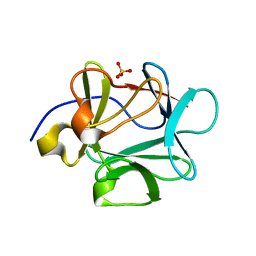
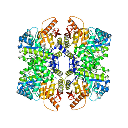
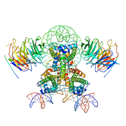
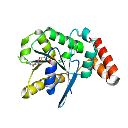
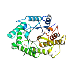
3FGM
 
 | |
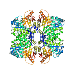
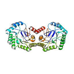
7FSC
 | | Structure of liver pyruvate kinase in complex with allosteric modulator 42 | | 分子名称: | 1,6-di-O-phosphono-beta-D-fructofuranose, MAGNESIUM ION, N-({4-[(3,4-dihydroxyphenyl)methyl]phenyl}methyl)benzenesulfonamide, ... | | 著者 | Lulla, A, Nilsson, O, Brear, P, Nain-Perez, A, Grotli, M, Hyvonen, M. | | 登録日 | 2022-12-18 | | 公開日 | 2023-04-12 | | 最終更新日 | 2024-05-22 | | 実験手法 | X-RAY DIFFRACTION (1.855 Å) | | 主引用文献 | Tuning liver pyruvate kinase activity up or down with a new class of allosteric modulators.
Eur.J.Med.Chem., 250, 2023
|
|
4GUE
 | | Structure of N-terminal kinase domain of RSK2 with flavonoid glycoside quercitrin | | 分子名称: | 2-(3,4-dihydroxyphenyl)-5,7-dihydroxy-4-oxo-4H-chromen-3-yl 6-deoxy-alpha-L-mannopyranoside, MAGNESIUM ION, Ribosomal protein S6 kinase alpha-3, ... | | 著者 | Derewenda, U, Utepbergenov, D, Szukalska, G, Derewenda, Z.S. | | 登録日 | 2012-08-29 | | 公開日 | 2013-01-30 | | 最終更新日 | 2023-09-13 | | 実験手法 | X-RAY DIFFRACTION (1.8 Å) | | 主引用文献 | Identification of quercitrin as an inhibitor of the p90 S6 ribosomal kinase (RSK): structure of its complex with the N-terminal domain of RSK2 at 1.8 A resolution.
Acta Crystallogr.,Sect.D, 69, 2013
|
|
4QFY
 | | Crystal structure of the tetrameric dGTP/dCTP-bound SAMHD1 (RN206) mutant catalytic core | | 分子名称: | 2'-DEOXYCYTIDINE-5'-TRIPHOSPHATE, 2'-DEOXYGUANOSINE-5'-TRIPHOSPHATE, Deoxynucleoside triphosphate triphosphohydrolase SAMHD1, ... | | 著者 | Koharudin, L.M.I, Wu, Y, DeLucia, M, Mehrens, J, Gronenborn, A.M, Ahn, J. | | 登録日 | 2014-05-22 | | 公開日 | 2014-10-15 | | 最終更新日 | 2023-09-20 | | 実験手法 | X-RAY DIFFRACTION (2.1 Å) | | 主引用文献 | Structural Basis of Allosteric Activation of Sterile alpha Motif and Histidine-Aspartate Domain-containing Protein 1 (SAMHD1) by Nucleoside Triphosphates.
J.Biol.Chem., 289, 2014
|
|
7FSB
 | | Structure of liver pyruvate kinase in complex with allosteric modulator 41 | | 分子名称: | 1,6-di-O-phosphono-beta-D-fructofuranose, MAGNESIUM ION, N-({4-[(3,4-dihydroxyphenyl)methyl]phenyl}methyl)-3,4-dihydroxybenzene-1-sulfonamide, ... | | 著者 | Lulla, A, Nilsson, O, Brear, P, Nain-Perez, A, Grotli, M, Hyvonen, M. | | 登録日 | 2022-12-18 | | 公開日 | 2023-04-12 | | 最終更新日 | 2024-05-22 | | 実験手法 | X-RAY DIFFRACTION (2.495 Å) | | 主引用文献 | Tuning liver pyruvate kinase activity up or down with a new class of allosteric modulators.
Eur.J.Med.Chem., 250, 2023
|
|
1HLE
 | | CRYSTAL STRUCTURE OF CLEAVED EQUINE LEUCOCYTE ELASTASE INHIBITOR DETERMINED AT 1.95 ANGSTROMS RESOLUTION | | 分子名称: | CALCIUM ION, HORSE LEUKOCYTE ELASTASE INHIBITOR | | 著者 | Baumann, U, Bode, W, Huber, R, Travis, J, Potempa, J. | | 登録日 | 1992-04-13 | | 公開日 | 1994-01-31 | | 最終更新日 | 2024-11-13 | | 実験手法 | X-RAY DIFFRACTION (1.95 Å) | | 主引用文献 | Crystal structure of cleaved equine leucocyte elastase inhibitor determined at 1.95 A resolution.
J.Mol.Biol., 226, 1992
|
|
1L9W
 | | CRYSTAL STRUCTURE OF 3-DEHYDROQUINASE FROM SALMONELLA TYPHI COMPLEXED WITH REACTION PRODUCT | | 分子名称: | 3-AMINO-4,5-DIHYDROXY-CYCLOHEX-1-ENECARBOXYLATE, 3-dehydroquinate dehydratase aroD | | 著者 | Lee, W.H, Perles, L.A, Nagem, R.A.P, Shrive, A.K, Hawkins, A, Sawyer, L, Polikarpov, I. | | 登録日 | 2002-03-26 | | 公開日 | 2003-03-26 | | 最終更新日 | 2024-11-06 | | 実験手法 | X-RAY DIFFRACTION (2.1 Å) | | 主引用文献 | Comparison of different crystal forms of 3-dehydroquinase from Salmonella typhi and its implication for the enzyme activity.
Acta Crystallogr.,Sect.D, 58, 2002
|
|
3M61
 | | Crystal structure of complex of urokinase and a upain-1 variant(W3A) in pH4.6 condition | | 分子名称: | Urokinase-type plasminogen activator, upain-1 W3A | | 著者 | Jiang, L, Yuan, C, Wind, T, Andreasen, P.A, Chen, L, Meehan, E.J, Huang, M. | | 登録日 | 2010-03-15 | | 公開日 | 2010-04-07 | | 最終更新日 | 2024-11-13 | | 実験手法 | X-RAY DIFFRACTION (1.68 Å) | | 主引用文献 | Crystal structure of complex of urokinase and a upain-1 variant(W3A) in pH4.6 condition
TO BE PUBLISHED
|
|
7FX2
 | | Crystal Structure of human FABP4 binding site mutated to that of FABP5 in complex with 2-[[6,6-difluoro-3-(4-methyl-1,3-thiazol-2-yl)-5,7-dihydro-4H-1-benzothiophen-2-yl]carbamoyl]cyclohexene-1-carboxylic acid, i.e. SMILES N(C(=O)C1=C(CCCC1)C(=O)O)C1=C(C2=NC(=CS2)C)C2=C(S1)CC(CC2)(F)F with IC50=0.0587459 microM | | 分子名称: | 2-{[(3M)-6,6-difluoro-3-(4-methyl-1,3-thiazol-2-yl)-4,5,6,7-tetrahydro-1-benzothiophen-2-yl]carbamoyl}cyclohex-1-ene-1-carboxylic acid, Fatty acid-binding protein, adipocyte | | 著者 | Ehler, A, Benz, J, Obst, U, Richter, H, Rudolph, M.G. | | 登録日 | 2023-04-27 | | 公開日 | 2023-06-14 | | 最終更新日 | 2025-08-13 | | 実験手法 | X-RAY DIFFRACTION (1.47 Å) | | 主引用文献 | A high-resolution data set of fatty acid-binding protein structures. III. Unexpectedly high occurrence of wrong ligands.
Acta Crystallogr D Struct Biol, 81, 2025
|
|
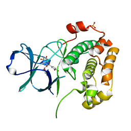
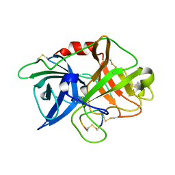
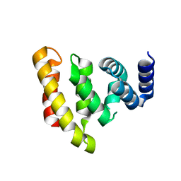
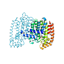
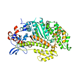
1HT3
 | |
7Y61
 | | Cryo-EM structure of the two CAF1LCs bound right-handed Di-tetrasome | | 分子名称: | Chromatin assembly factor 1 subunit A, Chromatin assembly factor 1 subunit B, Histone H3.1, ... | | 著者 | Liu, C.P, Yu, Z.Y, Yu, C, Xu, R.M. | | 登録日 | 2022-06-18 | | 公開日 | 2023-08-16 | | 最終更新日 | 2023-09-06 | | 実験手法 | ELECTRON MICROSCOPY (5.6 Å) | | 主引用文献 | Structural insights into histone binding and nucleosome assembly by chromatin assembly factor-1.
Science, 381, 2023
|
|
2VXG
 | | Crystal structure of the conserved C-terminal region of Ge-1 | | 分子名称: | CG6181-PA, ISOFORM A | | 著者 | Jinek, M, Eulalio, A, Lingel, A, Helms, S, Conti, E, Izaurralde, E. | | 登録日 | 2008-07-04 | | 公開日 | 2008-09-09 | | 最終更新日 | 2024-05-08 | | 実験手法 | X-RAY DIFFRACTION (1.9 Å) | | 主引用文献 | The C-Terminal Region of Ge-1 Presents Conserved Structural Features Required for P-Body Localization.
RNA, 14, 2008
|
|
7Y5V
 | | Cryo-EM structure of the dimeric human CAF1LC-H3-H4 complex | | 分子名称: | Chromatin assembly factor 1 subunit A, Chromatin assembly factor 1 subunit B, Histone H3.1, ... | | 著者 | Liu, C.P, Yu, C, Yu, Z.Y, Xu, R.M. | | 登録日 | 2022-06-17 | | 公開日 | 2023-08-16 | | 最終更新日 | 2023-09-06 | | 実験手法 | ELECTRON MICROSCOPY (6.1 Å) | | 主引用文献 | Structural insights into histone binding and nucleosome assembly by chromatin assembly factor-1.
Science, 381, 2023
|
|
3UWL
 | | Crystal structure of Enteroccocus faecalis thymidylate synthase (EfTS) in complex with 5-formyl tetrahydrofolate | | 分子名称: | 1,2-ETHANEDIOL, N-[4-({[(6S)-2-amino-5-formyl-4-oxo-3,4,5,6,7,8-hexahydropteridin-6-yl]methyl}amino)benzoyl]-L-glutamic acid, SULFATE ION, ... | | 著者 | Pozzi, C, Catalano, A, Cortesi, D, Luciani, R, Ferrari, S, Fritz, T, Costi, M.P, Mangani, S. | | 登録日 | 2011-12-02 | | 公開日 | 2012-08-29 | | 最終更新日 | 2024-10-30 | | 実験手法 | X-RAY DIFFRACTION (2.07 Å) | | 主引用文献 | The structure of Enterococcus faecalis thymidylate synthase provides clues about folate bacterial metabolism.
Acta Crystallogr.,Sect.D, 68, 2012
|
|
3V70
 | | Crystal Structure of Human GTPase IMAP family member 1 | | 分子名称: | GTPase IMAP family member 1, GUANOSINE-5'-DIPHOSPHATE, MAGNESIUM ION | | 著者 | Nedyalkova, L, Shen, Y, Tong, Y, Tempel, W, Mackenzie, F, Arrowsmith, C.H, Edwards, A.M, Bountra, C, Weigelt, J, Bochkarev, A, Andrews, D.W, Park, H, Structural Genomics Consortium (SGC) | | 登録日 | 2011-12-20 | | 公開日 | 2012-01-11 | | 最終更新日 | 2023-09-13 | | 実験手法 | X-RAY DIFFRACTION (2.206 Å) | | 主引用文献 | Crystal Structure of Human GTPase IMAP family member 1
to be published
|
|
1YV5
 | | Human farnesyl diphosphate synthase complexed with Mg and risedronate | | 分子名称: | 1-HYDROXY-2-(3-PYRIDINYL)ETHYLIDENE BIS-PHOSPHONIC ACID, Farnesyl pyrophosphate synthetase, MAGNESIUM ION, ... | | 著者 | Kavanagh, K.L, Guo, K, Von Delft, F, Arrowsmith, C, Sundstrom, M, Edwards, A, Oppermann, U, Structural Genomics Consortium (SGC) | | 登録日 | 2005-02-15 | | 公開日 | 2005-03-01 | | 最終更新日 | 2023-10-25 | | 実験手法 | X-RAY DIFFRACTION (2 Å) | | 主引用文献 | The molecular mechanism of nitrogen-containing bisphosphonates as antiosteoporosis drugs.
Proc.Natl.Acad.Sci.USA, 103, 2006
|
|
1W9K
 | | Dictyostelium discoideum Myosin II motor domain S456E with bound MgADP-BeFx | | 分子名称: | 1,2-ETHANEDIOL, ADENOSINE-5'-DIPHOSPHATE, BERYLLIUM TRIFLUORIDE ION, ... | | 著者 | Morris, C.A, Coureux, P.-D, Wells, A.L, Houdusse, A, Sweeney, H.L. | | 登録日 | 2004-10-13 | | 公開日 | 2006-03-16 | | 最終更新日 | 2023-12-13 | | 実験手法 | X-RAY DIFFRACTION (2.05 Å) | | 主引用文献 | Structure-Function Analysis of Myosin II Backdoor Mutants
To be Published
|
|
3V16
 | | An intramolecular pi-cation latch in phosphatidylinositol-specific phospholipase C from S.aureus controls substrate access to the active site | | 分子名称: | 1,2,3,4,5,6-HEXAHYDROXY-CYCLOHEXANE, 1-phosphatidylinositol phosphodiesterase, CHLORIDE ION | | 著者 | Goldstein, R.I, Cheng, J, Stec, B, Roberts, M.F. | | 登録日 | 2011-12-09 | | 公開日 | 2012-04-04 | | 最終更新日 | 2023-09-13 | | 実験手法 | X-RAY DIFFRACTION (2.05 Å) | | 主引用文献 | Structure of the S. aureus PI-Specific Phospholipase C Reveals Modulation of Active Site Access by a Titratable PI-Cation Latched Loop
Biochemistry, 51, 2012
|
|
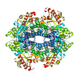
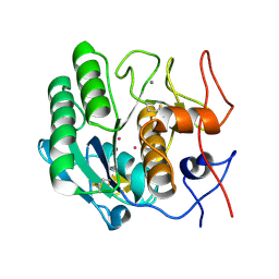
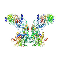
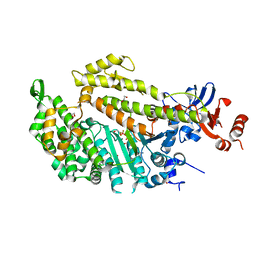
3MD2
 | |
2OJ0
 | | Crystal structure of the duplex form of the HIV-1(LAI) RNA dimerization initiation site MN soaked | | 分子名称: | 5'-R(*CP*UP*UP*GP*CP*UP*GP*AP*AP*GP*CP*GP*CP*GP*CP*AP*CP*GP*GP*CP*AP*AP*G)-3', MANGANESE (II) ION, POTASSIUM ION, ... | | 著者 | Ennifar, E, Walter, P, Dumas, P. | | 登録日 | 2007-01-12 | | 公開日 | 2007-12-25 | | 最終更新日 | 2023-12-27 | | 実験手法 | X-RAY DIFFRACTION (2.6 Å) | | 主引用文献 | Cation-dependent cleavage of the duplex form of the subtype-B HIV-1 RNA dimerization initiation site.
Nucleic Acids Res., 38, 2010
|
|
4P3C
 | | MT1-MMP:Fab complex (Form I) | | 分子名称: | 1,2-ETHANEDIOL, 4-(2-HYDROXYETHYL)-1-PIPERAZINE ETHANESULFONIC ACID, ACETATE ION, ... | | 著者 | Rozenberg, H, Udi, Y, Sagi, I. | | 登録日 | 2014-03-06 | | 公開日 | 2014-12-17 | | 最終更新日 | 2024-10-30 | | 実験手法 | X-RAY DIFFRACTION (1.943 Å) | | 主引用文献 | Inhibition mechanism of membrane metalloprotease by an exosite-swiveling conformational antibody.
Structure, 23, 2015
|
|
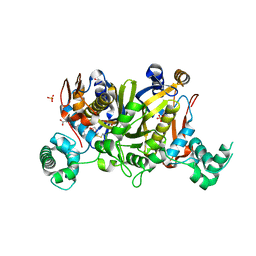
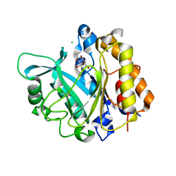
3MDX
 | |
1W3H
 | | The 3-dimensional structure of a thermostable mutant of a xylanase (Xyn10A) from Cellvibrio japonicus | | 分子名称: | 1,2-ETHANEDIOL, CALCIUM ION, ENDO-1,4-BETA-XYLANASE A PRECURSOR | | 著者 | Andrews, S, Taylor, E.J, Pell, G.N, Vincent, F, Ducros, V.M.A, Davies, G.J, Lakey, J.H, Glbert, H.J. | | 登録日 | 2004-07-15 | | 公開日 | 2004-09-30 | | 最終更新日 | 2024-11-13 | | 実験手法 | X-RAY DIFFRACTION (1.5 Å) | | 主引用文献 | The Use of Forced Protein Evolution to Investigate and Improve Stability of Family 10 Xylanases: The Production of Ca2+-Independent Stable Xylanases
J.Biol.Chem., 279, 2004
|
|
1HXA
 | | CRYSTAL STRUCTURE OF TEAS W273S FORM 2 | | 分子名称: | 1-HYDROXY-3,7,11-TRIMETHYLDODECA-2,6,10-TRIENE PHOSPHONIC ACID, 5-EPI-ARISTOLOCHENE SYNTHASE, MAGNESIUM ION | | 著者 | Starks, C.S, Rising, K.A, Chappell, J, Noel, J.P. | | 登録日 | 2001-01-12 | | 公開日 | 2003-06-24 | | 最終更新日 | 2023-08-09 | | 実験手法 | X-RAY DIFFRACTION (2.32 Å) | | 主引用文献 | Single Active Site Mutations Change the Specificity of a Sesquiterpene Cyclase
To be Published
|
|
1W9J
 | | Myosin II Dictyostelium discoideum motor domain S456Y bound with MgADP-AlF4 | | 分子名称: | 1,2-ETHANEDIOL, ADENOSINE-5'-DIPHOSPHATE, MAGNESIUM ION, ... | | 著者 | Morris, C.A, Coureux, P.-D, Wells, A.L, Houdusse, A, Sweeney, H.L. | | 登録日 | 2004-10-13 | | 公開日 | 2006-03-16 | | 最終更新日 | 2023-12-13 | | 実験手法 | X-RAY DIFFRACTION (2 Å) | | 主引用文献 | Structure-Function Analysis of Myosin II Backdoor Mutants
To be Published
|
|


