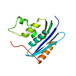
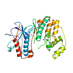
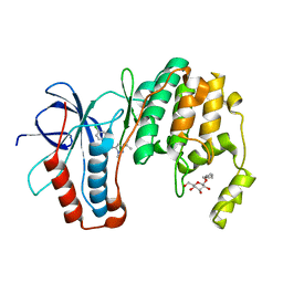
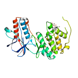
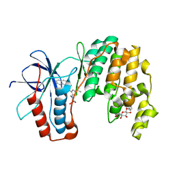
4Q0J
 
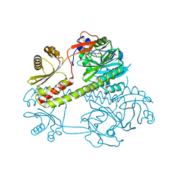 | | Deinococcus radiodurans BphP photosensory module | | 分子名称: | 3-[2-[(Z)-[3-(2-carboxyethyl)-5-[(Z)-(4-ethenyl-3-methyl-5-oxidanylidene-pyrrol-2-ylidene)methyl]-4-methyl-pyrrol-1-ium-2-ylidene]methyl]-5-[(Z)-[(3E)-3-ethylidene-4-methyl-5-oxidanylidene-pyrrolidin-2-ylidene]methyl]-4-methyl-1H-pyrrol-3-yl]propanoic acid, Bacteriophytochrome | | 著者 | Burgie, E.S, Vierstra, R.D. | | 登録日 | 2014-04-02 | | 公開日 | 2014-07-16 | | 最終更新日 | 2014-09-17 | | 実験手法 | X-RAY DIFFRACTION (2.747 Å) | | 主引用文献 | Crystallographic and Electron Microscopic Analyses of a Bacterial Phytochrome Reveal Local and Global Rearrangements during Photoconversion.
J.Biol.Chem., 289, 2014
|
|
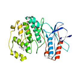
1KVA
 | | E. COLI RIBONUCLEASE HI D134A MUTANT | | 分子名称: | RIBONUCLEASE H | | 著者 | Kashiwagi, T, Jeanteur, D, Haruki, M, Katayanagi, K, Kanaya, S, Morikawa, K. | | 登録日 | 1996-10-04 | | 公開日 | 1997-03-12 | | 最終更新日 | 2021-11-03 | | 実験手法 | X-RAY DIFFRACTION (1.8 Å) | | 主引用文献 | Proposal for new catalytic roles for two invariant residues in Escherichia coli ribonuclease HI.
Protein Eng., 9, 1996
|
|
3NNV
 | | Crystal structure of P38 alpha in complex with DP437 | | 分子名称: | 1-{3-tert-butyl-1-[4-(hydroxymethyl)phenyl]-1H-pyrazol-5-yl}-3-naphthalen-1-ylurea, Mitogen-activated protein kinase 14 | | 著者 | Abendroth, J. | | 登録日 | 2010-06-24 | | 公開日 | 2010-09-15 | | 最終更新日 | 2023-12-27 | | 実験手法 | X-RAY DIFFRACTION (2.1 Å) | | 主引用文献 | Switch control pocket inhibitors of p38-MAP kinase. Durable type II inhibitors that do not require binding into the canonical ATP hinge region
Bioorg.Med.Chem.Lett., 20, 2010
|
|
5Z7B
 | |
1KVC
 | | E. COLI RIBONUCLEASE HI D134N MUTANT | | 分子名称: | RIBONUCLEASE H | | 著者 | Kashiwagi, T, Jeanteur, D, Haruki, M, Katayanagi, K, Kanaya, S, Morikawa, K. | | 登録日 | 1996-10-04 | | 公開日 | 1997-03-12 | | 最終更新日 | 2024-02-14 | | 実験手法 | X-RAY DIFFRACTION (1.9 Å) | | 主引用文献 | Proposal for new catalytic roles for two invariant residues in Escherichia coli ribonuclease HI.
Protein Eng., 9, 1996
|
|
3OD6
 | | Crystal structure of p38alpha Y323T active mutant | | 分子名称: | Mitogen-activated protein kinase 14, octyl beta-D-glucopyranoside | | 著者 | Livnah, O, Tzarum, N. | | 登録日 | 2010-08-11 | | 公開日 | 2011-01-12 | | 最終更新日 | 2024-02-21 | | 実験手法 | X-RAY DIFFRACTION (2.682 Å) | | 主引用文献 | Active mutants of the TCR-mediated p38alpha alternative activation site show changes in the phosphorylation lip and DEF site formation.
J.Mol.Biol., 405, 2011
|
|
4M38
 | |
5EYA
 | |
3FI4
 | | P38 kinase crystal structure in complex with RO4499 | | 分子名称: | (2S)-1-{[3-(2-chlorophenyl)-6-(2,4-difluorophenoxy)-1H-pyrazolo[3,4-d]pyrimidin-4-yl]amino}propan-2-ol, Mitogen-activated protein kinase 14 | | 著者 | Kuglstatter, A, Knapp, M, Dunten, P. | | 登録日 | 2008-12-10 | | 公開日 | 2009-12-22 | | 最終更新日 | 2023-09-06 | | 実験手法 | X-RAY DIFFRACTION (2.2 Å) | | 主引用文献 | Mapping Binding Pocket Volume: Potential Applications towards Ligand Design and Selectivity
To be Published
|
|
2LA6
 | | Solution NMR Structure of RRM domain of RNA-binding protein FUS from homo sapiens, Northeast Structural Genomics Consortium Target HR6430A | | 分子名称: | RNA-binding protein FUS | | 著者 | Liu, G, Xiao, R, Janjua, H, Ciccosanti, C, Wang, H, Lee, H, Acton, T.B, Everett, J.K, Huang, Y.J, Montelione, G.T, Northeast Structural Genomics Consortium (NESG) | | 登録日 | 2011-03-04 | | 公開日 | 2011-05-04 | | 最終更新日 | 2024-05-15 | | 実験手法 | SOLUTION NMR | | 主引用文献 | Northeast Structural Genomics Consortium Target HR6430A
To be Published
|
|
3FMN
 | |
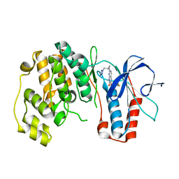
4QGX
 | | Crystal structure of the R132K:R111L:L121E mutant of Cellular Retinoic Acid Binding ProteinII complexed with a synthetic ligand (Merocyanine) at 1.47 angstrom resolution | | 分子名称: | (2E,4E,6E)-3-methyl-6-(1,3,3-trimethyl-1,3-dihydro-2H-indol-2-ylidene)hexa-2,4-dienal, Cellular retinoic acid-binding protein 2 | | 著者 | Nosrati, M, Yapici, I, Geiger, J.H. | | 登録日 | 2014-05-26 | | 公開日 | 2015-01-28 | | 最終更新日 | 2023-09-20 | | 実験手法 | X-RAY DIFFRACTION (1.471 Å) | | 主引用文献 | "Turn-on" protein fluorescence: in situ formation of cyanine dyes.
J.Am.Chem.Soc., 137, 2015
|
|
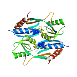
1G1U
 | | THE 2.5 ANGSTROM RESOLUTION CRYSTAL STRUCTURE OF THE RXRALPHA LIGAND BINDING DOMAIN IN TETRAMER IN THE ABSENCE OF LIGAND | | 分子名称: | RETINOIC ACID RECEPTOR RXR-ALPHA | | 著者 | Gampe Jr, R.T, Montana, V.G, Lambert, M.H, Wisely, G.B, Milburn, M.V, Xu, H.E. | | 登録日 | 2000-10-13 | | 公開日 | 2001-04-25 | | 最終更新日 | 2017-10-04 | | 実験手法 | X-RAY DIFFRACTION (2.5 Å) | | 主引用文献 | Structural basis for autorepression of retinoid X receptor by tetramer formation and the AF-2 helix.
Genes Dev., 14, 2000
|
|
4QFS
 | | Structure of AMPK in complex with Br2-A769662core activator and STAUROSPORINE inhibitor | | 分子名称: | 2-bromo-3-(4-bromophenyl)-4-hydroxy-6-oxo-6,7-dihydrothieno[2,3-b]pyridine-5-carbonitrile, 5'-AMP-activated protein kinase catalytic subunit alpha-1, 5'-AMP-activated protein kinase subunit beta-1, ... | | 著者 | Calabrese, M.F, Kurumbail, R.G. | | 登録日 | 2014-05-21 | | 公開日 | 2014-08-06 | | 最終更新日 | 2017-11-22 | | 実験手法 | X-RAY DIFFRACTION (3.55 Å) | | 主引用文献 | Structural Basis for AMPK Activation: Natural and Synthetic Ligands Regulate Kinase Activity from Opposite Poles by Different Molecular Mechanisms.
Structure, 22, 2014
|
|
3O8U
 | |
3FMK
 | |
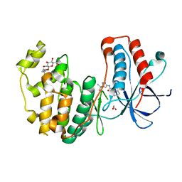
4QGV
 | | Crystal structure of the R132K:R111L mutant of Cellular Retinoic Acid Binding ProteinII complexed with a synthetic ligand (Merocyanine) at 1.73 angstrom resolution. | | 分子名称: | (2E,4E,6E)-3-methyl-6-(1,3,3-trimethyl-1,3-dihydro-2H-indol-2-ylidene)hexa-2,4-dienal, Cellular retinoic acid-binding protein 2 | | 著者 | Nosrati, M, Yapici, I, Geiger, J.H. | | 登録日 | 2014-05-25 | | 公開日 | 2015-01-28 | | 最終更新日 | 2023-09-20 | | 実験手法 | X-RAY DIFFRACTION (1.73 Å) | | 主引用文献 | "Turn-on" protein fluorescence: in situ formation of cyanine dyes.
J.Am.Chem.Soc., 137, 2015
|
|
4QH7
 | | LC8 - Ana2 (159-168) Complex | | 分子名称: | Anastral spindle 2, Dynein light chain 1, cytoplasmic | | 著者 | Slevin, L.K, Romes, E.R, Slep, K.C. | | 登録日 | 2014-05-27 | | 公開日 | 2014-06-11 | | 最終更新日 | 2023-09-20 | | 実験手法 | X-RAY DIFFRACTION (1.829 Å) | | 主引用文献 | The Mechanism of Dynein Light Chain LC8-mediated Oligomerization of the Ana2 Centriole Duplication Factor.
J.Biol.Chem., 289, 2014
|
|
3O8P
 | | Conformational plasticity of p38 MAP kinase DFG motif mutants in response to inhibitor binding | | 分子名称: | 1,2-ETHANEDIOL, 1-(5-TERT-BUTYL-2-METHYL-2H-PYRAZOL-3-YL)-3-(4-CHLORO-PHENYL)-UREA, Mitogen-activated protein kinase 14, ... | | 著者 | Namboodiri, H.V, Karpusas, M, Bukhtiyarova, M, Springman, E.B. | | 登録日 | 2010-08-03 | | 公開日 | 2010-09-01 | | 最終更新日 | 2023-09-06 | | 実験手法 | X-RAY DIFFRACTION (2.1 Å) | | 主引用文献 | Conformational plasticity of p38 MAP kinase DFG motif mutants in response to inhibitor binding
To be Published
|
|
3ODY
 | | Crystal structure of p38alpha Y323Q active mutant | | 分子名称: | Mitogen-activated protein kinase 14, octyl beta-D-glucopyranoside | | 著者 | Livnah, O, Tzarum, N. | | 登録日 | 2010-08-12 | | 公開日 | 2011-01-12 | | 最終更新日 | 2024-02-21 | | 実験手法 | X-RAY DIFFRACTION (2.2 Å) | | 主引用文献 | Active mutants of the TCR-mediated p38alpha alternative activation site show changes in the phosphorylation lip and DEF site formation.
J.Mol.Biol., 405, 2011
|
|
5ZZO
 | | Crystal structure of CcpE regulatory domain in complex with citrate from Staphyloccocus aureus | | 分子名称: | CITRATE ANION, LysR family transcriptional regulator | | 著者 | Chen, J, Wang, L, Shang, F, Xu, Y. | | 登録日 | 2018-06-04 | | 公開日 | 2018-06-20 | | 最終更新日 | 2023-11-22 | | 実験手法 | X-RAY DIFFRACTION (2.5 Å) | | 主引用文献 | Structural and Biochemical Analysis of the Citrate-Responsive Mechanism of the Regulatory Domain of Catabolite Control Protein E from Staphylococcus aureus
Biochemistry, 57, 2018
|
|
4QST
 | | Structure of the bromodomain of human ATPase family AAA domain-containing protein 2 (ATAD2) in complex with 1-methylquinolin 2-one | | 分子名称: | 1,2-ETHANEDIOL, 1-METHYLQUINOLIN-2(1H)-ONE, ATPase family AAA domain-containing protein 2, ... | | 著者 | Chaikuad, A, Felletar, I, von Delft, F, Arrowsmith, C.H, Edwards, A.M, Bountra, C, Knapp, S, Structural Genomics Consortium (SGC) | | 登録日 | 2014-07-06 | | 公開日 | 2014-07-23 | | 最終更新日 | 2023-09-20 | | 実験手法 | X-RAY DIFFRACTION (2.05 Å) | | 主引用文献 | Structure-based approaches towards identification of fragments for the low-druggability ATAD2 bromodomain
MedChemComm, 5, 2014
|
|
3S4Q
 | | P38 alpha kinase complexed with a pyrazolo-triazine based inhibitor | | 分子名称: | 3-[(6-benzoyl-5-methylpyrrolo[2,1-f][1,2,4]triazin-4-yl)amino]-N-cyclopropyl-4-methylbenzamide, Mitogen-activated protein kinase 14 | | 著者 | Sack, J.S. | | 登録日 | 2011-05-20 | | 公開日 | 2012-02-01 | | 最終更新日 | 2024-02-28 | | 実験手法 | X-RAY DIFFRACTION (2.27 Å) | | 主引用文献 | Discovery of pyrrolo[2,1-f][1,2,4]triazine C6-ketones as potent, orally active p38α MAP kinase inhibitors.
Bioorg.Med.Chem.Lett., 21, 2011
|
|
3P7A
 | | p38 inhibitor-bound | | 分子名称: | 1-[5-tert-butyl-2-(1,1-dioxidothiomorpholin-4-yl)thiophen-3-yl]-3-naphthalen-1-ylurea, Mitogen-activated protein kinase 14, octyl beta-D-glucopyranoside | | 著者 | Moffett, K.K, Namboodiri, H. | | 登録日 | 2010-10-12 | | 公開日 | 2011-10-12 | | 最終更新日 | 2024-02-21 | | 実験手法 | X-RAY DIFFRACTION (2.31 Å) | | 主引用文献 | Discovery of a novel class of non-ATP site DFG-out state p38 inhibitors utilizing computationally assisted virtual fragment-based drug design (vFBDD).
Bioorg.Med.Chem.Lett., 21, 2011
|
|
4R9P
 | | An Expansion to the Smad MH2-family: The structure of the N-MH2 expanded domain | | 分子名称: | RE28239p | | 著者 | Beich-Frandsen, M, Aragon, E, Llimargas, M, Benach, J, Riera, A, Macias, M.J. | | 登録日 | 2014-09-06 | | 公開日 | 2015-04-08 | | 最終更新日 | 2024-02-28 | | 実験手法 | X-RAY DIFFRACTION (1.592 Å) | | 主引用文献 | Structure of the N-terminal domain of the protein Expansion: an 'Expansion' to the Smad MH2
Acta Crystallogr.,Sect.D, 71, 2015
|
|


