Loading
PDBj
メニューPDBj@FacebookPDBj@TwitterPDBj@YouTubewwPDB FoundationwwPDB
RCSB PDBPDBeBMRBAdv. SearchSearch help

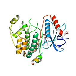
4FMQ
DownloadVisualize
BU of 4fmq by Molmil
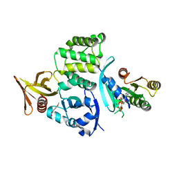
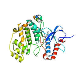
Crystal structure of human ERK2 complexed with a MAPK docking peptide
分子名称: MAPK DOCKING PEPTIDE, Mitogen-activated protein kinase 1, PHOSPHOAMINOPHOSPHONIC ACID-ADENYLATE ESTER
著者Gogl, G, Toeroe, I, Remenyi, A.
登録日2012-06-18
公開日2012-10-31
最終更新日2023-09-13
実験手法X-RAY DIFFRACTION (2.098 Å)
主引用文献Specificity of linear motifs that bind to a common mitogen-activated protein kinase docking groove.
Sci.Signal., 5, 2012
3TEI
DownloadVisualize
BU of 3tei by Molmil
Crystal structure of human ERK2 complexed with a MAPK docking peptide
分子名称: Mitogen-activated protein kinase 1, PHOSPHOAMINOPHOSPHONIC ACID-ADENYLATE ESTER, Ribosomal protein S6 kinase alpha-1
著者Gogl, G, Remenyi, A.
登録日2011-08-15
公開日2012-08-15
最終更新日2023-09-13
実験手法X-RAY DIFFRACTION (2.404 Å)
主引用文献Specificity of linear motifs that bind to a common mitogen-activated protein kinase docking groove.
Sci.Signal., 5, 2012
3PGX
DownloadVisualize
BU of 3pgx by Molmil
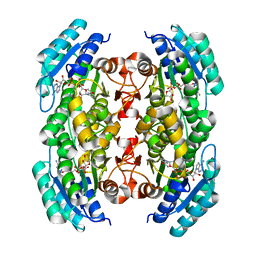
Crystal structure of a putative carveol dehydrogenase from Mycobacterium paratuberculosis bound to nicotinamide adenine dinucleotide
分子名称: NICOTINAMIDE-ADENINE-DINUCLEOTIDE, carveol dehydrogenase
著者Seattle Structural Genomics Center for Infectious Disease (SSGCID)
登録日2010-11-02
公開日2010-11-24
最終更新日2023-09-06
実験手法X-RAY DIFFRACTION (1.85 Å)
主引用文献Mycofactocin-associated mycobacterial dehydrogenases with non-exchangeable NAD cofactors.
Sci Rep, 7, 2017
3PXX
DownloadVisualize
BU of 3pxx by Molmil
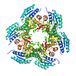
Crystal structure of carveol dehydrogenase from Mycobacterium avium bound to nicotinamide adenine dinucleotide
分子名称: Carveol dehydrogenase, NICOTINAMIDE-ADENINE-DINUCLEOTIDE
著者Seattle Structural Genomics Center for Infectious Disease (SSGCID)
登録日2010-12-10
公開日2010-12-29
最終更新日2024-02-21
実験手法X-RAY DIFFRACTION (2 Å)
主引用文献Mycofactocin-associated mycobacterial dehydrogenases with non-exchangeable NAD cofactors.
Sci Rep, 7, 2017
1BR3
DownloadVisualize
BU of 1br3 by Molmil
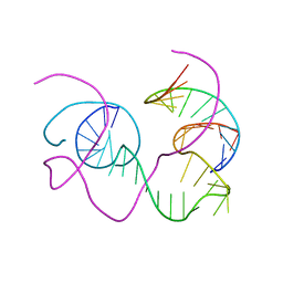
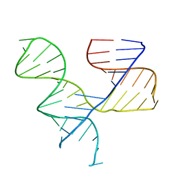
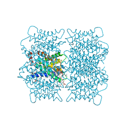
CRYSTAL STRUCTURE OF AN 82-NUCLEOTIDE RNA-DNA COMPLEX FORMED BY THE 10-23 DNA ENZYME
分子名称: DNA (10-23 DNA ENZYME), RNA (5'-R(*GP*GP*AP*CP*AP*GP*AP*UP*GP*GP*GP*AP*G)-3')
著者Nowakowski, J, Shim, P.J, Prasad, G.S, Stout, C.D, Joyce, G.F.
登録日1998-08-13
公開日1999-02-23
最終更新日2024-02-07
実験手法X-RAY DIFFRACTION (3 Å)
主引用文献Crystal structure of an 82-nucleotide RNA-DNA complex formed by the 10-23 DNA enzyme.
Nat.Struct.Biol., 6, 1999
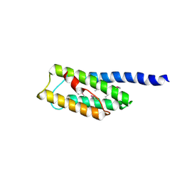
2MC3
DownloadVisualize
BU of 2mc3 by Molmil
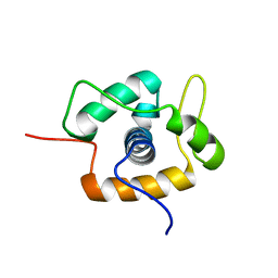
NMR solution structure of the winged-helix domain from MUS81 structure-specific endonuclease
分子名称: MUS81 endonuclease homolog (Yeast), isoform CRA_b
著者Harris, R, Fadden, A, Mcdonald, N.Q.
登録日2013-08-14
公開日2013-09-18
最終更新日2024-05-15
実験手法SOLUTION NMR
主引用文献A winged helix domain in human MUS81 binds DNA and modulates the endonuclease activity of MUS81 complexes.
Nucleic Acids Res., 41, 2013
7KTU
DownloadVisualize
BU of 7ktu by Molmil
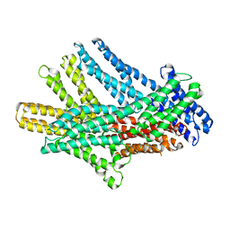
Cryogenic electron microscopy model of full-length human metavinculin H1'-parallel conformation 1
分子名称: metavinculin
著者Izard, T, Rangarajan, E.S.
登録日2020-11-24
公開日2021-01-27
最終更新日2024-03-06
実験手法ELECTRON MICROSCOPY (4.15 Å)
主引用文献The Cryogenic Electron Microscopy Structure of the Cell Adhesion Regulator Metavinculin Reveals an Isoform-Specific Kinked Helix in Its Cytoskeleton Binding Domain.
Int J Mol Sci, 22, 2021
7KTV
DownloadVisualize
BU of 7ktv by Molmil
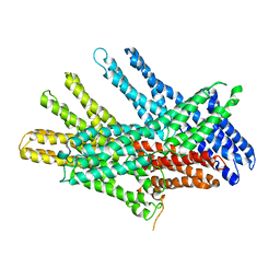
Cryogenic electron microscopy model of full-length human metavinculin H1' kinked conformation
分子名称: metavinculin
著者Izard, T, Rangarajan, E.S.
登録日2020-11-24
公開日2021-01-27
最終更新日2024-03-06
実験手法ELECTRON MICROSCOPY (4.5 Å)
主引用文献The Cryogenic Electron Microscopy Structure of the Cell Adhesion Regulator Metavinculin Reveals an Isoform-Specific Kinked Helix in Its Cytoskeleton Binding Domain.
Int J Mol Sci, 22, 2021
7KTT
DownloadVisualize
BU of 7ktt by Molmil
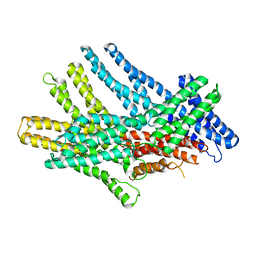
Cryogenic electron microscopy model of full-length human metavinculin
分子名称: metavinculin
著者Izard, T, Rangarajan, E.S.
登録日2020-11-24
公開日2021-01-27
最終更新日2024-03-06
実験手法ELECTRON MICROSCOPY (4.17 Å)
主引用文献The Cryogenic Electron Microscopy Structure of the Cell Adhesion Regulator Metavinculin Reveals an Isoform-Specific Kinked Helix in Its Cytoskeleton Binding Domain.
Int J Mol Sci, 22, 2021
7KTW
DownloadVisualize
BU of 7ktw by Molmil
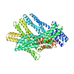
Cryogenic electron microscopy model of full-length human metavinculin H1'-parallel conformation 2
分子名称: metavinculin
著者Izard, T, Rangarajan, E.S.
登録日2020-11-24
公開日2021-01-27
最終更新日2024-03-06
実験手法ELECTRON MICROSCOPY (4.27 Å)
主引用文献The Cryogenic Electron Microscopy Structure of the Cell Adhesion Regulator Metavinculin Reveals an Isoform-Specific Kinked Helix in Its Cytoskeleton Binding Domain.
Int J Mol Sci, 22, 2021
6UZK
DownloadVisualize
BU of 6uzk by Molmil
Crystal Structure of the Ternary Complex of KRIT1 bound to both the Rap1 GTPase and HKi6
分子名称: 2-hydroxy-6-methoxynaphthalene-1-carbaldehyde, Krev interaction trapped protein 1, MAGNESIUM ION, ...
著者Gingras, A.R.
登録日2019-11-15
公開日2020-11-18
最終更新日2023-10-11
実験手法X-RAY DIFFRACTION (1.924 Å)
主引用文献Inhibition of the HEG1-KRIT1 interaction increases KLF4 and KLF2 expression in endothelial cells.
Faseb Bioadv, 3, 2021
1KH6
DownloadVisualize
BU of 1kh6 by Molmil
Crystal Structure of an RNA Tertiary Domain Essential to HCV IRES-mediated Translation Initiation.
分子名称: JIIIabc
著者Kieft, J.S, Zhou, K, Grech, A, Jubin, R, Doudna, J.A.
登録日2001-11-29
公開日2002-04-26
最終更新日2024-02-14
実験手法X-RAY DIFFRACTION (2.9 Å)
主引用文献Crystal structure of an RNA tertiary domain essential to HCV IRES-mediated translation initiation.
Nat.Struct.Biol., 9, 2002
1ELG
DownloadVisualize
BU of 1elg by Molmil
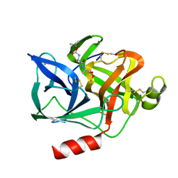
NATURE OF THE INACTIVATION OF ELASTASE BY N-PEPTIDYL-O-AROYL HYDROXYLAMINE AS A FUNCTION OF PH
分子名称: (TERT-BUTYLOXYCARBONYL)-ALANYL-ALANYL-AMINE, CALCIUM ION, PORCINE PANCREATIC ELASTASE
著者Ding, X, Rasmussen, B, Demuth, H.-U, Ringe, D, Steinmetz, A.C.U.
登録日1995-03-13
公開日1995-07-10
最終更新日2024-06-05
実験手法X-RAY DIFFRACTION (1.65 Å)
主引用文献Nature of the inactivation of elastase by N-peptidyl-O-aroyl hydroxylamine as a function of pH.
Biochemistry, 34, 1995
4GJ1
DownloadVisualize
BU of 4gj1 by Molmil
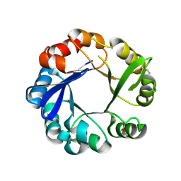
Crystal structure of 1-(5-phosphoribosyl)-5-[(5-phosphoribosylamino)methylideneamino] imidazole-4-carboxamide isomerase (hisA).
分子名称: 1-(5-phosphoribosyl)-5-[(5-phosphoribosylamino)methylideneamino] imidazole-4-carboxamide isomerase
著者Nocek, B, Gu, M, Kwon, K, Anderson, W.F, Joachimiak, A, CSGID, Center for Structural Genomics of Infectious Diseases (CSGID)
登録日2012-08-09
公開日2012-08-22
最終更新日2018-01-24
実験手法X-RAY DIFFRACTION (2.152 Å)
主引用文献Crystal structure of 1-(5-phosphoribosyl)-5-[(5-phosphoribosylamino)methylideneamino] imidazole-4-carboxamide isomerase (hisA).
To be Published
3N1B
DownloadVisualize
BU of 3n1b by Molmil
C-terminal domain of Vps54 subunit of the GARP complex
分子名称: Vacuolar protein sorting-associated protein 54
著者Perez-Victoria, F.J, Abascal-Palacios, G, Tascon, I, Kajava, A, Pioro, E.P, Bonifacino, J.S, Hierro, A.
登録日2010-05-15
公開日2010-07-14
最終更新日2017-11-08
実験手法X-RAY DIFFRACTION (2.398 Å)
主引用文献Structural basis for the wobbler mouse neurodegenerative disorder caused by mutation in the Vps54 subunit of the GARP complex.
Proc.Natl.Acad.Sci.USA, 107, 2010
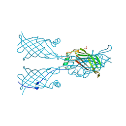
5W32
DownloadVisualize
BU of 5w32 by Molmil
Crystal structure of mutant CJ YCEI protein (CJ-N48C) with selenocysteine guest structure
分子名称: Putative periplasmic protein, SELENOCYSTEINE, SULFATE ION, ...
著者Huber, T.R, Snow, C.D.
登録日2017-06-07
公開日2018-01-03
最終更新日2023-10-04
実験手法X-RAY DIFFRACTION (2.6 Å)
主引用文献Installing Guest Molecules at Specific Sites within Scaffold Protein Crystals.
Bioconjug. Chem., 29, 2018
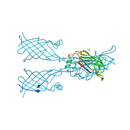
5W2Z
DownloadVisualize
BU of 5w2z by Molmil
Crystal structure of mutant CJ YCEI protein (CJ-N48C) with 5-mercapto-2-nitrobenzoic acid guest structure
分子名称: 5-MERCAPTO-2-NITRO-BENZOIC ACID, Putative periplasmic protein, SULFATE ION, ...
著者Huber, T.R, Snow, C.D.
登録日2017-06-07
公開日2018-01-03
最終更新日2023-10-04
実験手法X-RAY DIFFRACTION (2.8 Å)
主引用文献Installing Guest Molecules at Specific Sites within Scaffold Protein Crystals.
Bioconjug. Chem., 29, 2018
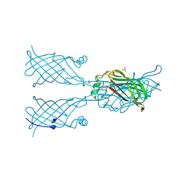
5W31
DownloadVisualize
BU of 5w31 by Molmil
Crystal structure of mutant CJ YCEI protein (CJ-N48C) with mercuribenzoic acid guest structure
分子名称: MERCURIBENZOIC ACID, Putative periplasmic protein, SULFATE ION, ...
著者Huber, T.R, Snow, C.D.
登録日2017-06-07
公開日2018-01-03
最終更新日2023-10-04
実験手法X-RAY DIFFRACTION (2.56 Å)
主引用文献Installing Guest Molecules at Specific Sites within Scaffold Protein Crystals.
Bioconjug. Chem., 29, 2018
8I4O
DownloadVisualize
BU of 8i4o by Molmil
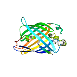
Design of a split green fluorescent protein for sensing and tracking an beta-amyloid
分子名称: Beta-amyloid, Split Green flourescent protein
著者Taegeun, Y, Jinsu, L, Jungmin, Y, Jungmin, C, Wondo, H, Song, J.J, Haksung, K.
登録日2023-01-20
公開日2023-11-29
最終更新日2023-12-13
実験手法X-RAY DIFFRACTION (3.1 Å)
主引用文献Engineering of a Fluorescent Protein for a Sensing of an Intrinsically Disordered Protein through Transition in the Chromophore State.
Jacs Au, 3, 2023
2H3L
DownloadVisualize
BU of 2h3l by Molmil
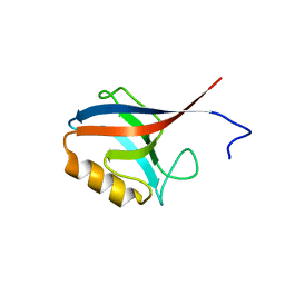
Crystal Structure of ERBIN PDZ
分子名称: LAP2 protein
著者Appleton, B.A, Zhang, Y, Wu, P, Yin, J.P, Hunziker, W, Skelton, N.J, Sidhu, S.S, Wiesmann, C.
登録日2006-05-22
公開日2006-06-13
最終更新日2023-08-30
実験手法X-RAY DIFFRACTION (1 Å)
主引用文献Comparative structural analysis of the Erbin PDZ domain and the first PDZ domain of ZO-1. Insights into determinants of PDZ domain specificity.
J.Biol.Chem., 281, 2006
2TMA
DownloadVisualize
BU of 2tma by Molmil
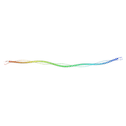
TROPOMYOSIN CRYSTAL STRUCTURE AND MUSCLE REGULATION. APPENDIX. CONSTRUCTION OF AN ATOMIC MODEL FOR TROPOMYOSIN AND IMPLICATIONS FOR INTERACTIONS WITH ACTIN
分子名称: TROPOMYOSIN
著者Phillips Jr, G.N, Cohen, C.
登録日1987-09-16
公開日1987-10-16
最終更新日2024-02-21
実験手法X-RAY DIFFRACTION (15 Å)
主引用文献Construction of an atomic model for tropomyosin and implications for interactions with actin.
J.Mol.Biol., 192, 1986
6OV2
DownloadVisualize
BU of 6ov2 by Molmil
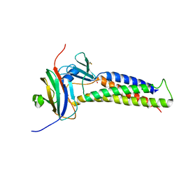
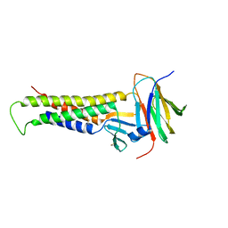
Crystal structure of human claudin-9 in complex with Clostridium perfringens entertoxin C-terminal domain in closed form
分子名称: Claudin-9, GLYCEROL, Heat-labile enterotoxin B chain
著者Vecchio, A.J, Stroud, R.M.
登録日2019-05-06
公開日2019-09-04
最終更新日2023-10-11
実験手法X-RAY DIFFRACTION (3.2 Å)
主引用文献Claudin-9 structures reveal mechanism for toxin-induced gut barrier breakdown.
Proc.Natl.Acad.Sci.USA, 116, 2019
2B6O
DownloadVisualize
BU of 2b6o by Molmil
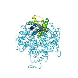
Electron crystallographic structure of lens Aquaporin-0 (AQP0) (lens MIP) at 1.9A resolution, in a closed pore state
分子名称: 1,2-DIMYRISTOYL-RAC-GLYCERO-3-PHOSPHOCHOLINE, Lens fiber major intrinsic protein
著者Gonen, T, Cheng, Y, Sliz, P, Hiroaki, Y, Fujiyoshi, Y, Harrison, S.C, Walz, T.
登録日2005-10-03
公開日2005-12-06
最終更新日2023-08-23
実験手法ELECTRON CRYSTALLOGRAPHY (1.9 Å)
主引用文献Lipid-protein interactions in double-layered two-dimensional AQP0 crystals.
Nature, 438, 2005
6OV3
DownloadVisualize
BU of 6ov3 by Molmil
Crystal structure of human claudin-9 in complex with Clostridium perfringens entertoxin C-terminal domain in open form
分子名称: Claudin-9, Heat-labile enterotoxin B chain
著者Vecchio, A.J, Stroud, R.M.
登録日2019-05-06
公開日2019-09-04
最終更新日2023-10-11
実験手法X-RAY DIFFRACTION (3.25 Å)
主引用文献Claudin-9 structures reveal mechanism for toxin-induced gut barrier breakdown.
Proc.Natl.Acad.Sci.USA, 116, 2019
2B6P
DownloadVisualize
BU of 2b6p by Molmil
X-ray structure of lens Aquaporin-0 (AQP0) (lens MIP) in an open pore state
分子名称: Lens fiber major intrinsic protein
著者Gonen, T, Cheng, Y, Sliz, P, Hiroaki, Y, Fujiyoshi, Y, Harrison, S.C, Walz, T.
登録日2005-10-03
公開日2005-12-06
最終更新日2024-02-14
実験手法X-RAY DIFFRACTION (2.4 Å)
主引用文献Lipid-protein interactions in double-layered two-dimensional AQP0 crystals.
Nature, 438, 2005

222624

件を2024-07-17に公開中

PDB statisticsPDBj update infoContact PDBjnumon