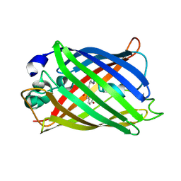
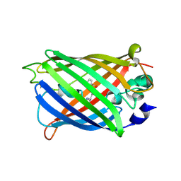
5M1Q
 
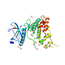
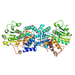
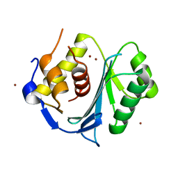 | | Crystal structure of the large terminase nuclease from thermophilic phage G20c with bound Zinc | | 分子名称: | Phage terminase large subunit, ZINC ION | | 著者 | Xu, R.G, Jenkins, H.T, Chechik, M, Blagova, E.V, Greive, S.J, Antson, A.A. | | 登録日 | 2016-10-09 | | 公開日 | 2016-10-26 | | 最終更新日 | 2024-05-01 | | 実験手法 | X-RAY DIFFRACTION (1.45 Å) | | 主引用文献 | Viral genome packaging terminase cleaves DNA using the canonical RuvC-like two-metal catalysis mechanism.
Nucleic Acids Res., 45, 2017
|
|
7A7W
 | |
6RNQ
 | |
7A8J
 | |
7A7Z
 | |
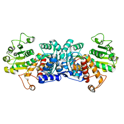
7AAJ
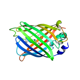
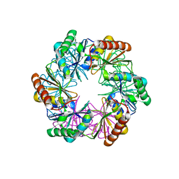
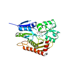 | | Human porphobilinogen deaminase in complex with cofactor | | 分子名称: | 3-[5-{[3-(2-carboxyethyl)-4-(carboxymethyl)-5-methyl-1H-pyrrol-2-yl]methyl}-4-(carboxymethyl)-1H-pyrrol-3-yl]propanoic acid, GLYCEROL, Porphobilinogen deaminase | | 著者 | Kallio, J.P, Bustad, H.J, Martinez, A. | | 登録日 | 2020-09-04 | | 公開日 | 2021-02-17 | | 最終更新日 | 2024-01-31 | | 実験手法 | X-RAY DIFFRACTION (1.8 Å) | | 主引用文献 | Characterization of porphobilinogen deaminase mutants reveals that arginine-173 is crucial for polypyrrole elongation mechanism.
Iscience, 24, 2021
|
|
7A8L
 | |
5LRN
 | |
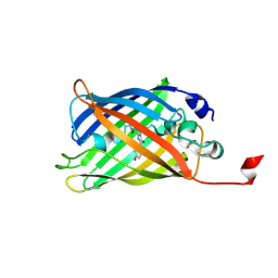
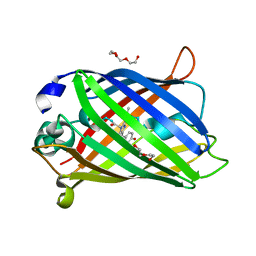
7AA1
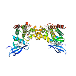
 | | Structural comparison of cellular retinoic acid binding proteins I and II in the presence and absence of natural and synthetic ligands | | 分子名称: | 4-[2-(5,5,8,8-tetramethyl-6,7-dihydroquinoxalin-2-yl)ethynyl]benzoic acid, Cellular retinoic acid-binding protein 2 | | 著者 | Tomlinson, C.W.E, Cornish, K.A.S, Pohl, E. | | 登録日 | 2020-09-02 | | 公開日 | 2021-02-17 | | 最終更新日 | 2024-01-31 | | 実験手法 | X-RAY DIFFRACTION (1.71 Å) | | 主引用文献 | Structure-functional relationship of cellular retinoic acid-binding proteins I and II interacting with natural and synthetic ligands.
Acta Crystallogr D Struct Biol, 77, 2021
|
|
7A7S
 | |
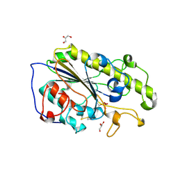
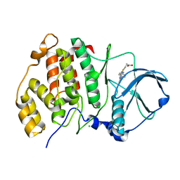
6ZN4
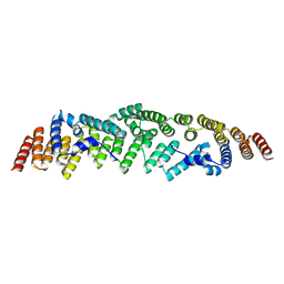
 | | MaeB malic enzyme domain apoprotein | | 分子名称: | MAGNESIUM ION, NADP-dependent malate dehydrogenase,Malate dehydrogenase | | 著者 | Lovering, A.L, Harding, C.J. | | 登録日 | 2020-07-06 | | 公開日 | 2021-02-17 | | 最終更新日 | 2024-01-31 | | 実験手法 | X-RAY DIFFRACTION (1.679 Å) | | 主引用文献 | A rotary mechanism for allostery in bacterial hybrid malic enzymes.
Nat Commun, 12, 2021
|
|
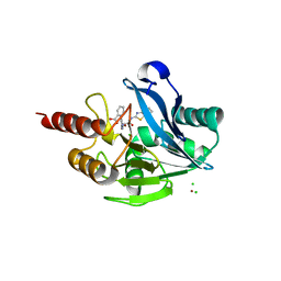
5M44
 | | Complex structure of human protein kinase CK2 catalytic subunit with a thieno[2,3-d]pyrimidin inhibitor crystallized under high-salt conditions | | 分子名称: | 3-[5-(4-methylphenyl)thieno[2,3-d]pyrimidin-4-yl]sulfanylpropanoic acid, CHLORIDE ION, Casein kinase II subunit alpha | | 著者 | Niefind, K, Bischoff, N, Yarmoluk, S.M, Bdzhola, V.G, Golub, A.G, Balanda, A.O, Prykhod'ko, A.O. | | 登録日 | 2016-10-18 | | 公開日 | 2017-01-18 | | 最終更新日 | 2024-01-17 | | 実験手法 | X-RAY DIFFRACTION (2.71 Å) | | 主引用文献 | Structural Hypervariability of the Two Human Protein Kinase CK2 Catalytic Subunit Paralogs Revealed by Complex Structures with a Flavonol- and a Thieno[2,3-d]pyrimidine-Based Inhibitor.
Pharmaceuticals, 10, 2017
|
|
7A8N
 | |
6S1B
 | | Crystal Structure of DYRK1A with small molecule inhibitor | | 分子名称: | 1,2-ETHANEDIOL, Dual specificity tyrosine-phosphorylation-regulated kinase 1A, SULFATE ION, ... | | 著者 | Sorrell, F.J, Henderson, S.H, Redondo, C, Burgess-Brown, N.A, von Delft, F, Arrowsmith, C.H, Bountra, C, Edwards, A.M, Elkins, J.M. | | 登録日 | 2019-06-18 | | 公開日 | 2019-06-26 | | 最終更新日 | 2024-01-24 | | 実験手法 | X-RAY DIFFRACTION (1.3 Å) | | 主引用文献 | Kinase Scaffold Repurposing in the Public Domain
To be published
|
|
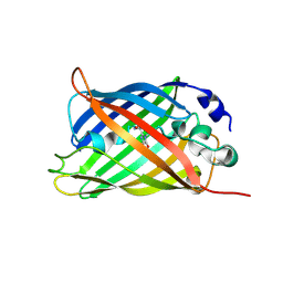
7A9Z
 | | Structural comparison of cellular retinoic acid binding protein I and II in the presence and absence of natural and synthetic ligands | | 分子名称: | 4-[2-(5,5,8,8-tetramethyl-6,7-dihydroquinoxalin-2-yl)ethynyl]benzoic acid, Cellular retinoic acid-binding protein 1 | | 著者 | Tomlinson, C.W.E, Cornish, K.A.S, Pohl, E. | | 登録日 | 2020-09-02 | | 公開日 | 2021-02-17 | | 最終更新日 | 2024-01-31 | | 実験手法 | X-RAY DIFFRACTION (2.41 Å) | | 主引用文献 | Structure-functional relationship of cellular retinoic acid-binding proteins I and II interacting with natural and synthetic ligands.
Acta Crystallogr D Struct Biol, 77, 2021
|
|
5LSC
 | | The structure of the metallo-beta-lactamase VIM-2 in complex with a triazolylthioacetamide inhibitor | | 分子名称: | 2-[5-[2-(1,3-benzothiazol-2-ylamino)-2-oxidanylidene-ethyl]sulfanyl-4~{H}-1,2,4-triazol-3-yl]benzoic acid, CHLORIDE ION, Metallo-beta-lactamase VIM-2-like protein, ... | | 著者 | Christopeit, T, Yang, K.-W, Yang, S.-K, Leiros, H.-K.S. | | 登録日 | 2016-08-25 | | 公開日 | 2016-11-09 | | 最終更新日 | 2024-01-17 | | 実験手法 | X-RAY DIFFRACTION (1.497 Å) | | 主引用文献 | The structure of the metallo-beta-lactamase VIM-2 in complex with a triazolylthioacetamide inhibitor.
Acta Crystallogr F Struct Biol Commun, 72, 2016
|
|
7A85
 | |
7A8E
 | |
6ZN7
 | | MaeB malic enzyme domain apoprotein | | 分子名称: | MAGNESIUM ION, NADP NICOTINAMIDE-ADENINE-DINUCLEOTIDE PHOSPHATE, NADP-dependent malate dehydrogenase,Malate dehydrogenase | | 著者 | Lovering, A.L, Harding, C.J. | | 登録日 | 2020-07-06 | | 公開日 | 2021-02-17 | | 最終更新日 | 2024-01-31 | | 実験手法 | X-RAY DIFFRACTION (1.67 Å) | | 主引用文献 | A rotary mechanism for allostery in bacterial hybrid malic enzymes.
Nat Commun, 12, 2021
|
|
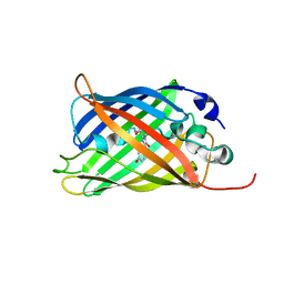
5LSR
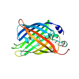
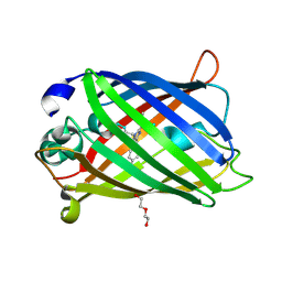
 | | Carboxysome shell protein CcmP from Synechococcus elongatus PCC 7942 | | 分子名称: | CcmP, THIOCYANATE ION | | 著者 | Larsson, A.M, Hasse, D, Valegard, K, Andersson, I. | | 登録日 | 2016-09-05 | | 公開日 | 2017-04-12 | | 最終更新日 | 2024-01-17 | | 実験手法 | X-RAY DIFFRACTION (1.65 Å) | | 主引用文献 | Crystal structures of beta-carboxysome shell protein CcmP: ligand binding correlates with the closed or open central pore.
J. Exp. Bot., 68, 2017
|
|
7A8K
 | |
6RSH
 | |
5LT4
 | |
7A80
 | |
6RSX
 | |


