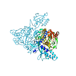
4XCX
 
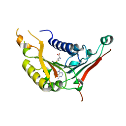 | | METHYLTRANSFERASE DOMAIN OF SMALL RNA 2'-O-METHYLTRANSFERASE | | 分子名称: | S-ADENOSYL-L-HOMOCYSTEINE, Small RNA 2'-O-methyltransferase | | 著者 | Walker, J.R, Zeng, H, Dong, A, Li, Y, Wernimont, A, Bountra, C, Arrowsmith, C.H, Edwards, A.M, Brown, P.J, Wu, H, Structural Genomics Consortium (SGC) | | 登録日 | 2014-12-18 | | 公開日 | 2015-01-14 | | 最終更新日 | 2023-09-27 | | 実験手法 | X-RAY DIFFRACTION (2.84 Å) | | 主引用文献 | Crystal structure of human C1ORF59 in complex with SAH
To be published
|
|
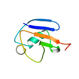
2MRP
 | |
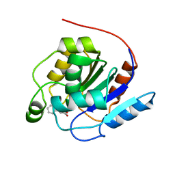
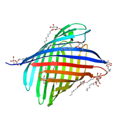
2X3V
 | | Structure of The F-BAR Domain of Mouse Syndapin I | | 分子名称: | PROTEIN KINASE C AND CASEIN KINASE SUBSTRATE IN NEURONS PROTEIN 1 | | 著者 | Ma, Q, Rao, Y, Vahedi-Faridi, A, Saenger, W, Haucke, V. | | 登録日 | 2010-01-27 | | 公開日 | 2010-04-07 | | 最終更新日 | 2024-05-08 | | 実験手法 | X-RAY DIFFRACTION (2.45 Å) | | 主引用文献 | Molecular Basis for SH3 Domain Regulation of F-Bar-Mediated Membrane Deformation.
Proc.Natl.Acad.Sci.USA, 107, 2010
|
|
7OEX
 | |
1PUT
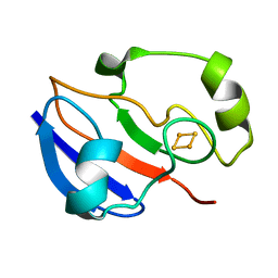 | | AN NMR-DERIVED MODEL FOR THE SOLUTION STRUCTURE OF OXIDIZED PUTIDAREDOXIN, A 2FE, 2-S FERREDOXIN FROM PSEUDOMONAS | | 分子名称: | FE2/S2 (INORGANIC) CLUSTER, PUTIDAREDOXIN | | 著者 | Pochapsky, T.C, Ye, X.M, Ratnaswamy, G, Lyons, T.A. | | 登録日 | 1994-07-09 | | 公開日 | 1994-09-30 | | 最終更新日 | 2024-05-01 | | 実験手法 | SOLUTION NMR | | 主引用文献 | An NMR-derived model for the solution structure of oxidized putidaredoxin, a 2-Fe, 2-S ferredoxin from Pseudomonas.
Biochemistry, 33, 1994
|
|
6WNF
 | |
7O9O
 | |
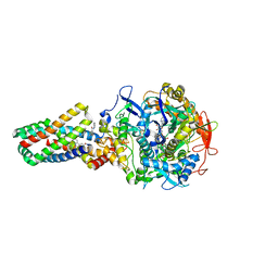
2ZI8
 | | Crystal structure of the HsaC extradiol dioxygenase from M. tuberculosis in complex with 3,4-dihydroxy-9,10-seconandrost-1,3,5(10)-triene-9,17-dione (DHSA) | | 分子名称: | 3,4-dihydroxy-9,10-secoandrosta-1(10),2,4-triene-9,17-dione, FE (II) ION, PROBABLE BIPHENYL-2,3-DIOL 1,2-DIOXYGENASE BPHC | | 著者 | D'Angelo, I, Yam, K.C, Eltis, L.D, Strynadka, N. | | 登録日 | 2008-02-13 | | 公開日 | 2009-02-24 | | 最終更新日 | 2023-11-01 | | 実験手法 | X-RAY DIFFRACTION (2.2 Å) | | 主引用文献 | Studies of a ring-cleaving dioxygenase illuminate the role of cholesterol metabolism in the pathogenesis of Mycobacterium tuberculosis.
Plos Pathog., 5, 2009
|
|
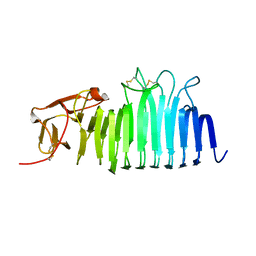
7NTN
 | | The structure of RRM domain of human TRMT2A at 2 A resolution | | 分子名称: | CHLORIDE ION, SODIUM ION, SULFATE ION, ... | | 著者 | Davydova, E, Janowski, R, Witzenberger, M, Niessing, D. | | 登録日 | 2021-03-10 | | 公開日 | 2022-01-19 | | 最終更新日 | 2024-06-19 | | 実験手法 | X-RAY DIFFRACTION (2.016 Å) | | 主引用文献 | Small-molecule modulators of TRMT2A decrease PolyQ aggregation and PolyQ-induced cell death.
Comput Struct Biotechnol J, 20, 2022
|
|
4GH3
 | |
2ZT6
 | |
2X9K
 | | Structure of a E.coli porin | | 分子名称: | OUTER MEMBRANE PROTEIN G, octyl beta-D-glucopyranoside | | 著者 | Korkmaz-Ozkan, F, Koster, S, Kuhlbrandt, W, Mantele, W, Yildiz, O. | | 登録日 | 2010-03-21 | | 公開日 | 2011-01-26 | | 最終更新日 | 2024-05-08 | | 実験手法 | X-RAY DIFFRACTION (2.18 Å) | | 主引用文献 | Correlation between the Ompg Secondary Structure and its Ph-Dependent Alterations Monitored by Ftir.
J.Mol.Biol., 401, 2010
|
|
3EC8
 | | The crystal structure of the RA domain of FLJ10324 (RADIL) | | 分子名称: | CHLORIDE ION, GLYCEROL, LEAD (II) ION, ... | | 著者 | Wisniewska, M, Lehtio, L, Andersson, J, Arrowsmith, C.H, Collins, R, Dahlgren, L.G, Edwards, A.M, Flodin, S, Flores, A, Graslund, S, Hammarstrom, M, Johansson, A, Johansson, I, Karlberg, T, Kotenyova, T, Moche, M, Nilsson, M.E, Nordlund, P, Nyman, T, Olesen, K, Persson, C, Sagemark, J, Schueler, H, Thorsell, A.G, Tresaugues, L, van den Berg, S, Weigelt, J, Welin, M, Wikstrom, M, Berglund, H, Structural Genomics Consortium (SGC) | | 登録日 | 2008-08-29 | | 公開日 | 2008-09-30 | | 最終更新日 | 2024-03-20 | | 実験手法 | X-RAY DIFFRACTION (2.6 Å) | | 主引用文献 | The crystal structure of the RA domain of FLJ10324 (RADIL)
to be published
|
|
2WS3
 | | Crystal structure of the E. coli succinate:quinone oxidoreductase (SQR) SdhD Tyr83Phe mutant | | 分子名称: | 2-METHYL-N-PHENYL-5,6-DIHYDRO-1,4-OXATHIINE-3-CARBOXAMIDE, FE2/S2 (INORGANIC) CLUSTER, FE3-S4 CLUSTER, ... | | 著者 | Ruprecht, J, Yankovskaya, V, Maklashina, E, Iwata, S, Cecchini, G. | | 登録日 | 2009-09-03 | | 公開日 | 2010-08-25 | | 最終更新日 | 2023-12-20 | | 実験手法 | X-RAY DIFFRACTION (3.2 Å) | | 主引用文献 | Succinate Dehydrogenase Activity
To be Published
|
|
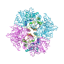
4PPH
 | | Crystal structure of conglutin gamma, a unique basic 7S globulin from lupine seeds | | 分子名称: | 1,2-ETHANEDIOL, 2-acetamido-2-deoxy-beta-D-glucopyranose, 2-acetamido-2-deoxy-beta-D-glucopyranose-(1-4)-2-acetamido-2-deoxy-beta-D-glucopyranose, ... | | 著者 | Czubinski, J, Barciszewski, J, Gilski, M, Lampart-Szczapa, E, Jaskolski, M. | | 登録日 | 2014-02-27 | | 公開日 | 2015-02-11 | | 最終更新日 | 2023-09-20 | | 実験手法 | X-RAY DIFFRACTION (2.009 Å) | | 主引用文献 | Structure of gamma-conglutin: insight into the quaternary structure of 7S basic globulins from legumes.
Acta Crystallogr.,Sect.D, 71, 2015
|
|
2N0Q
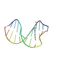 | | N2-dG-IQ modified DNA at the G1 position of the NarI recognition sequence | | 分子名称: | DNA_(5'-D(*CP*TP*CP*(IQG)P*GP*CP*GP*CP*CP*AP*TP*C)-3'), DNA_(5'-D(*GP*AP*TP*GP*GP*CP*GP*CP*CP*GP*AP*G)-3') | | 著者 | Stavros, K, Hawkins, E, Rizzo, C, Stone, M. | | 登録日 | 2015-03-11 | | 公開日 | 2015-08-19 | | 最終更新日 | 2024-05-01 | | 実験手法 | SOLUTION NMR | | 主引用文献 | Base-Displaced Intercalated Conformation of the 2-Amino-3-methylimidazo[4,5-f]quinoline N(2)-dG DNA Adduct Positioned at the Nonreiterated G(1) in the NarI Restriction Site.
Chem.Res.Toxicol., 28, 2015
|
|
5EOS
 | |
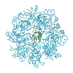
5EOW
 | | Crystal Structure of 6-Hydroxynicotinic Acid 3-Monooxygenase from Pseudomonas putida KT2440 | | 分子名称: | 6-hydroxynicotinate 3-monooxygenase, FLAVIN-ADENINE DINUCLEOTIDE | | 著者 | Yuen, M.E, Zhen, W, Gerwig, T.J, Story, R.W, Kopp, M, Nakamoto, K, Snider, M.J, Hicks, K.A. | | 登録日 | 2015-11-10 | | 公開日 | 2016-06-08 | | 最終更新日 | 2024-10-16 | | 実験手法 | X-RAY DIFFRACTION (2.1 Å) | | 主引用文献 | Structural and Biochemical Characterization of 6-Hydroxynicotinic Acid 3-Monooxygenase, A Novel Decarboxylative Hydroxylase Involved in Aerobic Nicotinate Degradation.
Biochemistry, 55, 2016
|
|
4XW1
 | |
2N2W
 | |
6WF3
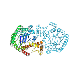
 | | Crystal structure of human Naa50 in complex with a cofactor derived inhibitor (compound 1) | | 分子名称: | ACE-MET-LEU-GLY-PRO-NH2, COENZYME A, N-alpha-acetyltransferase 50 | | 著者 | Greasley, S.E, Feng, J, Deng, Y.-L, Stewart, A.E. | | 登録日 | 2020-04-03 | | 公開日 | 2020-07-01 | | 最終更新日 | 2023-10-18 | | 実験手法 | X-RAY DIFFRACTION (2.291 Å) | | 主引用文献 | Characterization of SpecificN-alpha-Acetyltransferase 50 (Naa50) Inhibitors Identified Using a DNA Encoded Library.
Acs Med.Chem.Lett., 11, 2020
|
|
5X5H
 | |
1PLC
 | |
2ZVU
 | | Crystal structure of rat heme oxygenase-1 in complex with ferrous verdoheme | | 分子名称: | 5-OXA-PROTOPORPHYRIN IX CONTAINING FE, FORMIC ACID, Heme oxygenase 1 | | 著者 | Sato, H, Sugishima, M, Fukuyama, K, Noguchi, M. | | 登録日 | 2008-11-21 | | 公開日 | 2009-02-03 | | 最終更新日 | 2023-11-01 | | 実験手法 | X-RAY DIFFRACTION (2.2 Å) | | 主引用文献 | Crystal structure of rat haem oxygenase-1 in complex with ferrous verdohaem: presence of a hydrogen-bond network on the distal side
Biochem.J., 419, 2009
|
|
5ALS
 | | ligand complex structure of soluble epoxide hydrolase | | 分子名称: | BIFUNCTIONAL EPOXIDE HYDROLASE 2, N-CYCLOPENTYL-2-[4-(TRIFLUOROMETHYL)PHENYL]-3H-IMIDAZO[4,5-B]PYRIDINE-7-SULFONAMIDE, SULFATE ION | | 著者 | Oster, L, Tapani, S, Xue, Y, Kack, H. | | 登録日 | 2015-03-08 | | 公開日 | 2015-05-13 | | 最終更新日 | 2024-01-10 | | 実験手法 | X-RAY DIFFRACTION (2.57 Å) | | 主引用文献 | Successful Generation of Structural Information for Fragment-Based Drug Discovery.
Drug Discov Today, 20, 2015
|
|


