6XJ1
 
 | |
8X2T
 | |
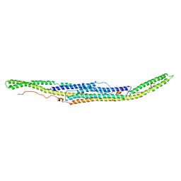
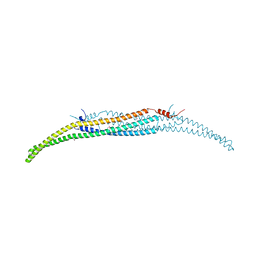
5I6R
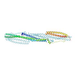
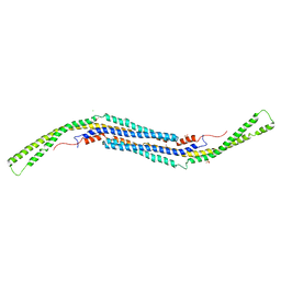
 | | Crystal Structure of srGAP2 F-BARx WT Form-1 | | 分子名称: | ACETATE ION, D-MALATE, SLIT-ROBO Rho GTPase-activating protein 2, ... | | 著者 | Sporny, M, Guez-Haddad, J, Isupov, M.N, Opatowsky, Y. | | 登録日 | 2016-02-16 | | 公開日 | 2017-08-30 | | 最終更新日 | 2024-05-08 | | 実験手法 | X-RAY DIFFRACTION (2.15 Å) | | 主引用文献 | Structural Basis for srGAP2 Membrane Interactions, and Antagonism by the Human Specific Paralog srGAP2C
To Be Published
|
|
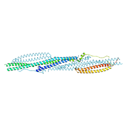
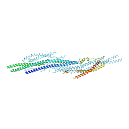
5C1F
 | | Structure of the Imp2 F-BAR domain | | 分子名称: | FORMIC ACID, Septation protein imp2 | | 著者 | Vander Kooi, C.W. | | 登録日 | 2015-06-13 | | 公開日 | 2016-01-27 | | 最終更新日 | 2019-12-25 | | 実験手法 | X-RAY DIFFRACTION (2.3551 Å) | | 主引用文献 | The Tubulation Activity of a Fission Yeast F-BAR Protein Is Dispensable for Its Function in Cytokinesis.
Cell Rep, 14, 2016
|
|
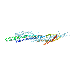
4DYL
 | | F-BAR domain of human FES tyrosine kinase | | 分子名称: | Tyrosine-protein kinase Fes/Fps | | 著者 | Ugochukwu, E, Salah, E, Elkins, J, Barr, A, Krojer, T, Filippakopoulos, P, Weigelt, J, Arrowsmith, C.H, Edwards, A, Bountra, C, von Delft, F, Knapp, S, Structural Genomics Consortium (SGC) | | 登録日 | 2012-02-29 | | 公開日 | 2012-04-04 | | 最終更新日 | 2024-02-28 | | 実験手法 | X-RAY DIFFRACTION (2.18 Å) | | 主引用文献 | F-BAR domain of human FES tyrosine kinase
TO BE PUBLISHED
|
|
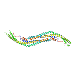
5I6J
 | |
3SYV
 | | Crystal structure of mPACSIN 3 F-BAR domain mutant | | 分子名称: | Protein kinase C and casein kinase II substrate protein 3 | | 著者 | Bai, X. | | 登録日 | 2011-07-18 | | 公開日 | 2012-05-16 | | 最終更新日 | 2024-03-20 | | 実験手法 | X-RAY DIFFRACTION (3.1 Å) | | 主引用文献 | Rigidity of wedge loop in PACSIN 3 protein is a key factor in dictating diameters of tubules
J.Biol.Chem., 287, 2012
|
|
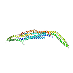
5I7D
 | |
4WPE
 | |
3QNI
 | |
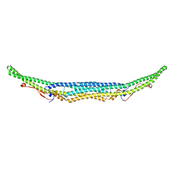
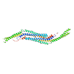
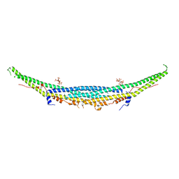
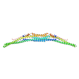
2EFK
 | | Crystal structure of the EFC domain of Cdc42-interacting protein 4 | | 分子名称: | Cdc42-interacting protein 4 | | 著者 | Shimada, A, Niwa, H, Chen, L, Liu, Z.-J, Wang, B.-C, Terada, T, Shirouzu, M, Yokoyama, S, RIKEN Structural Genomics/Proteomics Initiative (RSGI) | | 登録日 | 2007-02-23 | | 公開日 | 2007-05-29 | | 最終更新日 | 2011-07-13 | | 実験手法 | X-RAY DIFFRACTION (2.3 Å) | | 主引用文献 | Curved EFC/F-BAR-Domain Dimers Are Joined End to End into a Filament for Membrane Invagination in Endocytosis
Cell(Cambridge,Mass.), 129, 2007
|
|
4WPC
 | |
3I2W
 | |
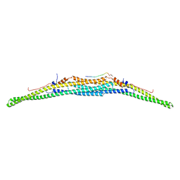
3LLL
 | | Crystal structure of mouse pacsin2 F-BAR domain | | 分子名称: | CALCIUM ION, CHLORIDE ION, Protein kinase C and casein kinase substrate in neurons protein 2, ... | | 著者 | Rudolph, M.G. | | 登録日 | 2010-01-29 | | 公開日 | 2010-05-26 | | 最終更新日 | 2023-09-06 | | 実験手法 | X-RAY DIFFRACTION (3.3 Å) | | 主引用文献 | A hinge in the distal end of the PACSIN 2 F-BAR domain may contribute to membrane-curvature sensing.
J.Mol.Biol., 400, 2010
|
|
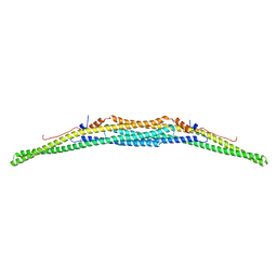
3ABH
 | | Crystal structure of the EFC/F-BAR domain of human PACSIN2/Syndapin II (2.0 A) | | 分子名称: | Protein kinase C and casein kinase substrate in neurons protein 2 | | 著者 | Shimada, A, Shirouzu, M, Hanawa-Suetsugu, K, Terada, T, Umehara, T, Suetsugu, S, Yamamoto, M, Yokoyama, S. | | 登録日 | 2009-12-11 | | 公開日 | 2010-04-14 | | 最終更新日 | 2024-04-03 | | 実験手法 | X-RAY DIFFRACTION (2 Å) | | 主引用文献 | Mapping of the basic amino-acid residues responsible for tubulation and cellular protrusion by the EFC/F-BAR domain of pacsin2/Syndapin II
Febs Lett., 584, 2010
|
|
3ACO
 | | Crystal structure of the EFC/F-BAR domain of human PACSIN2/Syndapin II (2.7 A) | | 分子名称: | CALCIUM ION, Protein kinase C and casein kinase substrate in neurons protein 2 | | 著者 | Shimada, A, Shirouzu, M, Hanawa-Suetsugu, K, Terada, T, Umehara, T, Suetsugu, S, Yamamoto, M, Yokoyama, S. | | 登録日 | 2010-01-07 | | 公開日 | 2010-04-14 | | 最終更新日 | 2011-07-13 | | 実験手法 | X-RAY DIFFRACTION (2.7 Å) | | 主引用文献 | Mapping of the basic amino-acid residues responsible for tubulation and cellular protrusion by the EFC/F-BAR domain of pacsin2/Syndapin II
Febs Lett., 584, 2010
|
|
2EFL
 | | Crystal structure of the EFC domain of formin-binding protein 17 | | 分子名称: | Formin-binding protein 1 | | 著者 | Shimada, A, Niwa, H, Terada, T, Shirouzu, M, Yokoyama, S, RIKEN Structural Genomics/Proteomics Initiative (RSGI) | | 登録日 | 2007-02-23 | | 公開日 | 2007-05-29 | | 最終更新日 | 2023-11-15 | | 実験手法 | X-RAY DIFFRACTION (2.61 Å) | | 主引用文献 | Curved EFC/F-BAR-Domain Dimers Are Joined End to End into a Filament for Membrane Invagination in Endocytosis
Cell(Cambridge,Mass.), 129, 2007
|
|
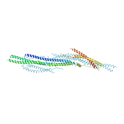
2X3V
 | | Structure of The F-BAR Domain of Mouse Syndapin I | | 分子名称: | PROTEIN KINASE C AND CASEIN KINASE SUBSTRATE IN NEURONS PROTEIN 1 | | 著者 | Ma, Q, Rao, Y, Vahedi-Faridi, A, Saenger, W, Haucke, V. | | 登録日 | 2010-01-27 | | 公開日 | 2010-04-07 | | 最終更新日 | 2024-05-08 | | 実験手法 | X-RAY DIFFRACTION (2.45 Å) | | 主引用文献 | Molecular Basis for SH3 Domain Regulation of F-Bar-Mediated Membrane Deformation.
Proc.Natl.Acad.Sci.USA, 107, 2010
|
|
7AAL
 | | Crystal structure of the F-BAR domain of PSTIPIP1, G258A mutant | | 分子名称: | Proline-serine-threonine phosphatase-interacting protein 1 | | 著者 | Manso, J.A, Alcon, P, Bayon, Y, Alonso, A, de Pereda, J.M. | | 登録日 | 2020-09-04 | | 公開日 | 2022-02-23 | | 最終更新日 | 2024-02-07 | | 実験手法 | X-RAY DIFFRACTION (1.97 Å) | | 主引用文献 | PSTPIP1-LYP phosphatase interaction: structural basis and implications for autoinflammatory disorders.
Cell.Mol.Life Sci., 79, 2022
|
|
7AAM
 | | Crystal structure of the F-BAR domain of PSTIPIP1 bound to the CTH domain of the phosphatase LYP | | 分子名称: | GLYCEROL, Proline-serine-threonine phosphatase-interacting protein 1, Tyrosine-protein phosphatase non-receptor type 22 | | 著者 | Manso, J.A, Alcon, P, Bayon, Y, Alonso, A, de Pereda, J.M. | | 登録日 | 2020-09-04 | | 公開日 | 2022-02-23 | | 最終更新日 | 2024-02-07 | | 実験手法 | X-RAY DIFFRACTION (2.15 Å) | | 主引用文献 | PSTPIP1-LYP phosphatase interaction: structural basis and implications for autoinflammatory disorders.
Cell.Mol.Life Sci., 79, 2022
|
|
7AAN
 | | Crystal structure of the F-BAR domain of PSTIPIP1 | | 分子名称: | Proline-serine-threonine phosphatase-interacting protein 1 | | 著者 | Manso, J.A, Alcon, P, Bayon, Y, Alonso, A, de Pereda, J.M. | | 登録日 | 2020-09-04 | | 公開日 | 2022-02-23 | | 最終更新日 | 2024-02-07 | | 実験手法 | X-RAY DIFFRACTION (2.14 Å) | | 主引用文献 | PSTPIP1-LYP phosphatase interaction: structural basis and implications for autoinflammatory disorders.
Cell.Mol.Life Sci., 79, 2022
|
|
3HAH
 | | Crystal structure of human PACSIN1 F-BAR domain (C2 lattice) | | 分子名称: | CALCIUM ION, human PACSIN1 F-BAR | | 著者 | Wang, Q, Navarro, M.V.A.S, Peng, G, Rajashankar, K.R, Sondermann, H. | | 登録日 | 2009-05-01 | | 公開日 | 2009-06-16 | | 最終更新日 | 2024-02-21 | | 実験手法 | X-RAY DIFFRACTION (2.77 Å) | | 主引用文献 | Molecular mechanism of membrane constriction and tubulation mediated by the F-BAR protein Pacsin/Syndapin.
Proc.Natl.Acad.Sci.USA, 106, 2009
|
|
3HAI
 | | Crystal structure of human PACSIN1 F-BAR domain (P21 lattice) | | 分子名称: | CALCIUM ION, human PACSIN1 F-BAR | | 著者 | Wang, Q, Navarro, M.V.A.S, Peng, G, Rajashankar, K.R, Sondermann, H. | | 登録日 | 2009-05-01 | | 公開日 | 2009-06-16 | | 最終更新日 | 2024-02-21 | | 実験手法 | X-RAY DIFFRACTION (2.881 Å) | | 主引用文献 | Molecular mechanism of membrane constriction and tubulation mediated by the F-BAR protein Pacsin/Syndapin.
Proc.Natl.Acad.Sci.USA, 106, 2009
|
|
3M3W
 | |
3Q0K
 | |


