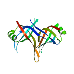
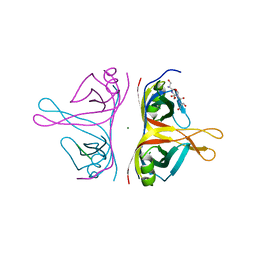
3KOJ
 
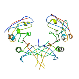
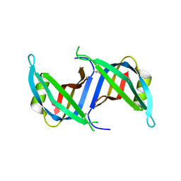 | | Crystal structure of the SSB domain of Q5N255_SYNP6 protein from Synechococcus sp. Northeast Structural Genomics Consortium Target SnR59a. | | 分子名称: | uncharacterized protein ycf41 | | 著者 | Vorobiev, S, Su, M, Seetharaman, J, Wang, H, Foote, L.E, Ciccosanti, C, Janjua, H, Xiao, R, Acton, T.B, Montelione, G.T, Tong, L, Hunt, J.F, Northeast Structural Genomics Consortium (NESG) | | 登録日 | 2009-11-13 | | 公開日 | 2009-11-24 | | 最終更新日 | 2021-10-13 | | 実験手法 | X-RAY DIFFRACTION (1.895 Å) | | 主引用文献 | Crystal structure of the SSB domain of Q5N255_SYNP6 protein from Synechococcus sp.
To be Published
|
|
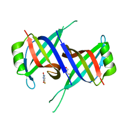
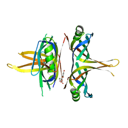
7R4W
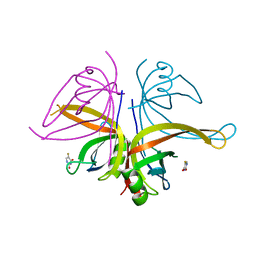
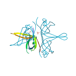 | | Single stranded DNA binding protein SSB M5 from Fervidobacterium gondwanense | | 分子名称: | ACETIC ACID, PHOSPHATE ION, Single-stranded DNA-binding protein | | 著者 | Hakansson, M, Svensson, L.A, Werbowy, O, Al-Karadaghi, S, Kaczorowski, T, Kaczorowska, A.K, Dorawa, S. | | 登録日 | 2022-02-09 | | 公開日 | 2023-08-23 | | 実験手法 | X-RAY DIFFRACTION (2.3 Å) | | 主引用文献 | Molecular characterization of a single stranded DNA binding protein from Fervidobacterium gondwanense
To Be Published
|
|
3K81
 | |
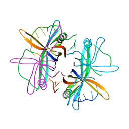
1UE6
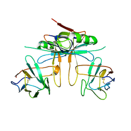
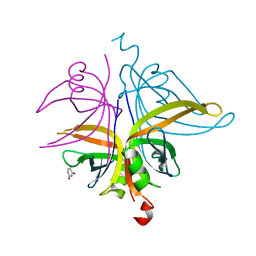
 | | Crystal structure of the single-stranded dna-binding protein from mycobacterium tuberculosis | | 分子名称: | Single-strand binding protein | | 著者 | Saikrishnan, K, Jeyakanthan, J, Venkatesh, J, Acharya, N, Sekar, K, Varshney, U, Vijayan, M, TB Structural Genomics Consortium (TBSGC) | | 登録日 | 2003-05-09 | | 公開日 | 2004-02-10 | | 最終更新日 | 2023-10-25 | | 実験手法 | X-RAY DIFFRACTION (2.7 Å) | | 主引用文献 | Structure of Mycobacterium tuberculosis single-stranded DNA-binding protein. Variability in quaternary structure and its implications
J.MOL.BIOL., 331, 2003
|
|
5XGT
 | |
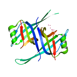
2DUD
 | |
7YM1
 | | Structure of SsbA protein in complex with the anticancer drug 5-fluorouracil | | 分子名称: | 5-FLUOROURACIL, GLYCEROL, Single-stranded DNA-binding protein | | 著者 | Huang, Y.H, Yang, P.C, Chiang, W.Y, Lin, E.S, Huang, C.Y. | | 登録日 | 2022-07-27 | | 公開日 | 2023-08-02 | | 最終更新日 | 2024-02-14 | | 実験手法 | X-RAY DIFFRACTION (2.36 Å) | | 主引用文献 | Crystal Structure of DNA Replication Protein SsbA Complexed with the Anticancer Drug 5-Fluorouracil.
Int J Mol Sci, 24, 2023
|
|
5GQO
 | |
8GW5
 | |
6RUP
 | | Human mitochondrial single-stranded DNA binding protein, SSBP1, at 2.1 A resolution - elucidated sequence | | 分子名称: | MAGNESIUM ION, SER-SER-SER-SER, Single-stranded DNA-binding protein, ... | | 著者 | Tarres-Sole, A, Chakraborty, A, Spelbrink, H.N, Delettre, C, Sola, M. | | 登録日 | 2019-05-28 | | 公開日 | 2019-10-09 | | 最終更新日 | 2024-01-24 | | 実験手法 | X-RAY DIFFRACTION (2.1 Å) | | 主引用文献 | Dominant mutations in mtDNA maintenance gene SSBP1 cause optic atrophy and foveopathy.
J.Clin.Invest., 130, 2020
|
|
1KAW
 | | STRUCTURE OF SINGLE STRANDED DNA BINDING PROTEIN (SSB) | | 分子名称: | SINGLE-STRANDED DNA BINDING PROTEIN | | 著者 | Raghunathan, S, Waksman, G. | | 登録日 | 1996-12-06 | | 公開日 | 1997-12-31 | | 最終更新日 | 2024-02-07 | | 実験手法 | X-RAY DIFFRACTION (2.9 Å) | | 主引用文献 | Crystal structure of the homo-tetrameric DNA binding domain of Escherichia coli single-stranded DNA-binding protein determined by multiwavelength x-ray diffraction on the selenomethionyl protein at 2.9-A resolution.
Proc.Natl.Acad.Sci.USA, 94, 1997
|
|
7VUM
 | | Crystal structure of SSB complexed with que | | 分子名称: | 3,5,7,3',4'-PENTAHYDROXYFLAVONE, Single-stranded DNA-binding protein | | 著者 | Lin, E.S, Huang, Y.H, Huang, C.Y. | | 登録日 | 2021-11-03 | | 公開日 | 2022-03-09 | | 最終更新日 | 2023-11-29 | | 実験手法 | X-RAY DIFFRACTION (2.319 Å) | | 主引用文献 | A Complexed Crystal Structure of a Single-Stranded DNA-Binding Protein with Quercetin and the Structural Basis of Flavonol Inhibition Specificity.
Int J Mol Sci, 23, 2022
|
|
3EIV
 | |
4MZ9
 | | Revised structure of E. coli SSB | | 分子名称: | Single-stranded DNA-binding protein | | 著者 | Oakley, A.J. | | 登録日 | 2013-09-29 | | 公開日 | 2013-12-18 | | 最終更新日 | 2023-09-20 | | 実験手法 | X-RAY DIFFRACTION (2.2 Å) | | 主引用文献 | Intramolecular binding mode of the C-terminus of Escherichia coli single-stranded DNA binding protein determined by nuclear magnetic resonance spectroscopy.
Nucleic Acids Res., 42, 2014
|
|
2VW9
 | | Single stranded DNA binding protein complex from Helicobacter pylori | | 分子名称: | POLY-DT, SINGLE-STRANDED DNA BINDING PROTEIN | | 著者 | Chan, K.-W, Wang, C.-H, Lee, Y.-J, Sun, Y.-J. | | 登録日 | 2008-06-18 | | 公開日 | 2009-06-02 | | 最終更新日 | 2023-12-13 | | 実験手法 | X-RAY DIFFRACTION (2.3 Å) | | 主引用文献 | Single-Stranded DNA-Binding Protein Complex from Helicobacter Pylori Suggests an Ssdna-Binding Surface.
J.Mol.Biol., 388, 2009
|
|
3PGZ
 | |
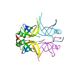
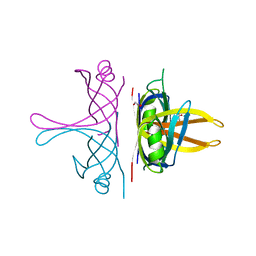
3K80
 | | Structure of essential protein from Trypanosoma brucei | | 分子名称: | Antibody, MP18 RNA editing complex protein | | 著者 | Wu, M. | | 登録日 | 2009-10-13 | | 公開日 | 2010-11-17 | | 最終更新日 | 2023-09-06 | | 実験手法 | X-RAY DIFFRACTION (2.4 Å) | | 主引用文献 | Structures of a key interaction protein from the Trypanosoma brucei editosome in complex with single domain antibodies.
J.Struct.Biol., 174, 2011
|
|
7DEP
 | | S. aureus SsbB with 5-FU | | 分子名称: | 5-FLUOROURACIL, Single-stranded DNA-binding protein | | 著者 | Lin, E.S, Huang, Y.H, Huang, C.Y. | | 登録日 | 2020-11-04 | | 公開日 | 2020-12-23 | | 最終更新日 | 2023-11-29 | | 実験手法 | X-RAY DIFFRACTION (3.094 Å) | | 主引用文献 | Crystal structure of the single-stranded DNA-binding protein SsbB in complex with the anticancer drug 5-fluorouracil: Extension of the 5-fluorouracil interactome to include the oligonucleotide/oligosaccharide-binding fold protein
Biochem.Biophys.Res.Commun., 534, 2020
|
|
7D8J
 | | S. aureus SsbB with 5-FU | | 分子名称: | 5-FLUOROURACIL, Single-stranded DNA-binding protein | | 著者 | Lin, E.S, Huang, Y.H, Huang, C.Y. | | 登録日 | 2020-10-08 | | 公開日 | 2020-12-23 | | 最終更新日 | 2023-11-29 | | 実験手法 | X-RAY DIFFRACTION (2.88 Å) | | 主引用文献 | Crystal structure of the single-stranded DNA-binding protein SsbB in complex with the anticancer drug 5-fluorouracil: Extension of the 5-fluorouracil interactome to include the oligonucleotide/oligosaccharide-binding fold protein
Biochem.Biophys.Res.Commun., 534, 2020
|
|
6JDG
 | |
1EQQ
 | | SINGLE STRANDED DNA BINDING PROTEIN AND SSDNA COMPLEX | | 分子名称: | 5'-R(*(5MU)P*(5MU)P*(5MU))-3', SINGLE STRANDED DNA BINDING PROTEIN | | 著者 | Matsumoto, T, Morimoto, Y, Shibata, N, Yasuoka, N, Shimamoto, N. | | 登録日 | 2000-04-06 | | 公開日 | 2003-09-23 | | 最終更新日 | 2024-02-07 | | 実験手法 | X-RAY DIFFRACTION (3.2 Å) | | 主引用文献 | Roles of functional loops and the C-terminal segment of a single-stranded DNA binding protein elucidated by X-Ray structure analysis
J.Biochem.(Tokyo), 127, 2000
|
|
1EYG
 | |
6IRQ
 | |
5ODP
 | |
5ODN
 | |


