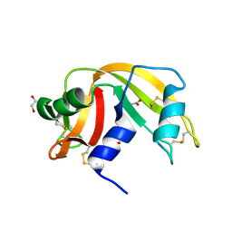
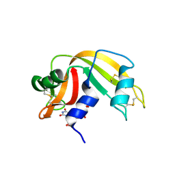
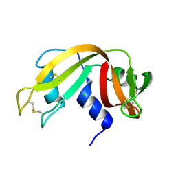
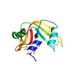
8C3B
 
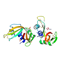 | | X-ray structure of RNase A upon reaction with a Ruthenium(II)-arene Complexed with Glycosylated Carbene Ligands (5) | | 分子名称: | (1,3-dimethyl-2~{H}-imidazol-2-yl)-oxidanyl-oxidanylidene-ruthenium, (1,3-dimethylimidazol-1-ium-2-yl)-tetrakis(oxidanyl)ruthenium, (1,3-dimethylimidazol-1-ium-2-yl)-tris(oxidanyl)ruthenium, ... | | 著者 | Ferraro, G, Merlino, A. | | 登録日 | 2022-12-23 | | 公開日 | 2024-01-10 | | 最終更新日 | 2024-07-24 | | 実験手法 | X-RAY DIFFRACTION (1.24 Å) | | 主引用文献 | Ruthenium(II)–Arene Complexes with Glycosylated NHC-Carbene Co-Ligands: Synthesis, Hydrolytic Behavior, and Binding to Biological Molecules
Organometallics, 42, 2023
|
|
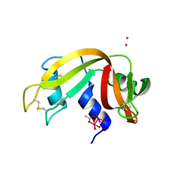
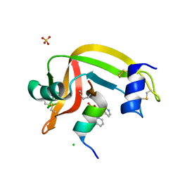
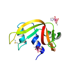
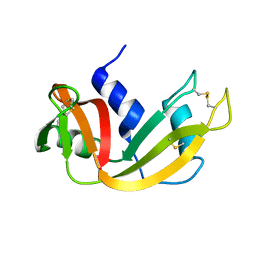
1RUV
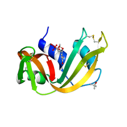 | | RIBONUCLEASE A-URIDINE VANADATE COMPLEX: HIGH RESOLUTION RESOLUTION X-RAY STRUCTURE (1.3 A) | | 分子名称: | RIBONUCLEASE A, TERTIARY-BUTYL ALCOHOL, URIDINE-2',3'-VANADATE | | 著者 | Ladner, J.E, Wladkowski, B, Svensson, L.A, Sjolin, L, Gilliland, G.L. | | 登録日 | 1995-07-27 | | 公開日 | 1997-04-01 | | 最終更新日 | 2024-06-05 | | 実験手法 | X-RAY DIFFRACTION (1.25 Å) | | 主引用文献 | X-ray structure of a ribonuclease A-uridine vanadate complex at 1.3 A resolution.
Acta Crystallogr.,Sect.D, 53, 1997
|
|
7RSA
 | |
7P8R
 | |
3QL1
 | |
3I6H
 | | Ribonuclease A by LB nanotemplate method before high X-Ray dose on ESRF ID14-2 beamline | | 分子名称: | CHLORIDE ION, Ribonuclease pancreatic | | 著者 | Pechkova, E, Tripathi, S.K, Ravelli, R, McSweeney, S, Nicolini, C. | | 登録日 | 2009-07-07 | | 公開日 | 2010-07-07 | | 最終更新日 | 2023-11-01 | | 実験手法 | X-RAY DIFFRACTION (1.3 Å) | | 主引用文献 | Atomic structure and radiation resistance of langmuir-blodgett protein crystals
To be Published
|
|
3I6F
 | | Ribonuclease A by Classical hanging drop method before high X-Ray dose on ESRF ID14-2 beamline | | 分子名称: | CHLORIDE ION, Ribonuclease pancreatic | | 著者 | Pechkova, E, Tripathi, S.K, Ravelli, R, McSweeney, S, Nicolini, C. | | 登録日 | 2009-07-07 | | 公開日 | 2010-07-07 | | 最終更新日 | 2023-11-01 | | 実験手法 | X-RAY DIFFRACTION (1.3 Å) | | 主引用文献 | Atomic structure and radiation resistance of langmuir-blodgett protein crystals
To be Published
|
|
3I67
 | | Ribonuclease A by LB nanotemplate method after high X-Ray dose on ESRF ID14-2 beamline | | 分子名称: | CHLORIDE ION, Ribonuclease pancreatic | | 著者 | Pechkova, E, Tripathi, S.K, Ravelli, R, McSweeney, S, Nicolini, C. | | 登録日 | 2009-07-06 | | 公開日 | 2010-07-07 | | 最終更新日 | 2023-11-01 | | 実験手法 | X-RAY DIFFRACTION (1.3 Å) | | 主引用文献 | Atomic structure and radiation resistance of langmuir-blodgett protein crystals
To be Published
|
|
3I6J
 | | Ribonuclease A by Classical hanging drop method after high X-Ray dose on ESRF ID14-2 beamline | | 分子名称: | CHLORIDE ION, Ribonuclease pancreatic | | 著者 | Pechkova, E, Tripathi, S.K, Ravelli, R, McSweeney, S, Nicolini, C. | | 登録日 | 2009-07-07 | | 公開日 | 2010-07-07 | | 最終更新日 | 2023-11-01 | | 実験手法 | X-RAY DIFFRACTION (1.3 Å) | | 主引用文献 | Atomic structure and radiation resistance of langmuir-blodgett protein crystals
To be Published
|
|
7QQ0
 | |
2QCA
 | | A New Crystal Form of Bovine Pancreatic RNase A in Complex with 2'-Deoxyguanosine-5'-monophosphate | | 分子名称: | 2'-DEOXYGUANOSINE-5'-MONOPHOSPHATE, Ribonuclease pancreatic | | 著者 | Larson, S.B, Day, J.S, Cudney, R, McPherson, A, Center for High-Throughput Structural Biology (CHTSB) | | 登録日 | 2007-06-19 | | 公開日 | 2007-07-03 | | 最終更新日 | 2023-08-30 | | 実験手法 | X-RAY DIFFRACTION (1.33 Å) | | 主引用文献 | A new crystal form of bovine pancreatic RNase A in complex with 2'-deoxyguanosine-5'-monophosphate.
Acta Crystallogr.,Sect.F, 63, 2007
|
|
4K7L
 | | Crystal structure of RNase S variant (K7C/Q11C) | | 分子名称: | Ribonuclease pancreatic, SULFATE ION | | 著者 | Genz, M, Straeter, N. | | 登録日 | 2013-04-17 | | 公開日 | 2014-05-14 | | 最終更新日 | 2023-09-20 | | 実験手法 | X-RAY DIFFRACTION (1.38 Å) | | 主引用文献 | Crystal structure of RNase S with a [Hg(Cys2)] metal center in the S-peptide as a template for structure-based design of artificial metalloenzymes using peptide-protein complementation
To be Published
|
|
2P49
 | | Complex of a camelid single-domain vhh antibody fragment with RNASE A at 1.4A resolution: native mono_1 crystal form | | 分子名称: | ANTIBODY CAB-RN05, PHOSPHATE ION, Ribonuclease pancreatic | | 著者 | Tereshko, V, Uysal, S, Margalef, K, Koide, A, Kossiakoff, A.A, Koide, S. | | 登録日 | 2007-03-11 | | 公開日 | 2007-08-28 | | 最終更新日 | 2023-08-30 | | 実験手法 | X-RAY DIFFRACTION (1.38 Å) | | 主引用文献 | Exploring the capacity of minimalist protein interfaces: interface energetics and affinity maturation to picomolar KD of a single-domain antibody with a flat paratope.
J.Mol.Biol., 373, 2007
|
|
3DXG
 | | Ribonuclease A- uridine 5' phosphate complex | | 分子名称: | Ribonuclease pancreatic, URIDINE-5'-MONOPHOSPHATE | | 著者 | Tsirkone, V.G, Dossi, K, Leonidas, D.D, Zographos, S.E, Oikonomakos, N.G, Kontou, M. | | 登録日 | 2008-07-24 | | 公開日 | 2009-06-16 | | 最終更新日 | 2023-11-01 | | 実験手法 | X-RAY DIFFRACTION (1.39 Å) | | 主引用文献 | Inhibitor design to Ribonuclease A: The binding of two 5'phosphate uridine analogues
Acta Crystallogr.,Sect.F, 2009
|
|
4O37
 | | seminsynthetic RNase S1-15-3Pl-7/11 | | 分子名称: | CHLORIDE ION, Ribonuclease pancreatic, S-peptide, ... | | 著者 | Genz, M, Strater, N. | | 登録日 | 2013-12-18 | | 公開日 | 2014-05-07 | | 最終更新日 | 2023-12-06 | | 実験手法 | X-RAY DIFFRACTION (1.4 Å) | | 主引用文献 | An Artificial Imine Reductase based on the Ribonuclease S Scaffold
Chem.Cat.Chem, 2014
|
|
2BLP
 | |
6XVX
 | |
2BLZ
 | |
7QHR
 | |
1RPG
 | | STRUCTURES OF RNASE A COMPLEXED WITH 3'-CMP AND D(CPA): ACTIVE SITE CONFORMATION AND CONSERVED WATER MOLECULES | | 分子名称: | (4S)-2-METHYL-2,4-PENTANEDIOL, 2'-DEOXYCYTIDINE-2'-DEOXYADENOSINE-3',5'-MONOPHOSPHATE, RIBONUCLEASE A | | 著者 | Zegers, I, Wyns, L, Palmer, R. | | 登録日 | 1994-08-29 | | 公開日 | 1994-12-20 | | 最終更新日 | 2024-06-05 | | 実験手法 | X-RAY DIFFRACTION (1.4 Å) | | 主引用文献 | The structures of RNase A complexed with 3'-CMP and d(CpA): active site conformation and conserved water molecules.
Protein Sci., 3, 1994
|
|
3DIB
 | | Crystal structure of bovine pancreatic ribonuclease A variant (I106A) | | 分子名称: | CHLORIDE ION, Ribonuclease pancreatic, SULFATE ION | | 著者 | Kurpiewska, K, Font, J, Ribo, M, Vilanova, M, Lewinski, K. | | 登録日 | 2008-06-20 | | 公開日 | 2008-07-15 | | 最終更新日 | 2023-11-01 | | 実験手法 | X-RAY DIFFRACTION (1.4 Å) | | 主引用文献 | X-ray crystallographic studies of RNase A variants engineered at the most destabilizing positions of the main hydrophobic core: further insight into protein stability
Proteins, 77, 2009
|
|
3DXH
 | | Ribonuclease A uridine 5' diphosphate complex | | 分子名称: | Ribonuclease pancreatic, URIDINE-5'-DIPHOSPHATE | | 著者 | Tsirkone, V.G, Dossi, K, Leonidas, D.D, Zographos, S.E, Oikonomakos, N.G, Kontou, M. | | 登録日 | 2008-07-24 | | 公開日 | 2009-06-16 | | 最終更新日 | 2023-11-01 | | 実験手法 | X-RAY DIFFRACTION (1.4 Å) | | 主引用文献 | Inhibitor design to Ribonuclease A: The binding of two 5'phosphate uridine analogues
Acta Crystallogr.,Sect.F, 2009
|
|
1EIE
 | |
1EID
 | |
1EIC
 | |


