3S9C
 
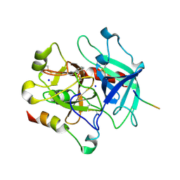
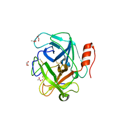 | | Russell's viper venom serine proteinase, RVV-V in complex with the fragment (residues 1533-1546) of human factor V | | 分子名称: | ACETATE ION, Coagulation factor V, Vipera russelli proteinase RVV-V gamma, ... | | 著者 | Nakayama, D, Ben Ammar, Y, Takeda, S. | | 登録日 | 2011-06-01 | | 公開日 | 2011-09-07 | | 最終更新日 | 2023-11-01 | | 実験手法 | X-RAY DIFFRACTION (1.8 Å) | | 主引用文献 | Structural basis of coagulation factor V recognition for cleavage by RVV-V
Febs Lett., 585, 2011
|
|
9CTH
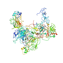
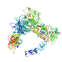 | | Preliminary map of the Prothrombin-prothrombinase complex on nano discs | | 分子名称: | 2-acetamido-2-deoxy-beta-D-glucopyranose, Activated Factor V (FVa) heavy chain, Activated Factor V (FVa) light chain, ... | | 著者 | Stojanovski, B.M, Mohammed, B.M, Di Cera, E. | | 登録日 | 2024-07-25 | | 公開日 | 2024-08-07 | | 最終更新日 | 2024-08-14 | | 実験手法 | ELECTRON MICROSCOPY (6.47 Å) | | 主引用文献 | The Prothrombin-Prothrombinase Interaction.
Subcell Biochem, 104, 2024
|
|
8TN9
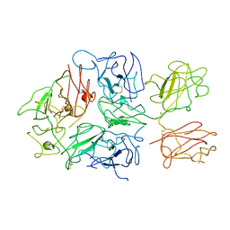
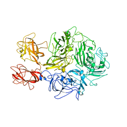 | | Structural architecture of the acidic region of the B domain of coagulation factor V | | 分子名称: | 2-acetamido-2-deoxy-beta-D-glucopyranose, Coagulation factor V | | 著者 | Mohammed, B.M, Basore, K, Summers, B, Pelc, L.A, Di Cera, E. | | 登録日 | 2023-08-01 | | 公開日 | 2023-10-04 | | 最終更新日 | 2024-03-13 | | 実験手法 | ELECTRON MICROSCOPY (3.05 Å) | | 主引用文献 | Structural architecture of the acidic region of the B domain of coagulation factor V.
J.Thromb.Haemost., 22, 2024
|
|
8FDG
 | |
7TPP
 | |
3P70
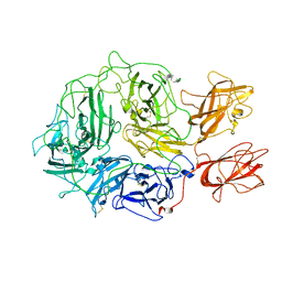
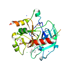
 | | Structural basis of thrombin-mediated factor V activation: essential role of the hirudin-like sequence Glu666-Glu672 for processing at the heavy chain-B domain junction | | 分子名称: | 2-acetamido-2-deoxy-beta-D-glucopyranose, 2-acetamido-2-deoxy-beta-D-glucopyranose-(1-4)-2-acetamido-2-deoxy-beta-D-glucopyranose, BENZAMIDINE, ... | | 著者 | Corral-Rodriguez, M.A, Bock, P.E, Hernandez-Carvajal, E, Gutierrez-Gallego, R, Fuentes-Prior, P. | | 登録日 | 2010-10-11 | | 公開日 | 2011-09-28 | | 最終更新日 | 2023-09-06 | | 実験手法 | X-RAY DIFFRACTION (2.55 Å) | | 主引用文献 | Structural basis of thrombin-mediated factor V activation: the Glu666-Glu672 sequence is critical for processing at the heavy chain-B domain junction.
Blood, 117, 2011
|
|
3P6Z
 | | Structural basis of thrombin mediated factor V activation: essential role of the hirudin-like sequence Glu666-Glu672 for processing at the heavy chain-B domain junction | | 分子名称: | (4S)-2-METHYL-2,4-PENTANEDIOL, 2-acetamido-2-deoxy-beta-D-glucopyranose, CHLORIDE ION, ... | | 著者 | Corral-Rodriguez, M.A, Bock, P.E, Hernandez-Carvajal, E, Gutierrez-Gallego, R, Fuentes-Prior, P. | | 登録日 | 2010-10-11 | | 公開日 | 2011-06-15 | | 最終更新日 | 2024-03-13 | | 実験手法 | X-RAY DIFFRACTION (1.7 Å) | | 主引用文献 | Structural basis of thrombin-mediated factor V activation: the Glu666-Glu672 sequence is critical for processing at the heavy chain-B domain junction.
Blood, 117, 2011
|
|
1CZT
 | | CRYSTAL STRUCTURE OF THE C2 DOMAIN OF HUMAN COAGULATION FACTOR V | | 分子名称: | PROTEIN (COAGULATION FACTOR V) | | 著者 | Macedo-Ribeiro, S, Bode, W, Huber, R, Kane, W.H, Fuentes-Prior, P. | | 登録日 | 1999-09-07 | | 公開日 | 1999-11-26 | | 最終更新日 | 2023-08-09 | | 実験手法 | X-RAY DIFFRACTION (1.87 Å) | | 主引用文献 | Crystal structures of the membrane-binding C2 domain of human coagulation factor V.
Nature, 402, 1999
|
|
1CZV
 | | CRYSTAL STRUCTURE OF THE C2 DOMAIN OF HUMAN COAGULATION FACTOR V: DIMERIC CRYSTAL FORM | | 分子名称: | PROTEIN (COAGULATION FACTOR V) | | 著者 | Macedo-Ribeiro, S, Bode, W, Huber, R, Kane, W.H, Fuentes-Prior, P. | | 登録日 | 1999-09-07 | | 公開日 | 1999-11-26 | | 最終更新日 | 2023-08-09 | | 実験手法 | X-RAY DIFFRACTION (2.4 Å) | | 主引用文献 | Crystal structures of the membrane-binding C2 domain of human coagulation factor V.
Nature, 402, 1999
|
|
7KVE
 | |
7KXY
 | |
7KVF
 | |
1CZS
 | | CRYSTAL STRUCTURE OF THE C2 DOMAIN OF HUMAN COAGULATION FACTOR V: COMPLEX WITH PHENYLMERCURY | | 分子名称: | PHENYLMERCURY, PROTEIN (COAGULATION FACTOR V) | | 著者 | Macedo-Ribeiro, S, Bode, W, Huber, R, Kane, W.H, Fuentes-Prior, P. | | 登録日 | 1999-09-07 | | 公開日 | 1999-11-26 | | 最終更新日 | 2011-07-13 | | 実験手法 | X-RAY DIFFRACTION (1.9 Å) | | 主引用文献 | Crystal structures of the membrane-binding C2 domain of human coagulation factor V.
Nature, 402, 1999
|
|


