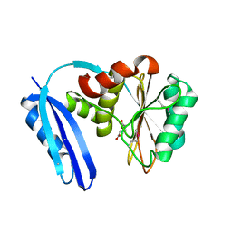
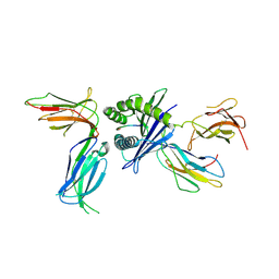
3TH5
 
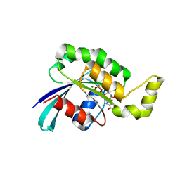 | | Crystal structure of wild-type RAC1 | | 分子名称: | MAGNESIUM ION, PHOSPHOAMINOPHOSPHONIC ACID-GUANYLATE ESTER, Ras-related C3 botulinum toxin substrate 1 | | 著者 | Ha, B.H, Boggon, T.J. | | 登録日 | 2011-08-18 | | 公開日 | 2012-07-18 | | 最終更新日 | 2023-09-13 | | 実験手法 | X-RAY DIFFRACTION (2.3 Å) | | 主引用文献 | Exome sequencing identifies recurrent somatic RAC1 mutations in melanoma.
Nat.Genet., 44, 2012
|
|
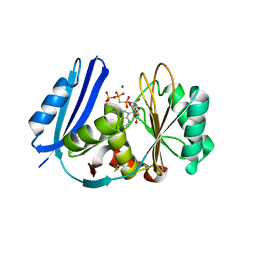
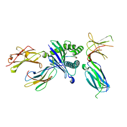
3SBE
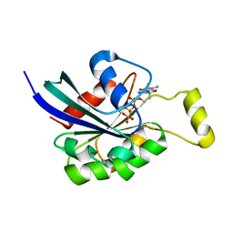 | | Crystal structure of RAC1 P29S mutant | | 分子名称: | MAGNESIUM ION, PHOSPHOAMINOPHOSPHONIC ACID-GUANYLATE ESTER, Ras-related C3 botulinum toxin substrate 1 | | 著者 | Ha, B.H, Boggon, T.J. | | 登録日 | 2011-06-03 | | 公開日 | 2012-07-18 | | 最終更新日 | 2024-02-28 | | 実験手法 | X-RAY DIFFRACTION (2.6 Å) | | 主引用文献 | Exome sequencing identifies recurrent somatic RAC1 mutations in melanoma.
Nat.Genet., 44, 2012
|
|
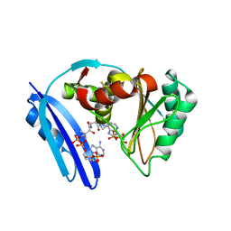
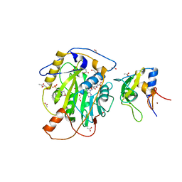
3SBD
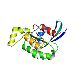 | | Crystal structure of RAC1 P29S mutant | | 分子名称: | MAGNESIUM ION, PHOSPHOAMINOPHOSPHONIC ACID-GUANYLATE ESTER, Ras-related C3 botulinum toxin substrate 1 | | 著者 | Ha, B.H, Boggon, T.J. | | 登録日 | 2011-06-03 | | 公開日 | 2012-07-18 | | 最終更新日 | 2023-09-13 | | 実験手法 | X-RAY DIFFRACTION (2.1 Å) | | 主引用文献 | Exome sequencing identifies recurrent somatic RAC1 mutations in melanoma.
Nat.Genet., 44, 2012
|
|
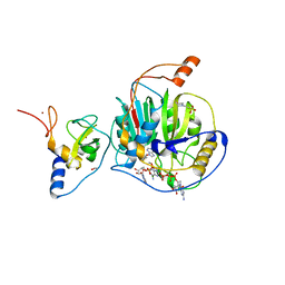
5TV5
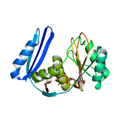 | | BioW from Aquifex aeoulicus | | 分子名称: | 6-carboxyhexanoate--CoA ligase | | 著者 | Estrada, P, Manandhar, M, Dong, S.-H, Deveryshetty, J, Agarwal, V, Cronan, J.E, Nair, S.K. | | 登録日 | 2016-11-08 | | 公開日 | 2016-12-07 | | 最終更新日 | 2017-05-31 | | 実験手法 | X-RAY DIFFRACTION (2.5 Å) | | 主引用文献 | The pimeloyl-CoA synthetase BioW defines a new fold for adenylate-forming enzymes.
Nat. Chem. Biol., 13, 2017
|
|
5TV6
 | | A. aeolicus BioW with pimelate | | 分子名称: | 6-carboxyhexanoate--CoA ligase, PIMELIC ACID | | 著者 | Estrada, P, Manandhar, M, Dong, S.-H, Deveryshetty, J, Agarwal, V, Cronan, J.E, Nair, S.K. | | 登録日 | 2016-11-08 | | 公開日 | 2016-12-07 | | 最終更新日 | 2017-05-31 | | 実験手法 | X-RAY DIFFRACTION (2.456 Å) | | 主引用文献 | The pimeloyl-CoA synthetase BioW defines a new fold for adenylate-forming enzymes.
Nat. Chem. Biol., 13, 2017
|
|
5TV8
 | | A. aeolicus BioW with AMP-CPP and pimelate | | 分子名称: | 6-carboxyhexanoate--CoA ligase, DIPHOSPHOMETHYLPHOSPHONIC ACID ADENOSYL ESTER, MAGNESIUM ION, ... | | 著者 | Estrada, P, Manandhar, M, Dong, S.-H, Deveryshetty, J, Agarwal, V, Cronan, J.E, Nair, S.K. | | 登録日 | 2016-11-08 | | 公開日 | 2016-12-07 | | 最終更新日 | 2017-05-31 | | 実験手法 | X-RAY DIFFRACTION (2.55 Å) | | 主引用文献 | The pimeloyl-CoA synthetase BioW defines a new fold for adenylate-forming enzymes.
Nat. Chem. Biol., 13, 2017
|
|
5TVA
 | | A. aeolicus BioW with AMP and CoA | | 分子名称: | 6-carboxyhexanoate--CoA ligase, ADENOSINE MONOPHOSPHATE, COENZYME A | | 著者 | Estrada, P, Manandhar, M, Dong, S.-H, Deveryshetty, J, Agarwal, V, Cronan, J.E, Nair, S.K. | | 登録日 | 2016-11-08 | | 公開日 | 2016-12-07 | | 最終更新日 | 2017-05-31 | | 実験手法 | X-RAY DIFFRACTION (2.25 Å) | | 主引用文献 | The pimeloyl-CoA synthetase BioW defines a new fold for adenylate-forming enzymes.
Nat. Chem. Biol., 13, 2017
|
|
1BUI
 | | Structure of the ternary microplasmin-staphylokinase-microplasmin complex: a proteinase-cofactor-substrate complex in action | | 分子名称: | L-alpha-glutamyl-N-{(1S)-4-{[amino(iminio)methyl]amino}-1-[(1S)-2-chloro-1-hydroxyethyl]butyl}glycinamide, Plasminogen, Staphylokinase | | 著者 | Parry, M.A.A, Fernandez-Catalan, C, Bergner, A, Huber, R, Hopfner, K, Schlott, B, Guehrs, K, Bode, W. | | 登録日 | 1998-09-04 | | 公開日 | 1999-09-02 | | 最終更新日 | 2023-08-09 | | 実験手法 | X-RAY DIFFRACTION (2.65 Å) | | 主引用文献 | The ternary microplasmin-staphylokinase-microplasmin complex is a proteinase-cofactor-substrate complex in action.
Nat.Struct.Biol., 5, 1998
|
|
6PA1
 | | Killer cell immunoglobulin-like receptor 2DL2 in complex with HLA-C*07:02 | | 分子名称: | ARG-TYR-ARG-PRO-GLY-THR-VAL-ALA-LEU, Beta-2-microglobulin, HLA class I histocompatibility antigen, ... | | 著者 | Moradi, S, Rossjohn, J, Vivian, J.P. | | 登録日 | 2019-06-11 | | 公開日 | 2020-12-16 | | 最終更新日 | 2023-10-11 | | 実験手法 | X-RAY DIFFRACTION (3.01 Å) | | 主引用文献 | Structural plasticity of KIR2DL2 and KIR2DL3 enables altered docking geometries atop HLA-C.
Nat Commun, 12, 2021
|
|
6PAG
 | | Killer cell immunoglobulin-like receptor 2DL3 in complex with HLA-C*07:02 | | 分子名称: | ARG-TYR-ARG-PRO-GLY-THR-VAL-ALA-LEU, Beta-2-microglobulin, HLA class I histocompatibility antigen, ... | | 著者 | Moradi, S, Rossjohn, J, Vivian, J.P. | | 登録日 | 2019-06-11 | | 公開日 | 2020-12-16 | | 最終更新日 | 2023-10-11 | | 実験手法 | X-RAY DIFFRACTION (2.501 Å) | | 主引用文献 | Structural plasticity of KIR2DL2 and KIR2DL3 enables altered docking geometries atop HLA-C.
Nat Commun, 12, 2021
|
|
7LW4
 | | Structure of SARS-CoV-2 nsp16/nsp10 complex in presence of S-adenosyl-L-homocysteine (SAH) | | 分子名称: | 1,2-ETHANEDIOL, 2'-O-methyltransferase, 2-(N-MORPHOLINO)-ETHANESULFONIC ACID, ... | | 著者 | Gupta, Y.K, Viswanathan, T, Misra, A, Qi, S. | | 登録日 | 2021-02-27 | | 公開日 | 2021-05-05 | | 最終更新日 | 2023-10-18 | | 実験手法 | X-RAY DIFFRACTION (2.5 Å) | | 主引用文献 | A metal ion orients SARS-CoV-2 mRNA to ensure accurate 2'-O methylation of its first nucleotide.
Nat Commun, 12, 2021
|
|
7LW3
 | | Structure of SARS-CoV-2 nsp16/nsp10 complex in presence of Cap-1 analog (m7GpppAmU) and SAH | | 分子名称: | 1,2-ETHANEDIOL, 2'-O-methyltransferase, 2-(N-MORPHOLINO)-ETHANESULFONIC ACID, ... | | 著者 | Gupta, Y.K, Viswanathan, T, Misra, A, Qi, S. | | 登録日 | 2021-02-27 | | 公開日 | 2021-05-05 | | 最終更新日 | 2023-10-18 | | 実験手法 | X-RAY DIFFRACTION (2.3 Å) | | 主引用文献 | A metal ion orients SARS-CoV-2 mRNA to ensure accurate 2'-O methylation of its first nucleotide.
Nat Commun, 12, 2021
|
|
1QRZ
 | | CATALYTIC DOMAIN OF PLASMINOGEN | | 分子名称: | PLASMINOGEN | | 著者 | Peisach, E, Wang, J, de los Santos, T, Reich, E, Ringe, D. | | 登録日 | 1999-06-16 | | 公開日 | 1999-10-14 | | 最終更新日 | 2021-11-03 | | 実験手法 | X-RAY DIFFRACTION (2 Å) | | 主引用文献 | Crystal structure of the proenzyme domain of plasminogen.
Biochemistry, 38, 1999
|
|


