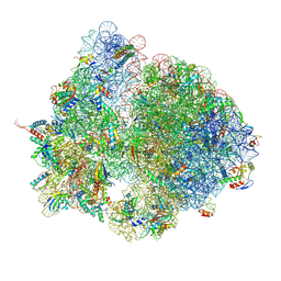
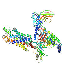
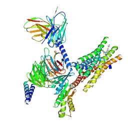
8XXH
 
 | | Structure of CXCR2 bound to CXCL2 (CXCR2-CXCL2-Go Full map) | | Descriptor: | Antibody fragment ScFv16, C-X-C chemokine receptor type 2, C-X-C motif chemokine 2, ... | | Authors: | Sano, F.K, Saha, S, Sharma, S, Ganguly, M, Shihoya, W, Nureki, O, Shukla, A.K, Banerjee, R. | | Deposit date: | 2024-01-18 | | Release date: | 2025-01-15 | | Last modified: | 2025-04-09 | | Method: | ELECTRON MICROSCOPY (2.8 Å) | | Cite: | Molecular basis of promiscuous chemokine binding and structural mimicry at the C-X-C chemokine receptor, CXCR2.
Mol.Cell, 85, 2025
|
|
7QJB
 | |
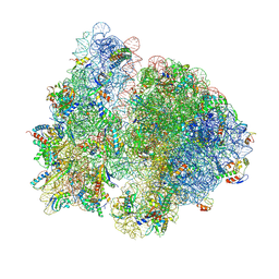
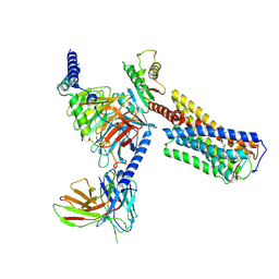
8XXR
 | | Structure of CXCR2 bound to CXCL6 (CXCR2-CXCL6-Go Full map) | | Descriptor: | Antibody fragment ScFv16, C-X-C chemokine receptor type 2, Guanine nucleotide-binding protein G(I)/G(S)/G(O) subunit gamma-2, ... | | Authors: | Sano, F.K, Saha, S, Sharma, S, Ganguly, M, Shihoya, W, Nureki, O, Shukla, A.K, Banerjee, R. | | Deposit date: | 2024-01-18 | | Release date: | 2025-01-15 | | Last modified: | 2025-04-09 | | Method: | ELECTRON MICROSCOPY (3.17 Å) | | Cite: | Molecular basis of promiscuous chemokine binding and structural mimicry at the C-X-C chemokine receptor, CXCR2.
Mol.Cell, 85, 2025
|
|
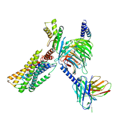
8XX3
 | | Structure of CXCR2 bound to CXCL3 (CXCR2-CXCL3-Go Full map) | | Descriptor: | Antibody fragment ScFv16, C-X-C chemokine receptor type 2, C-X-C motif chemokine 3, ... | | Authors: | Sano, F.K, Saha, S, Sharma, S, Ganguly, M, Shihoya, W, Nureki, O, Shukla, A.K, Banerjee, R. | | Deposit date: | 2024-01-17 | | Release date: | 2025-01-15 | | Last modified: | 2025-04-09 | | Method: | ELECTRON MICROSCOPY (3.38 Å) | | Cite: | Molecular basis of promiscuous chemokine binding and structural mimicry at the C-X-C chemokine receptor, CXCR2.
Mol.Cell, 85, 2025
|
|
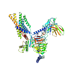
8XX6
 | | Structure of CXCR2 bound to CXCL8 (CXCR2-CXCL8-Go Full map) | | Descriptor: | Antibody fragment ScFv16, C-X-C chemokine receptor type 2, Guanine nucleotide-binding protein G(I)/G(S)/G(O) subunit gamma-2, ... | | Authors: | Sano, F.K, Saha, S, Sharma, S, Ganguly, M, Shihoya, W, Nureki, O, Shukla, A.K, Banerjee, R. | | Deposit date: | 2024-01-17 | | Release date: | 2025-01-15 | | Last modified: | 2025-04-09 | | Method: | ELECTRON MICROSCOPY (2.99 Å) | | Cite: | Molecular basis of promiscuous chemokine binding and structural mimicry at the C-X-C chemokine receptor, CXCR2.
Mol.Cell, 85, 2025
|
|
8XQF
 | | Cryo-EM structure of human monomeric APJR-Gi complex with apelin-13. | | Descriptor: | Apelin-13, G protein subunit alpha i1, Guanine nucleotide-binding protein G(I)/G(S)/G(O) subunit gamma-2, ... | | Authors: | Yue, Y, Liu, L.E, Wu, L.J, Xu, F. | | Deposit date: | 2024-01-05 | | Release date: | 2025-01-22 | | Last modified: | 2025-06-18 | | Method: | ELECTRON MICROSCOPY (3.13 Å) | | Cite: | Structural insights into the regulation of monomeric and dimeric apelin receptor.
Nat Commun, 16, 2025
|
|
8XXX
 | | Structure of CXCR2 bound to CXCL6 (Composite map) | | Descriptor: | Antibody fragment ScFv16, C-X-C chemokine receptor type 2, C-X-C motif chemokine 6, ... | | Authors: | Sano, F.K, Saha, S, Sharma, S, Ganguly, M, Shihoya, W, Nureki, O, Shukla, A.K, Banerjee, R. | | Deposit date: | 2024-01-19 | | Release date: | 2025-01-15 | | Last modified: | 2025-04-09 | | Method: | ELECTRON MICROSCOPY (3.17 Å) | | Cite: | Molecular basis of promiscuous chemokine binding and structural mimicry at the C-X-C chemokine receptor, CXCR2.
Mol.Cell, 85, 2025
|
|
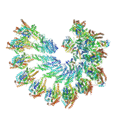
7QIN
 | | In situ structure of actomyosin complex in skeletal sarcomere | | Descriptor: | ADENOSINE-5'-DIPHOSPHATE, Actin, alpha skeletal muscle, ... | | Authors: | Wang, Z, Grange, M, Pospich, S, Wagner, T, Kho, A.L, Gautel, M, Raunser, S. | | Deposit date: | 2021-12-15 | | Release date: | 2022-02-16 | | Last modified: | 2022-03-02 | | Method: | ELECTRON MICROSCOPY (6.6 Å) | | Cite: | Structures from intact myofibrils reveal mechanism of thin filament regulation through nebulin.
Science, 375, 2022
|
|
8XWV
 | | Structure of CXCR2 bound to CXCL1 (CXCR2-CXCL1-Go Full map) | | Descriptor: | Antibody fragment ScFv16, C-X-C chemokine receptor type 2, Growth-regulated alpha protein, ... | | Authors: | Sano, F.K, Saha, S, Sharma, S, Ganguly, M, Shihoya, W, Nureki, O, Shukla, A.K, Banerjee, R. | | Deposit date: | 2024-01-16 | | Release date: | 2025-01-22 | | Last modified: | 2025-04-09 | | Method: | ELECTRON MICROSCOPY (3.07 Å) | | Cite: | Molecular basis of promiscuous chemokine binding and structural mimicry at the C-X-C chemokine receptor, CXCR2.
Mol.Cell, 85, 2025
|
|
8XQE
 | | Cryo-EM structure of human dimeric APJR-Gi complex with apelin-13. | | Descriptor: | Apelin-13, G protein subunit alpha i1, Guanine nucleotide-binding protein G(I)/G(S)/G(O) subunit gamma-2, ... | | Authors: | Yue, Y, Liu, L.E, Wu, L.J, Xu, F. | | Deposit date: | 2024-01-05 | | Release date: | 2025-01-22 | | Last modified: | 2025-07-02 | | Method: | ELECTRON MICROSCOPY (3.48 Å) | | Cite: | Structural insights into the regulation of monomeric and dimeric apelin receptor.
Nat Commun, 16, 2025
|
|
7QIM
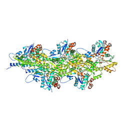 | | In situ structure of nebulin bound to actin filament in skeletal sarcomere | | Descriptor: | ACTS protein, ADENOSINE-5'-DIPHOSPHATE, MAGNESIUM ION, ... | | Authors: | Wang, Z, Grange, M, Pospich, S, Wagner, T, Kho, A.L, Gautel, M, Raunser, S. | | Deposit date: | 2021-12-15 | | Release date: | 2022-03-16 | | Method: | ELECTRON MICROSCOPY (4.5 Å) | | Cite: | Structures from intact myofibrils reveal mechanism of thin filament regulation through nebulin.
Science, 375, 2022
|
|
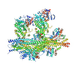
7QGH
 | | Structure of the E. coli disome - collided 70S ribosome | | Descriptor: | 16S rRNA, 23S rRNA, 30S ribosomal protein S1, ... | | Authors: | Kratzat, H, Buschauer, R, Berninghausen, O, Beckmann, R. | | Deposit date: | 2021-12-08 | | Release date: | 2022-03-16 | | Last modified: | 2024-11-20 | | Method: | ELECTRON MICROSCOPY (4.48 Å) | | Cite: | Ribosome collisions induce mRNA cleavage and ribosome rescue in bacteria.
Nature, 603, 2022
|
|
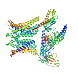
3VLG
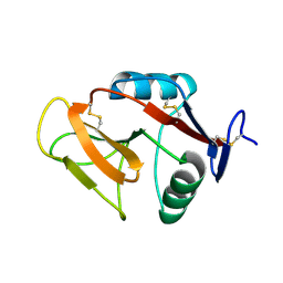 | | Crystal structure of the W150A mutant LOX-1 CTLD showing impaired OxLDL binding | | Descriptor: | Oxidized low-density lipoprotein receptor 1 | | Authors: | Nakano, S, Sugihara, M, Yamada, R, Katayanagi, K, Tate, S. | | Deposit date: | 2011-12-01 | | Release date: | 2012-04-18 | | Last modified: | 2024-10-30 | | Method: | X-RAY DIFFRACTION (2.3 Å) | | Cite: | Structural implication for the impaired binding of W150A mutant LOX-1 to oxidized low density lipoprotein, OxLDL
Biochim.Biophys.Acta, 1824, 2012
|
|
7QG8
 | | Structure of the collided E. coli disome - VemP-stalled 70S ribosome | | Descriptor: | 16S rRNA, 23S rRNA, 30S ribosomal protein S10, ... | | Authors: | Kratzat, H, Buschauer, R, Berninghausen, O, Beckmann, R. | | Deposit date: | 2021-12-07 | | Release date: | 2022-03-16 | | Last modified: | 2024-11-20 | | Method: | ELECTRON MICROSCOPY (3.97 Å) | | Cite: | Ribosome collisions induce mRNA cleavage and ribosome rescue in bacteria.
Nature, 603, 2022
|
|
8Y63
 | | Cryo-EM structure of the C20:0 ceramide-bound FPR2-Gi complex | | Descriptor: | Cer(d18:0/20:0), Guanine nucleotide-binding protein G(I)/G(S)/G(O) subunit gamma-2, Guanine nucleotide-binding protein G(I)/G(S)/G(T) subunit beta-1, ... | | Authors: | Sun, J.P, Jiang, C.T, Kong, W, Yu, X, Cai, K, Guo, L.L. | | Deposit date: | 2024-02-01 | | Release date: | 2025-02-05 | | Last modified: | 2025-06-18 | | Method: | ELECTRON MICROSCOPY (3.2 Å) | | Cite: | Metabolic signaling of ceramides through the FPR2 receptor inhibits adipocyte thermogenesis.
Science, 388, 2025
|
|
8Y62
 | | Cryo-EM structure of the C16:0 ceramide-bound FPR2-Gi complex | | Descriptor: | Guanine nucleotide-binding protein G(I)/G(S)/G(O) subunit gamma-2, Guanine nucleotide-binding protein G(I)/G(S)/G(T) subunit beta-1, Guanine nucleotide-binding protein G(i) subunit alpha-1, ... | | Authors: | Sun, J.P, Jiang, C.T, Kong, W, Yu, X, Cai, K, Guo, L.L. | | Deposit date: | 2024-02-01 | | Release date: | 2025-02-05 | | Last modified: | 2025-06-25 | | Method: | ELECTRON MICROSCOPY (3.2 Å) | | Cite: | Metabolic signaling of ceramides through the FPR2 receptor inhibits adipocyte thermogenesis.
Science, 388, 2025
|
|
8Y01
 | | Cryo-EM structure of Medium-wave-sensitive opsin 1 | | Descriptor: | Guanine nucleotide-binding protein G(I)/G(S)/G(O) subunit gamma-2, Guanine nucleotide-binding protein G(I)/G(S)/G(T) subunit beta-1, Guanine nucleotide-binding protein G(i) subunit alpha-1, ... | | Authors: | Peng, Q, Jiang, H.H, Cheng, X.Y, Li, J, Zhang, J. | | Deposit date: | 2024-01-21 | | Release date: | 2025-02-12 | | Method: | ELECTRON MICROSCOPY (2.48 Å) | | Cite: | Cryo-EM structure of Medium-wave-sensitive opsin 1
To Be Published
|
|
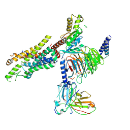
8Y51
 | | Cryo-EM structure of the BRS3-Gq complex | | Descriptor: | Bombesin receptor subtype-3, Guanine nucleotide-binding protein G(I)/G(S)/G(O) subunit gamma-2, Guanine nucleotide-binding protein G(I)/G(S)/G(T) subunit beta-1, ... | | Authors: | Li, C, Xu, Y, Yin, W, Xu, H.E. | | Deposit date: | 2024-01-31 | | Release date: | 2025-02-12 | | Method: | ELECTRON MICROSCOPY (3.3 Å) | | Cite: | Structural basis of self-activation and ligand recognition of human bombesin receptor subtype-3
To Be Published
|
|
8Y0N
 | | Structure of CXCR3 in complex with VUF11418 and Go (Full map) | | Descriptor: | Antibody fragment ScFv16, C-X-C chemokine receptor type 3, Guanine nucleotide-binding protein G(I)/G(S)/G(O) subunit gamma-2, ... | | Authors: | Sano, F.K, Saha, S, Sharma, S, Ganguly, M, Shihoya, W, Nureki, O, Shukla, A.K, Banerjee, R. | | Deposit date: | 2024-01-22 | | Release date: | 2025-02-26 | | Last modified: | 2025-04-09 | | Method: | ELECTRON MICROSCOPY (3.07 Å) | | Cite: | Structural visualization of small molecule recognition by CXCR3 uncovers dual-agonism in the CXCR3-CXCR7 system.
Nat Commun, 16, 2025
|
|
8XYK
 | | Structure of CXCR3 in complex with VUF10661 and Go (Full map) | | Descriptor: | (3~{S})-~{N}-[(2~{S})-6-azanyl-1-(2,2-diphenylethylamino)-1-oxidanylidene-hexan-2-yl]-2-(4-oxidanylidene-4-phenyl-butanoyl)-3,4-dihydro-1~{H}-isoquinoline-3-carboxamide, Antibody fragment ScFv16, C-X-C chemokine receptor type 3, ... | | Authors: | Sano, F.K, Saha, S, Sharma, S, Ganguly, M, Shihoya, W, Nureki, O, Shukla, A.K, Banerjee, R. | | Deposit date: | 2024-01-19 | | Release date: | 2025-02-26 | | Last modified: | 2025-04-09 | | Method: | ELECTRON MICROSCOPY (3.03 Å) | | Cite: | Structural visualization of small molecule recognition by CXCR3 uncovers dual-agonism in the CXCR3-CXCR7 system.
Nat Commun, 16, 2025
|
|
8XXZ
 | | Structure of CXCR3 in the apo-state (Full map) | | Descriptor: | Antibody fragment ScFv16, C-X-C chemokine receptor type 3, Guanine nucleotide-binding protein G(I)/G(S)/G(O) subunit gamma-2, ... | | Authors: | Sano, F.K, Saha, S, Sharma, S, Ganguly, M, Shihoya, W, Nureki, O, Shukla, A.K, Banerjee, R. | | Deposit date: | 2024-01-19 | | Release date: | 2025-02-26 | | Last modified: | 2025-04-09 | | Method: | ELECTRON MICROSCOPY (3.3 Å) | | Cite: | Structural visualization of small molecule recognition by CXCR3 uncovers dual-agonism in the CXCR3-CXCR7 system.
Nat Commun, 16, 2025
|
|
8Y6W
 | | TUG-1375 and 4-CMTB-bound human FFA2 in complex with Gi | | Descriptor: | (2R,4R)-2-(2-chlorophenyl)-3-[4-(3,5-dimethyl-1,2-oxazol-4-yl)phenyl]carbonyl-1,3-thiazolidine-4-carboxylic acid, (2~{S})-2-(4-chlorophenyl)-3-methyl-~{N}-(1,3-thiazol-2-yl)butanamide, Free fatty acid receptor 2, ... | | Authors: | Kugawa, M, Kawakami, K, Kise, R, Kobayashi, K, Kojima, A, Inoue, W, Fukuda, M, Inoue, A, Kato, H.E. | | Deposit date: | 2024-02-03 | | Release date: | 2025-04-02 | | Last modified: | 2025-04-09 | | Method: | ELECTRON MICROSCOPY (3.19 Å) | | Cite: | Structural insights into lipid chain-length selectivity and allosteric regulation of FFA2.
Nat Commun, 16, 2025
|
|
3SVP
 | | Structure of rat neuronal nitric oxide synthase heme domain in complex with 6-(((3R,4R)-4-(2-((2,2-Difluoro-2-(3-chloro-5-fluorophenyl)ethyl)amino)ethoxy)pyrrolidin-3-yl)methyl)-4-methylpyridin-2-amine | | Descriptor: | 5,6,7,8-TETRAHYDROBIOPTERIN, 6-{[(3R,4R)-4-(2-{[2-(3-chloro-5-fluorophenyl)-2,2-difluoroethyl]amino}ethoxy)pyrrolidin-3-yl]methyl}-4-methylpyridin-2-amine, ACETATE ION, ... | | Authors: | Li, H, Poulos, T.L. | | Deposit date: | 2011-07-12 | | Release date: | 2011-09-28 | | Last modified: | 2023-09-13 | | Method: | X-RAY DIFFRACTION (2.05 Å) | | Cite: | Improved Synthesis of Chiral Pyrrolidine Inhibitors and Their Binding Properties to Neuronal Nitric Oxide Synthase.
J.Med.Chem., 54, 2011
|
|
8Y02
 | | Cryo-EM structure of Short-wave-sensitive opsin 1 | | Descriptor: | Guanine nucleotide-binding protein G(I)/G(S)/G(O) subunit gamma-2, Guanine nucleotide-binding protein G(I)/G(S)/G(T) subunit beta-1, Guanine nucleotide-binding protein G(i) subunit alpha-1, ... | | Authors: | Peng, Q, Jiang, H.H, Cheng, X.Y, Li, J, Zhang, J. | | Deposit date: | 2024-01-21 | | Release date: | 2025-06-11 | | Method: | ELECTRON MICROSCOPY (2.61 Å) | | Cite: | Cryo-EM structure of Short-wave-sensitive opsin 1
To Be Published
|
|
3T0O
 | | Crystal Structure Analysis of Human RNase T2 | | Descriptor: | 2-acetamido-2-deoxy-beta-D-glucopyranose, Ribonuclease T2 | | Authors: | Thorn, A, Kraetzner, R, Steinfeld, R, Sheldrick, G. | | Deposit date: | 2011-07-20 | | Release date: | 2012-07-11 | | Last modified: | 2024-11-20 | | Method: | X-RAY DIFFRACTION (1.59 Å) | | Cite: | Structure and activity of the only human RNase T2.
Nucleic Acids Res., 40, 2012
|
|


