3THJ
 
 | |
7BA9
 | | Cys-42-tethered stabilizer 11 of 14-3-3(sigma)/ERa PPI | | Descriptor: | 14-3-3 protein sigma, 2-methyl-2-(4-methylphenoxy)-~{N}-(2-sulfanylethyl)propanamide, Estrogen receptor, ... | | Authors: | Sijbesma, E, Ottmann, C. | | Deposit date: | 2020-12-15 | | Release date: | 2021-07-07 | | Last modified: | 2024-10-23 | | Method: | X-RAY DIFFRACTION (1.48 Å) | | Cite: | Exploration of a 14-3-3 PPI Pocket by Covalent Fragments as Stabilizers.
Acs Med.Chem.Lett., 12, 2021
|
|
1O6L
 | | Crystal structure of an activated Akt/protein kinase B (PKB-PIF chimera) ternary complex with AMP-PNP and GSK3 peptide | | Descriptor: | GLYCOGEN SYNTHASE KINASE-3 BETA, MANGANESE (II) ION, PHOSPHOAMINOPHOSPHONIC ACID-ADENYLATE ESTER, ... | | Authors: | Yang, J, Cron, P, Good, V.M, Thompson, V, Hemmings, B.A, Barford, D. | | Deposit date: | 2002-10-08 | | Release date: | 2002-11-19 | | Last modified: | 2024-11-20 | | Method: | X-RAY DIFFRACTION (1.6 Å) | | Cite: | Crystal Structure of an Activated Akt/Protein Kinase B Ternary Complex with Gsk-3 Peptide and AMP-Pnp
Nat.Struct.Biol., 9, 2002
|
|
1AVL
 | |
7BAA
 | | Cys-42-tethered stabilizer 12 of 14-3-3(sigma)/ERa PPI | | Descriptor: | 14-3-3 protein sigma, 2-(4-bromanyl-3-methoxy-phenoxy)-2-methyl-~{N}-(2-sulfanylethyl)propanamide, Estrogen receptor, ... | | Authors: | Sijbesma, E, Ottmann, C. | | Deposit date: | 2020-12-15 | | Release date: | 2021-07-07 | | Last modified: | 2024-11-20 | | Method: | X-RAY DIFFRACTION (1.1 Å) | | Cite: | Exploration of a 14-3-3 PPI Pocket by Covalent Fragments as Stabilizers.
Acs Med.Chem.Lett., 12, 2021
|
|
7B9T
 | | Cys-45-tethered stabilizer 5 of 14-3-3(sigma)/ERa PPI | | Descriptor: | 14-3-3 protein sigma, 2-(4-chlorophenyl)sulfanyl-~{N}-(3-sulfanylpropyl)ethanamide, Estrogen receptor, ... | | Authors: | Sijbesma, E, Ottmann, C. | | Deposit date: | 2020-12-14 | | Release date: | 2021-07-07 | | Last modified: | 2024-10-09 | | Method: | X-RAY DIFFRACTION (1.15 Å) | | Cite: | Exploration of a 14-3-3 PPI Pocket by Covalent Fragments as Stabilizers.
Acs Med.Chem.Lett., 12, 2021
|
|
7BJK
 | |
1VAS
 | | ATOMIC MODEL OF A PYRIMIDINE DIMER SPECIFIC EXCISION REPAIR ENZYME COMPLEXED WITH A DNA SUBSTRATE: STRUCTURAL BASIS FOR DAMAGED DNA RECOGNITION | | Descriptor: | DNA (5'-D(*AP*TP*CP*GP*CP*GP*TP*TP*GP*CP*GP*CP*T)-3'), DNA (5'-D(*TP*AP*GP*CP*GP*CP*AP*AP*CP*GP*CP*GP*A)-3'), PROTEIN (T4 ENDONUCLEASE V (E.C.3.1.25.1)) | | Authors: | Vassylyev, D.G, Kashiwagi, T, Mikami, Y, Ariyoshi, M, Iwai, S, Ohtsuka, E, Morikawa, K. | | Deposit date: | 1995-09-08 | | Release date: | 1996-01-31 | | Last modified: | 2024-02-14 | | Method: | X-RAY DIFFRACTION (2.75 Å) | | Cite: | Atomic model of a pyrimidine dimer excision repair enzyme complexed with a DNA substrate: structural basis for damaged DNA recognition.
Cell(Cambridge,Mass.), 83, 1995
|
|
4ITU
 | |
7BA6
 | | Cys-42-tethered stabilizer 8 of 14-3-3(sigma)/ERa PPI | | Descriptor: | 14-3-3 protein sigma, 2-[3,5-bis(fluoranyl)phenoxy]-2-methyl-~{N}-(2-sulfanylethyl)propanamide, Estrogen receptor, ... | | Authors: | Sijbesma, E, Ottmann, C. | | Deposit date: | 2020-12-15 | | Release date: | 2021-07-07 | | Last modified: | 2024-11-20 | | Method: | X-RAY DIFFRACTION (1.4 Å) | | Cite: | Exploration of a 14-3-3 PPI Pocket by Covalent Fragments as Stabilizers.
Acs Med.Chem.Lett., 12, 2021
|
|
7BAB
 | | Cys-42-tethered stabilizer 13 of 14-3-3(sigma)/ERa PPI | | Descriptor: | 14-3-3 protein sigma, 2-[2-chloranyl-4-[oxidanyl(oxidanylidene)-$l^{4}-azanyl]phenoxy]-2-methyl-~{N}-(2-sulfanylethyl)propanamide, Estrogen receptor, ... | | Authors: | Sijbesma, E, Ottmann, C. | | Deposit date: | 2020-12-15 | | Release date: | 2021-07-07 | | Last modified: | 2024-10-23 | | Method: | X-RAY DIFFRACTION (1.3 Å) | | Cite: | Exploration of a 14-3-3 PPI Pocket by Covalent Fragments as Stabilizers.
Acs Med.Chem.Lett., 12, 2021
|
|
6EIH
 | |
2WQ2
 | |
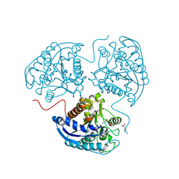
3TEV
 | | The crystal structure of glycosyl hydrolase from Deinococcus radiodurans R1 | | Descriptor: | Glycosyl hyrolase, family 3 | | Authors: | Chang, C, Hatzos-Skintges, C, Kohler, M, Clancy, S, Joachimiak, A, Midwest Center for Structural Genomics (MCSG) | | Deposit date: | 2011-08-15 | | Release date: | 2011-08-31 | | Last modified: | 2024-11-27 | | Method: | X-RAY DIFFRACTION (2.3 Å) | | Cite: | The crystal structure of glycosyl hydrolase from Deinococcus radiodurans R1
To be Published
|
|
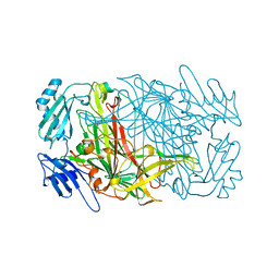
7BV7
 | | INTS3 complexed with INTS6 | | Descriptor: | Integrator complex subunit 3, Integrator complex subunit 6 | | Authors: | Jia, Y, Bharath, S.R, Song, H. | | Deposit date: | 2020-04-09 | | Release date: | 2021-07-14 | | Last modified: | 2024-05-29 | | Method: | X-RAY DIFFRACTION (2.4 Å) | | Cite: | Crystal structure of the INTS3/INTS6 complex reveals the functional importance of INTS3 dimerization in DSB repair.
Cell Discov, 7, 2021
|
|
5AEM
 | | Structure of t131 N-terminal TPR array | | Descriptor: | TRANSCRIPTION FACTOR TAU 131 KDA SUBUNIT | | Authors: | Taylor, N.M.I, Muller, C.W. | | Deposit date: | 2015-01-05 | | Release date: | 2015-06-24 | | Last modified: | 2024-05-08 | | Method: | X-RAY DIFFRACTION (3.4 Å) | | Cite: | Architecture of TFIIIC and its role in RNA polymerase III pre-initiation complex assembly.
Nat Commun, 6, 2015
|
|
4IXQ
 | | RT fs X-ray diffraction of Photosystem II, dark state | | Descriptor: | 1,2-DI-O-ACYL-3-O-[6-DEOXY-6-SULFO-ALPHA-D-GLUCOPYRANOSYL]-SN-GLYCEROL, 1,2-DIPALMITOYL-PHOSPHATIDYL-GLYCEROLE, 1,2-DISTEAROYL-MONOGALACTOSYL-DIGLYCERIDE, ... | | Authors: | Kern, J, Alonso-Mori, R, Tran, R, Hattne, J, Gildea, R.J, Echols, N, Gloeckner, C, Hellmich, J, Laksmono, H, Sierra, R.G, Lassalle-Kaiser, B, Koroidov, S, Lampe, A, Han, G, Gul, S, DiFiore, D, Milathianaki, D, Fry, A.R, Miahnahri, A, Schafer, D.W, Messerschmidt, M, Seibert, M.M, Koglin, J.E, Sokaras, D, Weng, T.-C, Sellberg, J, Latimer, M.J, Grosse-Kunstleve, R.W, Zwart, P.H, White, W.E, Glatzel, P, Adams, P.D, Bogan, M.J, Williams, G.J, Boutet, S, Messinger, J, Zouni, A, Sauter, N.K, Yachandra, V.K, Bergmann, U, Yano, J. | | Deposit date: | 2013-01-27 | | Release date: | 2013-02-20 | | Last modified: | 2024-10-30 | | Method: | X-RAY DIFFRACTION (5.7 Å) | | Cite: | Simultaneous femtosecond X-ray spectroscopy and diffraction of photosystem II at room temperature.
Science, 340, 2013
|
|
1CU3
 | | T4 LYSOZYME MUTANT V87M | | Descriptor: | 2-HYDROXYETHYL DISULFIDE, CHLORIDE ION, LYSOZYME | | Authors: | Gassner, N.C, Baase, W.A, Lindstrom, J.D, Lu, J, Matthews, B.W. | | Deposit date: | 1999-08-20 | | Release date: | 1999-11-10 | | Last modified: | 2024-02-07 | | Method: | X-RAY DIFFRACTION (2.12 Å) | | Cite: | Methionine and alanine substitutions show that the formation of wild-type-like structure in the carboxy-terminal domain of T4 lysozyme is a rate-limiting step in folding.
Biochemistry, 38, 1999
|
|
1CEO
 | |
7B9M
 | | Cys-45-tethered stabilizer 3 of 14-3-3(sigma)/ERa PPI | | Descriptor: | 14-3-3 protein sigma, 2-(2-cyanophenyl)sulfanyl-~{N}-(2-sulfanylethyl)benzamide, Estrogen receptor, ... | | Authors: | Sijbesma, E, Ottmann, C. | | Deposit date: | 2020-12-14 | | Release date: | 2021-07-07 | | Last modified: | 2024-11-13 | | Method: | X-RAY DIFFRACTION (1.7 Å) | | Cite: | Exploration of a 14-3-3 PPI Pocket by Covalent Fragments as Stabilizers.
Acs Med.Chem.Lett., 12, 2021
|
|
1CF3
 | | GLUCOSE OXIDASE FROM APERGILLUS NIGER | | Descriptor: | 2-acetamido-2-deoxy-beta-D-glucopyranose, FLAVIN-ADENINE DINUCLEOTIDE, PROTEIN (GLUCOSE OXIDASE), ... | | Authors: | Hecht, H.J, Kalisz, H. | | Deposit date: | 1999-03-23 | | Release date: | 1999-03-26 | | Last modified: | 2024-10-30 | | Method: | X-RAY DIFFRACTION (1.9 Å) | | Cite: | 1.8 and 1.9 A resolution structures of the Penicillium amagasakiense and Aspergillus niger glucose oxidases as a basis for modelling substrate complexes.
Acta Crystallogr.,Sect.D, 55, 1999
|
|
7B9R
 | | Cys-45-tethered stabilizer 4 of 14-3-3(sigma)/ERa PPI | | Descriptor: | 14-3-3 protein sigma, 2-(2-cyanophenyl)sulfanyl-~{N}-(3-sulfanylpropyl)benzamide, Estrogen receptor, ... | | Authors: | Sijbesma, E, Ottmann, C. | | Deposit date: | 2020-12-14 | | Release date: | 2021-07-07 | | Last modified: | 2024-11-20 | | Method: | X-RAY DIFFRACTION (1.15 Å) | | Cite: | Exploration of a 14-3-3 PPI Pocket by Covalent Fragments as Stabilizers.
Acs Med.Chem.Lett., 12, 2021
|
|
7BA3
 | | Cys-42-tethered stabilizer 6 of 14-3-3(sigma)/ERa PPI | | Descriptor: | 14-3-3 protein sigma, 2-(4-bromanylphenoxy)-2-methyl-~{N}-(2-sulfanylethyl)propanamide, Estrogen receptor, ... | | Authors: | Sijbesma, E, Ottmann, C. | | Deposit date: | 2020-12-15 | | Release date: | 2021-07-07 | | Last modified: | 2024-11-13 | | Method: | X-RAY DIFFRACTION (1.4 Å) | | Cite: | Exploration of a 14-3-3 PPI Pocket by Covalent Fragments as Stabilizers.
Acs Med.Chem.Lett., 12, 2021
|
|
3P4T
 | | Crystal structure of a putative acyl-CoA dehydrogenase from Mycobacterium smegmatis | | Descriptor: | 1,2-ETHANEDIOL, Putative acyl-CoA dehydrogenase, [(2R,3S,4R,5R)-5-(6-amino-9H-purin-9-yl)-3,4-dihydroxytetrahydrofuran-2-yl]methyl (2R,3S,4S)-5-[(4aS,10aR)-7,8-dimethyl-2,4-dioxo-1,3,4,4a,5,10a-hexahydrobenzo[g]pteridin-10(2H)-yl]-2,3,4-trihydroxypentyl dihydrogen diphosphate | | Authors: | Seattle Structural Genomics Center for Infectious Disease (SSGCID) | | Deposit date: | 2010-10-07 | | Release date: | 2010-10-20 | | Last modified: | 2023-09-06 | | Method: | X-RAY DIFFRACTION (1.7 Å) | | Cite: | Increasing the structural coverage of tuberculosis drug targets.
Tuberculosis (Edinb), 95, 2015
|
|
1CUP
 | | METHIONINE CORE MUTANT OF T4 LYSOZYME | | Descriptor: | 2-HYDROXYETHYL DISULFIDE, CHLORIDE ION, LYSOZYME | | Authors: | Gassner, N.C, Baase, W.A, Lindstrom, J.D, Lu, J, Matthews, B.W. | | Deposit date: | 1999-08-20 | | Release date: | 1999-11-10 | | Last modified: | 2024-02-07 | | Method: | X-RAY DIFFRACTION (1.89 Å) | | Cite: | Methionine and alanine substitutions show that the formation of wild-type-like structure in the carboxy-terminal domain of T4 lysozyme is a rate-limiting step in folding.
Biochemistry, 38, 1999
|
|


