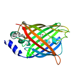
Loading
PDBj
MenuPDBj@FacebookPDBj@TwitterPDBj@YouTubewwPDB FoundationwwPDB
RCSB PDBPDBeBMRBAdv. SearchSearch help
PDB: 524 results

7O7D
DownloadVisualize
BU of 7o7d by Molmil
Crystal structure of rsEGFP2 mutant V151A in the fluorescent on-state determined by synchrotron radiation at 100K
Descriptor: Green fluorescent protein
Authors:Woodhouse, J, Adam, V, Hadjidemetriou, K, Colletier, J.-P, Weik, M.
Deposit date:2021-04-13
Release date:2022-07-27
Last modified:2024-01-31
Method:X-RAY DIFFRACTION (1.4 Å)
Cite:Rational Control of Off-State Heterogeneity in a Photoswitchable Fluorescent Protein Provides Switching Contrast Enhancement.
Chemphyschem, 23, 2022
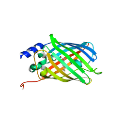
2B3P
DownloadVisualize
BU of 2b3p by Molmil
Crystal structure of a superfolder green fluorescent protein
Descriptor: ACETIC ACID, CADMIUM ION, green fluorescent protein
Authors:Pedelacq, J.D, Cabantous, S, Tran, T.H, Terwilliger, T.C, Waldo, G.S.
Deposit date:2005-09-20
Release date:2005-11-08
Last modified:2023-11-15
Method:X-RAY DIFFRACTION (1.4 Å)
Cite:Engineering and characterization of a superfolder green fluorescent protein.
Nat.Biotechnol., 24, 2006
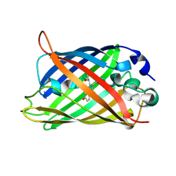
6KRG
DownloadVisualize
BU of 6krg by Molmil
Crystal structure of sfGFP Y182TMSiPhe
Descriptor: CHLORIDE ION, GLYCEROL, Green fluorescent protein, ...
Authors:Sun, J.P, Wang, J.Y, Zhu, Z.L, He, Q.T, Xiao, P.
Deposit date:2019-08-21
Release date:2020-09-09
Last modified:2023-11-22
Method:X-RAY DIFFRACTION (1.4 Å)
Cite:DeSiphering receptor core-induced and ligand-dependent conformational changes in arrestin via genetic encoded trimethylsilyl 1 H-NMR probe.
Nat Commun, 11, 2020
2FWQ
DownloadVisualize
BU of 2fwq by Molmil
Reduced enolate chromophore intermediate for Y66H GFP variant
Descriptor: Green fluorescent protein, MAGNESIUM ION
Authors:Barondeau, D.P, Tainer, J.A, Getzoff, E.D.
Deposit date:2006-02-02
Release date:2006-03-14
Last modified:2023-11-15
Method:X-RAY DIFFRACTION (1.4 Å)
Cite:Structural evidence for an enolate intermediate in GFP fluorophore biosynthesis.
J.Am.Chem.Soc., 128, 2006
3DQ9
DownloadVisualize
BU of 3dq9 by Molmil
Structure of the Yellow Fluorescent Protein Citrine Frozen at 1500 Atmospheres Number 1: Structure 15 in a Series of 26 High Pressure Structures
Descriptor: Green fluorescent protein
Authors:Barstow, B, Kim, C.U.
Deposit date:2008-07-09
Release date:2008-09-23
Last modified:2023-11-15
Method:X-RAY DIFFRACTION (1.4 Å)
Cite:Alteration of citrine structure by hydrostatic pressure explains the accompanying spectral shift.
Proc.Natl.Acad.Sci.Usa, 105, 2008
3DQD
DownloadVisualize
BU of 3dqd by Molmil
Structure of the Yellow Fluorescent Protein Citrine Frozen at 1250 Atmospheres Number 2: Structure 12 in a Series of 26 High Pressure Structures
Descriptor: Green fluorescent protein
Authors:Barstow, B, Kim, C.U.
Deposit date:2008-07-09
Release date:2008-09-23
Last modified:2023-11-15
Method:X-RAY DIFFRACTION (1.4 Å)
Cite:Alteration of citrine structure by hydrostatic pressure explains the accompanying spectral shift.
Proc.Natl.Acad.Sci.Usa, 105, 2008
3DQK
DownloadVisualize
BU of 3dqk by Molmil
Structure of the Yellow Fluorescent Protein Citrine Frozen at 1000 Atmospheres Number 2: Structure 6 in a Series of 26 High Pressure Structures
Descriptor: Green fluorescent protein
Authors:Barstow, B, Kim, C.U.
Deposit date:2008-07-09
Release date:2008-09-23
Last modified:2023-11-15
Method:X-RAY DIFFRACTION (1.4 Å)
Cite:Alteration of citrine structure by hydrostatic pressure explains the accompanying spectral shift.
Proc.Natl.Acad.Sci.Usa, 105, 2008
2HRS
DownloadVisualize
BU of 2hrs by Molmil
Crystal structure of L42H V224H design intermediate for GFP metal ion reporter
Descriptor: 1,2-ETHANEDIOL, Green fluorescent protein, MAGNESIUM ION
Authors:Barondeau, D.P, Tubbs, J.L, Tainer, J.A, Getzoff, E.D.
Deposit date:2006-07-20
Release date:2008-04-08
Last modified:2023-11-15
Method:X-RAY DIFFRACTION (1.4 Å)
Cite:Iterative Structure-Based Design of a Green Fluorescent Protein Metal Ion Reporter
To be Published
3AI5
DownloadVisualize
BU of 3ai5 by Molmil
Crystal structure of yeast enhanced green fluorescent protein-ubiquitin fusion protein
Descriptor: 1,2-ETHANEDIOL, yeast enhanced green fluorescent protein,Ubiquitin
Authors:Suzuki, N, Wakatsuki, S, Kawasaki, M.
Deposit date:2010-05-10
Release date:2010-09-29
Last modified:2023-11-15
Method:X-RAY DIFFRACTION (1.4 Å)
Cite:Crystallization of small proteins assisted by green fluorescent protein
Acta Crystallogr.,Sect.D, 66, 2010
2DUG
DownloadVisualize
BU of 2dug by Molmil
crystal structure of a green fluorescent protein S65T/H148N at pH 5
Descriptor: Green fluorescent protein
Authors:Shu, X, Remington, S.J.
Deposit date:2006-07-23
Release date:2007-07-10
Last modified:2021-11-10
Method:X-RAY DIFFRACTION (1.4 Å)
Cite:Ultrafast excited-state dynamics in the green fluorescent protein variant S65T/H148D. 1. Mutagenesis and structural studies.
Biochemistry, 46, 2007
3SST
DownloadVisualize
BU of 3sst by Molmil
Engineered low-affinity halide-binding protein derived from YFP: chloride complex
Descriptor: CHLORIDE ION, Green fluorescent protein
Authors:Wang, W, Grimley, J.S, Beese, L.S, Hellinga, H.W.
Deposit date:2011-07-08
Release date:2012-07-11
Last modified:2023-11-15
Method:X-RAY DIFFRACTION (1.403 Å)
Cite:Determination of engineered chloride-binding site structures in fluorescent proteins reveals principles of halide recognition
To be Published
5DPH
DownloadVisualize
BU of 5dph by Molmil
sfGFP mutant - 149 p-cyano-L-phenylalanine
Descriptor: CARBON DIOXIDE, Green fluorescent protein, MAGNESIUM ION
Authors:Dippel, A.B, Olenginski, G.M, Maurici, N, Liskov, M.T, Brewer, S.H, Phillips-Piro, C.M.
Deposit date:2015-09-12
Release date:2016-01-13
Last modified:2023-11-15
Method:X-RAY DIFFRACTION (1.42 Å)
Cite:Probing the effectiveness of spectroscopic reporter unnatural amino acids: a structural study.
Acta Crystallogr D Struct Biol, 72, 2016
3DQI
DownloadVisualize
BU of 3dqi by Molmil
Structure of the Yellow Fluorescent Protein Citrine Frozen at 1000 Atmospheres Number 4: Structure 8 in a Series of 26 High Pressure Structures
Descriptor: Green fluorescent protein
Authors:Barstow, B, Kim, C.U.
Deposit date:2008-07-09
Release date:2008-09-23
Last modified:2023-11-15
Method:X-RAY DIFFRACTION (1.42 Å)
Cite:Alteration of citrine structure by hydrostatic pressure explains the accompanying spectral shift.
Proc.Natl.Acad.Sci.Usa, 105, 2008
3DQU
DownloadVisualize
BU of 3dqu by Molmil
Structure of the Yellow Fluorescent Protein Citrine Frozen at 2000 Atmospheres Number 1: Structure 20 in a Series of 26 High Pressure Structures
Descriptor: Green fluorescent protein
Authors:Barstow, B, Kim, C.U.
Deposit date:2008-07-09
Release date:2008-09-23
Last modified:2023-11-15
Method:X-RAY DIFFRACTION (1.42 Å)
Cite:Alteration of citrine structure by hydrostatic pressure explains the accompanying spectral shift.
Proc.Natl.Acad.Sci.Usa, 105, 2008
7PNN
DownloadVisualize
BU of 7pnn by Molmil
mVenus released from fusion protein.
Descriptor: CHLORIDE ION, mVenus
Authors:Bloch, Y, Savvides, S.N.
Deposit date:2021-09-07
Release date:2021-11-03
Last modified:2024-01-31
Method:X-RAY DIFFRACTION (1.43 Å)
Cite:mVenus released from fusion protein.
To Be Published
3DQE
DownloadVisualize
BU of 3dqe by Molmil
Structure of the Yellow Fluorescent Protein Citrine Frozen at 1250 Atmospheres Number 1: Structure 11 in a Series of 26 High Pressure Structures
Descriptor: Green fluorescent protein
Authors:Barstow, B, Kim, C.U.
Deposit date:2008-07-09
Release date:2008-09-23
Last modified:2023-11-15
Method:X-RAY DIFFRACTION (1.43 Å)
Cite:Alteration of citrine structure by hydrostatic pressure explains the accompanying spectral shift.
Proc.Natl.Acad.Sci.Usa, 105, 2008
1KYS
DownloadVisualize
BU of 1kys by Molmil
Crystal Structure of a Zn-bound Green Fluorescent Protein Biosensor
Descriptor: Green Fluorescent Protein, ZINC ION
Authors:Barondeau, D.P, Kassmann, C.J, Tainer, J.A, Getzoff, E.D.
Deposit date:2002-02-05
Release date:2002-04-10
Last modified:2023-11-15
Method:X-RAY DIFFRACTION (1.44 Å)
Cite:Structural chemistry of a green fluorescent protein Zn biosensor.
J.Am.Chem.Soc., 124, 2002
3DQA
DownloadVisualize
BU of 3dqa by Molmil
Structure of the Yellow Fluorescent Protein Citrine Frozen at 1250 Atmospheres Number 4: Structure 14 in a Series of 26 High Pressure Structures
Descriptor: Green fluorescent protein
Authors:Barstow, B, Kim, C.U.
Deposit date:2008-07-09
Release date:2008-09-23
Last modified:2023-11-15
Method:X-RAY DIFFRACTION (1.44 Å)
Cite:Alteration of citrine structure by hydrostatic pressure explains the accompanying spectral shift.
Proc.Natl.Acad.Sci.Usa, 105, 2008
3DQM
DownloadVisualize
BU of 3dqm by Molmil
Structure of the Yellow Fluorescent Protein Citrine Frozen at 750 Atmospheres: Structure 4 in a Series of 26 High Pressure Structures
Descriptor: Green fluorescent protein
Authors:Barstow, B, Kim, C.U.
Deposit date:2008-07-09
Release date:2008-09-23
Last modified:2023-11-15
Method:X-RAY DIFFRACTION (1.44 Å)
Cite:Alteration of citrine structure by hydrostatic pressure explains the accompanying spectral shift.
Proc.Natl.Acad.Sci.Usa, 105, 2008
3DQN
DownloadVisualize
BU of 3dqn by Molmil
Structure of the Yellow Fluorescent Protein Citrine Frozen at 500 Atmospheres: Structure 3 in a Series of 26 High Pressure Structures
Descriptor: Green fluorescent protein
Authors:Barstow, B, Kim, C.U.
Deposit date:2008-07-09
Release date:2008-09-23
Last modified:2023-11-15
Method:X-RAY DIFFRACTION (1.44 Å)
Cite:Alteration of citrine structure by hydrostatic pressure explains the accompanying spectral shift.
Proc.Natl.Acad.Sci.Usa, 105, 2008
3SSL
DownloadVisualize
BU of 3ssl by Molmil
Engineered high-affinity halide-binding protein derived from YFP: iodide complex
Descriptor: Green fluorescent protein, IODIDE ION
Authors:Wang, W, Grimley, J.S, Beese, L.S, Hellinga, H.W.
Deposit date:2011-07-08
Release date:2012-07-11
Last modified:2023-11-15
Method:X-RAY DIFFRACTION (1.449 Å)
Cite:Determination of engineered chloride-binding site structures in fluorescent proteins reveals principles of halide recognition
To be published
6PFT
DownloadVisualize
BU of 6pft by Molmil
rsEGFP2 with a chlorinated chromophore in the non-fluorescent off-state
Descriptor: Green fluorescent protein, SULFATE ION
Authors:Chang, J, Romei, M.G, Boxer, S.G.
Deposit date:2019-06-22
Release date:2019-08-07
Last modified:2023-11-15
Method:X-RAY DIFFRACTION (1.45 Å)
Cite:Structural Evidence of Photoisomerization Pathways in Fluorescent Proteins.
J.Am.Chem.Soc., 141, 2019
5DTX
DownloadVisualize
BU of 5dtx by Molmil
Crystal structure of rsEGFP2 in the fluorescent on-state
Descriptor: Green fluorescent protein
Authors:Adam, V, Martins, A.
Deposit date:2015-09-18
Release date:2016-01-20
Last modified:2024-01-10
Method:X-RAY DIFFRACTION (1.45 Å)
Cite:Rational design of ultrastable and reversibly photoswitchable fluorescent proteins for super-resolution imaging of the bacterial periplasm.
Sci Rep, 6, 2016
1Q4A
DownloadVisualize
BU of 1q4a by Molmil
S65T Q80R Green Fluorescent Protein (GFP) pH 8.5
Descriptor: Green Fluorescent Protein
Authors:Jain, R.K, Ranganathan, R.
Deposit date:2003-08-02
Release date:2004-02-03
Last modified:2023-11-15
Method:X-RAY DIFFRACTION (1.45 Å)
Cite:Local complexity of amino acid interactions in a protein core.
Proc.Natl.Acad.Sci.USA, 101, 2004
4H48
DownloadVisualize
BU of 4h48 by Molmil
1.45 angstrom CyPet Structure at pH7.0
Descriptor: 2-AMINO-2-HYDROXYMETHYL-PROPANE-1,3-DIOL, Green fluorescent protein
Authors:Hu, X.-J, Liu, R.
Deposit date:2012-09-17
Release date:2013-09-18
Last modified:2023-12-06
Method:X-RAY DIFFRACTION (1.45 Å)
Cite:Structure insight of the fluorescent state of CyPet
To be Published

221716

PDB entries from 2024-06-26

PDB statisticsPDBj update infoContact PDBjnumon