Loading
PDBj
MenuPDBj@FacebookPDBj@TwitterPDBj@YouTubewwPDB FoundationwwPDB
RCSB PDBPDBeBMRBAdv. SearchSearch help
PDB: 591 results

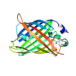
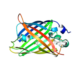
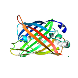
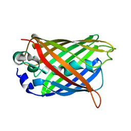
2DUF
DownloadVisualize
BU of 2duf by Molmil
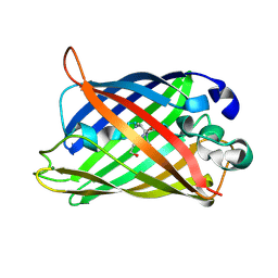
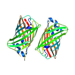
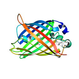
crystal structure of a green fluorescent protein variant S65T/H148D at pH 5.6
Descriptor: Green fluorescent protein
Authors:Shu, X, Remington, S.J.
Deposit date:2006-07-23
Release date:2007-07-10
Last modified:2021-11-10
Method:X-RAY DIFFRACTION (1.5 Å)
Cite:Ultrafast excited-state dynamics in the green fluorescent protein variant S65T/H148D. 1. Mutagenesis and structural studies.
Biochemistry, 46, 2007
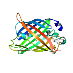
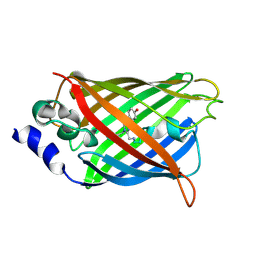
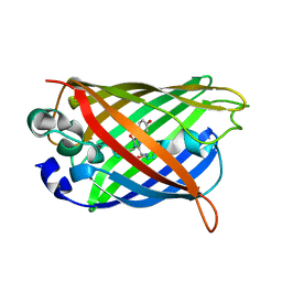
2QRF
DownloadVisualize
BU of 2qrf by Molmil
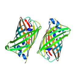
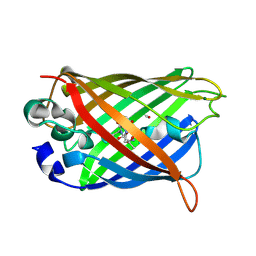
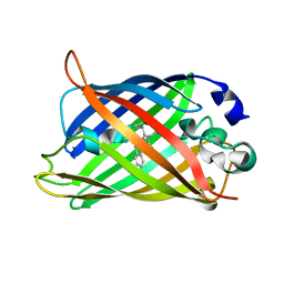
Green Fluorescent Protein: Cyclized-only Intermediate of Chromophore Maturation in the Q183E variant
Descriptor: 1,2-ETHANEDIOL, Green fluorescent protein, MAGNESIUM ION
Authors:Wood, T.I, Barondeau, D.P, Hitomi, C, Tainer, J.A, Getzoff, E.D.
Deposit date:2007-07-28
Release date:2009-04-21
Last modified:2023-11-15
Method:X-RAY DIFFRACTION (1.5 Å)
Cite:Green Fluorescent Protein Chromophore - Cyclized Intermediate
To be Published
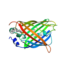
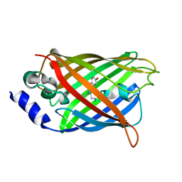
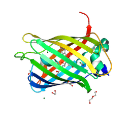
1YJ2
DownloadVisualize
BU of 1yj2 by Molmil
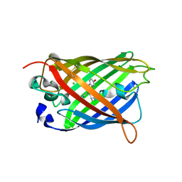
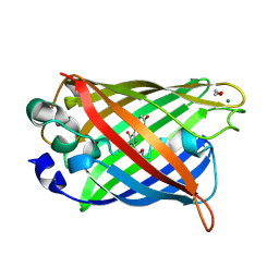
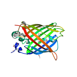
Cyclized, non-dehydrated post-translational product for S65A Y66S H148G GFP variant
Descriptor: 1,2-ETHANEDIOL, Green Fluorescent Protein, MAGNESIUM ION
Authors:Barondeau, D.P, Kassmann, C.J, Tainer, J.A, Getzoff, E.D.
Deposit date:2005-01-13
Release date:2005-02-15
Last modified:2021-10-20
Method:X-RAY DIFFRACTION (1.5 Å)
Cite:Understanding GFP Chromophore Biosynthesis: Controlling Backbone Cyclization and Modifying Post-translational Chemistry.
Biochemistry, 44, 2005
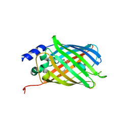
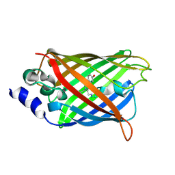
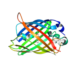
6UN7
DownloadVisualize
BU of 6un7 by Molmil
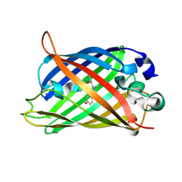
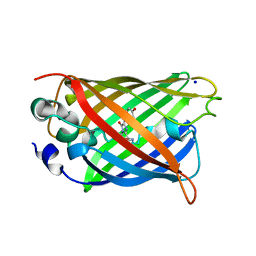
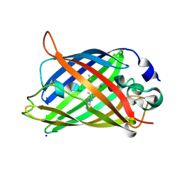
Crystal structure of green fluorescent protein (GFP); S65T, Y66(3-OMeY); ih circular permutant (50-51)
Descriptor: Green fluorescent protein
Authors:Lin, C.-Y, Boxer, S.G.
Deposit date:2019-10-10
Release date:2020-10-14
Last modified:2023-11-15
Method:X-RAY DIFFRACTION (1.5 Å)
Cite:Unusual Spectroscopic and Electric Field Sensitivity of Chromophores with Short Hydrogen Bonds: GFP and PYP as Model Systems.
J.Phys.Chem.B, 124, 2020
6UHJ
DownloadVisualize
BU of 6uhj by Molmil
X-ray Structure of C148 mGFP
Descriptor: C148 mGFP
Authors:Winegar, P.W, Hayes, O.G, McMillan, J.R, Figg, C.A, Focia, P.J, Mirkin, C.A.
Deposit date:2019-09-27
Release date:2020-03-18
Last modified:2023-11-15
Method:X-RAY DIFFRACTION (1.5 Å)
Cite:DNA-Directed Protein Packing within Single Crystals.
Chem, 6, 2020
6UN6
DownloadVisualize
BU of 6un6 by Molmil
Crystal structure of green fluorescent protein (GFP); S65T, Y66(3-NO2Y); ih circular permutant (50-51)
Descriptor: Green fluorescent protein
Authors:Lin, C.-Y, Boxer, S.G.
Deposit date:2019-10-10
Release date:2020-10-14
Last modified:2023-11-15
Method:X-RAY DIFFRACTION (1.5 Å)
Cite:Unusual Spectroscopic and Electric Field Sensitivity of Chromophores with Short Hydrogen Bonds: GFP and PYP as Model Systems.
J.Phys.Chem.B, 124, 2020
6QUH
DownloadVisualize
BU of 6quh by Molmil
GHK tagged GFP variant crystal form II at 1.34A wavelength
Descriptor: (4S)-2-METHYL-2,4-PENTANEDIOL, CALCIUM ION, COPPER (II) ION, ...
Authors:Huyton, T, Gorlich, D.
Deposit date:2019-02-27
Release date:2020-05-27
Last modified:2022-12-07
Method:X-RAY DIFFRACTION (1.5 Å)
Cite:The copper(II)-binding tripeptide GHK, a valuable crystallization and phasing tag for macromolecular crystallography.
Acta Crystallogr D Struct Biol, 76, 2020
1H6R
DownloadVisualize
BU of 1h6r by Molmil
The oxidized state of a redox sensitive variant of green fluorescent protein
Descriptor: CHLORIDE ION, GREEN FLUORESCENT PROTEIN
Authors:Ostergaard, H, Henriksen, A, Hansen, F.G, Winther, J.R.
Deposit date:2001-06-22
Release date:2001-11-15
Last modified:2023-12-13
Method:X-RAY DIFFRACTION (1.5 Å)
Cite:Shedding Light on Disulfide Bond Formation: Engineering a Redox Switch in Green Fluorescent Protein
Embo J., 20, 2001
1QYF
DownloadVisualize
BU of 1qyf by Molmil
Crystal structure of matured green fluorescent protein R96A variant
Descriptor: 1,2-ETHANEDIOL, MAGNESIUM ION, green-fluorescent protein
Authors:Barondeau, D.P, Putnam, C.D, Kassmann, C.J, Tainer, J.A, Getzoff, E.D.
Deposit date:2003-09-10
Release date:2003-09-30
Last modified:2023-11-15
Method:X-RAY DIFFRACTION (1.5 Å)
Cite:Mechanism and energetics of green fluorescent protein chromophore synthesis revealed by trapped intermediate structures.
Proc.Natl.Acad.Sci.Usa, 100, 2003
5DTZ
DownloadVisualize
BU of 5dtz by Molmil
Crystal structure of rsFolder in the fluorescent on-state
Descriptor: DI(HYDROXYETHYL)ETHER, Green fluorescent protein
Authors:El Khatib, M, Colletier, J.P, Adam, V.
Deposit date:2015-09-18
Release date:2016-01-27
Last modified:2024-01-10
Method:X-RAY DIFFRACTION (1.5 Å)
Cite:Rational design of ultrastable and reversibly photoswitchable fluorescent proteins for super-resolution imaging of the bacterial periplasm.
Sci Rep, 6, 2016
5DTY
DownloadVisualize
BU of 5dty by Molmil
Crystal structure of rsEGFP2 in the non-fluorescent off-state
Descriptor: Green fluorescent protein
Authors:Adam, V, Dos-Santos Martins, A.
Deposit date:2015-09-18
Release date:2016-01-20
Method:X-RAY DIFFRACTION (1.5 Å)
Cite:Rational design of ultrastable and reversibly photoswitchable fluorescent proteins for super-resolution imaging of the bacterial periplasm.
Sci Rep, 6, 2016
2Y0G
DownloadVisualize
BU of 2y0g by Molmil
X-ray structure of Enhanced Green Fluorescent Protein (EGFP)
Descriptor: GREEN FLUORESCENT PROTEIN
Authors:Royant, A, Noirclerc-Savoye, M.
Deposit date:2010-12-02
Release date:2011-04-27
Last modified:2023-12-20
Method:X-RAY DIFFRACTION (1.5 Å)
Cite:Stabilizing Role of Glutamic Acid 222 in the Structure of Enhanced Green Fluorescent Protein.
J.Struct.Biol., 174, 2011
1KYR
DownloadVisualize
BU of 1kyr by Molmil
Crystal Structure of a Cu-bound Green Fluorescent Protein Zn Biosensor
Descriptor: COPPER (II) ION, Green Fluorescent Protein, MAGNESIUM ION
Authors:Barondeau, D.P, Kassmann, C.J, Tainer, J.A, Getzoff, E.D.
Deposit date:2002-02-05
Release date:2002-04-10
Last modified:2023-11-15
Method:X-RAY DIFFRACTION (1.5 Å)
Cite:Structural chemistry of a green fluorescent protein Zn biosensor.
J.Am.Chem.Soc., 124, 2002
4P1Q
DownloadVisualize
BU of 4p1q by Molmil
GREEN FLUORESCENT PROTEIN E222H VARIANT
Descriptor: Green fluorescent protein, SODIUM ION
Authors:Klein, M, Carius, Y, Auerbach, D, Franz, S, Jung, G, Lancaster, C.R.D.
Deposit date:2014-02-27
Release date:2014-07-16
Last modified:2023-11-15
Method:X-RAY DIFFRACTION (1.5 Å)
Cite:Replacement of Highly Conserved E222 by the Photostable Non-photoconvertible Histidine in GFP.
Chembiochem, 15, 2014
3W1D
DownloadVisualize
BU of 3w1d by Molmil
Structure of a pressure sensitive YFP variant YFP-G3
Descriptor: Green Fluorescent protein
Authors:Imada, K, Yoshizawa, K, Kinoshita, M, Watanabe, T.M.
Deposit date:2012-11-14
Release date:2013-10-02
Last modified:2023-12-06
Method:X-RAY DIFFRACTION (1.5 Å)
Cite:Glycine insertion makes yellow fluorescent protein sensitive to hydrostatic pressure.
Plos One, 8, 2013
1S6Z
DownloadVisualize
BU of 1s6z by Molmil
Enhanced Green Fluorescent Protein Containing the Y66L Substitution
Descriptor: CHLORIDE ION, green fluorescent protein
Authors:Rosenow, M.A, Huffman, H.A, Phail, M.E, Wachter, R.M.
Deposit date:2004-01-28
Release date:2004-05-04
Last modified:2023-11-15
Method:X-RAY DIFFRACTION (1.5 Å)
Cite:The Crystal Structure of the Y66L Variant of Green Fluorescent Protein Supports a Cyclization-Oxidation-Dehydration Mechanism for Chromophore Maturation(,).
Biochemistry, 43, 2004
1Z1Q
DownloadVisualize
BU of 1z1q by Molmil
Y66L Variant of Enhanced Green Fluorescent Protein with 374-nm Absorbing Chromophore
Descriptor: Green Fluorescent Protein, SODIUM ION
Authors:Rosenow, M.A, Patel, H.N, Wachter, R.M.
Deposit date:2005-03-04
Release date:2005-06-21
Last modified:2023-11-15
Method:X-RAY DIFFRACTION (1.5 Å)
Cite:Oxidative Chemistry in the GFP Active Site Leads to Covalent Cross-Linking of a Modified Leucine Side Chain with a Histidine Imidazole: Implications for the Mechanism of Chromophore Formation.
Biochemistry, 44, 2005
5J2O
DownloadVisualize
BU of 5j2o by Molmil
Crystal structure of the cyan fluorescence protein Cerulean S175G mutant
Descriptor: Green fluorescent protein
Authors:Park, S.-W, Kang, S, Yoon, T.-S.
Deposit date:2016-03-29
Release date:2016-04-13
Last modified:2023-11-15
Method:X-RAY DIFFRACTION (1.5 Å)
Cite:Crystal structure of the cyan fluorescent protein Cerulean-S175G
Acta Crystallogr.,Sect.F, 72, 2016
3DQO
DownloadVisualize
BU of 3dqo by Molmil
Structure of the Yellow Fluorescent Protein Citrine Frozen at 1 Atmosphere Number 2: Structure 2 in a Series of 26 High Pressure Structures
Descriptor: Green fluorescent protein
Authors:Barstow, B, Kim, C.U.
Deposit date:2008-07-09
Release date:2008-09-23
Last modified:2023-11-15
Method:X-RAY DIFFRACTION (1.5 Å)
Cite:Alteration of citrine structure by hydrostatic pressure explains the accompanying spectral shift.
Proc.Natl.Acad.Sci.Usa, 105, 2008
3DPX
DownloadVisualize
BU of 3dpx by Molmil
Structure of the Yellow Fluorescent Protein Citrine Frozen at 5000 Atmospheres: Structure 26 in a Series of 26 High Pressure Structures
Descriptor: Green fluorescent protein
Authors:Barstow, B, Kim, C.U.
Deposit date:2008-07-09
Release date:2008-09-23
Last modified:2023-11-15
Method:X-RAY DIFFRACTION (1.5 Å)
Cite:Alteration of citrine structure by hydrostatic pressure explains the accompanying spectral shift.
Proc.Natl.Acad.Sci.Usa, 105, 2008
3DQ5
DownloadVisualize
BU of 3dq5 by Molmil
Structure of the Yellow Fluorescent Protein Citrine Frozen at 1960 Atmospheres: Structure 19 in a Series of 26 High Pressure Structures
Descriptor: Green fluorescent protein
Authors:Barstow, B, Kim, C.U.
Deposit date:2008-07-09
Release date:2008-09-23
Last modified:2023-11-15
Method:X-RAY DIFFRACTION (1.5 Å)
Cite:Alteration of citrine structure by hydrostatic pressure explains the accompanying spectral shift.
Proc.Natl.Acad.Sci.Usa, 105, 2008
1JBZ
DownloadVisualize
BU of 1jbz by Molmil
CRYSTAL STRUCTURE ANALYSIS OF A DUAL-WAVELENGTH EMISSION GREEN FLUORESCENT PROTEIN VARIANT AT HIGH PH
Descriptor: 1,2-ETHANEDIOL, GREEN FLUORESCENT PROTEIN, MAGNESIUM ION
Authors:Hanson, G.T, McAnaney, T.B, Park, E.S, Rendell, M.E.P, Yarbrough, D.K, Chu, S, Xi, L, Boxer, S.G, Montrose, M.H, Remington, S.J.
Deposit date:2001-06-07
Release date:2003-01-07
Last modified:2023-11-15
Method:X-RAY DIFFRACTION (1.5 Å)
Cite:Green Fluorescent Protein Variants as Ratiometric Dual Emission pH Sensors. 1. Structural Characterization and Preliminary Application.
Biochemistry, 41, 2002
1YHH
DownloadVisualize
BU of 1yhh by Molmil
Uncyclized precursor structure of S65A Y66S G67A GFP variant
Descriptor: green fluorescent protein
Authors:Barondeau, D.P, Kassmann, C.J, Tainer, J.A, Getzoff, E.D.
Deposit date:2005-01-07
Release date:2005-02-15
Last modified:2024-02-14
Method:X-RAY DIFFRACTION (1.5 Å)
Cite:Understanding GFP Chromophore Biosynthesis: Controlling Backbone Cyclization and Modifying Post-translational Chemistry.
Biochemistry, 44, 2005
3CD9
DownloadVisualize
BU of 3cd9 by Molmil
Development of a family of redox-sensitive green fluorescent protein indicators for use in relatively oxidizing subcellular environments
Descriptor: Green fluorescent protein
Authors:Lohman, J.R.
Deposit date:2008-02-26
Release date:2008-08-19
Last modified:2021-10-20
Method:X-RAY DIFFRACTION (1.502 Å)
Cite:Development of a family of redox-sensitive green fluorescent protein indicators for use in relatively oxidizing subcellular environments
Biochemistry, 47, 2008
6PFR
DownloadVisualize
BU of 6pfr by Molmil
rsEGFP2 with a chlorinated chromophore in the fluorescent on-state
Descriptor: Green fluorescent protein
Authors:Chang, J, Romei, M.G, Boxer, S.G.
Deposit date:2019-06-22
Release date:2019-08-07
Last modified:2023-11-15
Method:X-RAY DIFFRACTION (1.51 Å)
Cite:Structural Evidence of Photoisomerization Pathways in Fluorescent Proteins.
J.Am.Chem.Soc., 141, 2019

221716

PDB entries from 2024-06-26

PDB statisticsPDBj update infoContact PDBjnumon