6QGS
 
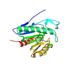 | | Crystal structure of APT1 bound to palmitic acid. | | 分子名称: | Acyl-protein thioesterase 1, CHLORIDE ION, PALMITIC ACID | | 著者 | Audagnotto, M, Marcaida, M.J, Ho, S, Pojer, F, Van der Goot, G, Dal Peraro, M. | | 登録日 | 2019-01-12 | | 公開日 | 2020-02-05 | | 最終更新日 | 2024-01-24 | | 実験手法 | X-RAY DIFFRACTION (2.755 Å) | | 主引用文献 | Palmitoylated acyl protein thioesterase APT2 deforms membranes to extract substrate acyl chains.
Nat.Chem.Biol., 2021
|
|
6QGQ
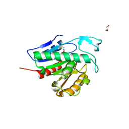 | | Crystal structure of APT1 C2S mutant bound to palmitic acid. | | 分子名称: | Acyl-protein thioesterase 1, GLYCEROL, PALMITIC ACID | | 著者 | Audagnotto, M, Marcaida, M.J, Ho, S, Pojer, F, Van der Goot, G, Dal Peraro, M. | | 登録日 | 2019-01-12 | | 公開日 | 2020-02-05 | | 最終更新日 | 2024-01-24 | | 実験手法 | X-RAY DIFFRACTION (2.601 Å) | | 主引用文献 | Palmitoylated acyl protein thioesterase APT2 deforms membranes to extract substrate acyl chains.
Nat.Chem.Biol., 2021
|
|
6QGO
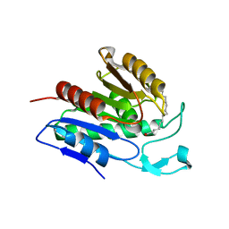 | | Crystal structure of APT1 S119A mutant bound to palmitic acid. | | 分子名称: | Acyl-protein thioesterase 1, PALMITIC ACID | | 著者 | Audagnotto, M, Marcaida, M.J, Ho, S, Pojer, F, Van der Goot, G, Dal Peraro, M. | | 登録日 | 2019-01-12 | | 公開日 | 2020-02-05 | | 最終更新日 | 2024-01-24 | | 実験手法 | X-RAY DIFFRACTION (2.599 Å) | | 主引用文献 | Palmitoylated acyl protein thioesterase APT2 deforms membranes to extract substrate acyl chains.
Nat.Chem.Biol., 2021
|
|
6QGN
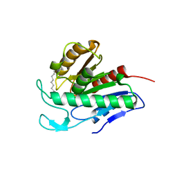 | | Crystal structure of APT1 bound to 2-Bromopalmitate | | 分子名称: | 2-Bromopalmitic acid, Acyl-protein thioesterase 1 | | 著者 | Marcaida, M.J, Audagnotto, M, Ho, S, Pojer, F, Van der Goot, G, Dal Peraro, M. | | 登録日 | 2019-01-12 | | 公開日 | 2020-02-05 | | 最終更新日 | 2024-01-24 | | 実験手法 | X-RAY DIFFRACTION (2.099 Å) | | 主引用文献 | Palmitoylated acyl protein thioesterase APT2 deforms membranes to extract substrate acyl chains.
Nat.Chem.Biol., 2021
|
|
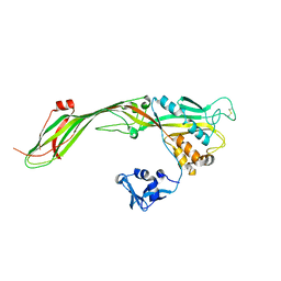
3C0N
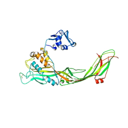 | | Crystal structure of the proaerolysin mutant Y221G at 2.2 A | | 分子名称: | Aerolysin | | 著者 | Pernot, L, Schiltz, M, Thurnheer, S, Burr, S.E, van der Goot, G. | | 登録日 | 2008-01-21 | | 公開日 | 2008-02-12 | | 最終更新日 | 2023-11-01 | | 実験手法 | X-RAY DIFFRACTION (2.2 Å) | | 主引用文献 | Molecular assembly of the aerolysin pore reveals a swirling membrane-insertion mechanism.
Nat.Chem.Biol., 9, 2013
|
|
3C0O
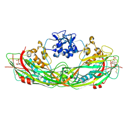 | | Crystal structure of the proaerolysin mutant Y221G complexed with mannose-6-phosphate | | 分子名称: | 6-O-phosphono-alpha-D-mannopyranose, ACETATE ION, Aerolysin | | 著者 | Pernot, L, Schiltz, M, Thurnheer, S, Burr, S.E, van der Goot, G. | | 登録日 | 2008-01-21 | | 公開日 | 2008-02-12 | | 最終更新日 | 2023-11-01 | | 実験手法 | X-RAY DIFFRACTION (2.5 Å) | | 主引用文献 | Molecular assembly of the aerolysin pore reveals a swirling membrane-insertion mechanism.
Nat.Chem.Biol., 9, 2013
|
|
3C0M
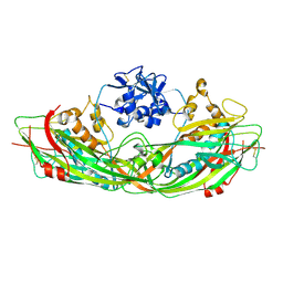 | | Crystal structure of the proaerolysin mutant Y221G | | 分子名称: | Aerolysin | | 著者 | Pernot, L, Schiltz, M, Thurnheer, S, Burr, S.E, van der Goot, G. | | 登録日 | 2008-01-21 | | 公開日 | 2008-02-12 | | 最終更新日 | 2023-11-01 | | 実験手法 | X-RAY DIFFRACTION (2.88 Å) | | 主引用文献 | Molecular assembly of the aerolysin pore reveals a swirling membrane-insertion mechanism.
Nat.Chem.Biol., 9, 2013
|
|
3G4O
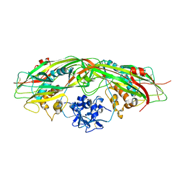 | |
3G4N
 | |


