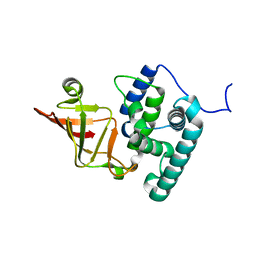
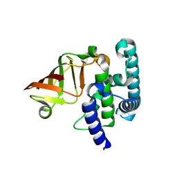
2HZ5
 
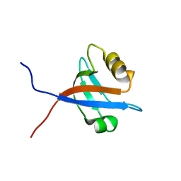
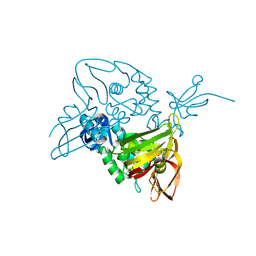
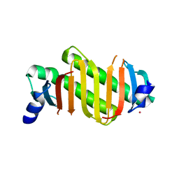 | | Crystal structure of human dynein light chain Dnlc2A | | 分子名称: | CESIUM ION, Dynein light chain 2A, cytoplasmic | | 著者 | Liu, J.-F, Wang, Z.-X, Wang, X.-Q, Tang, Q, An, X.-M, Gui, L.-L, Liang, D.-C. | | 登録日 | 2006-08-08 | | 公開日 | 2007-08-14 | | 最終更新日 | 2024-03-13 | | 実験手法 | X-RAY DIFFRACTION (2.1 Å) | | 主引用文献 | Crystal structure of human dynein light chain Dnlc2A: Structural insights into the interaction with IC74
Biochem.Biophys.Res.Commun., 349, 2006
|
|
4N8V
 | |
7V8G
 | |
7V8E
 | |
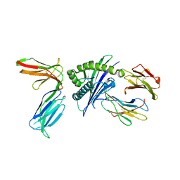
7V8F
 | | Crystal structure of UBE2L3 bound to HOIP RING1 domain. | | 分子名称: | E3 ubiquitin-protein ligase RNF31, Ubiquitin-conjugating enzyme E2 L3, ZINC ION | | 著者 | Liu, J, Wang, Y, Pan, L. | | 登録日 | 2021-08-22 | | 公開日 | 2022-03-30 | | 最終更新日 | 2023-11-29 | | 実験手法 | X-RAY DIFFRACTION (1.66 Å) | | 主引用文献 | Mechanistic insights into the subversion of the linear ubiquitin chain assembly complex by the E3 ligase IpaH1.4 of Shigella flexneri.
Proc.Natl.Acad.Sci.USA, 119, 2022
|
|
7V8H
 | |
8PE9
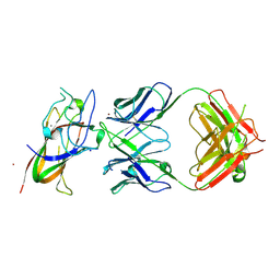 | | Complex between DDR1 DS-like domain and PRTH-101 Fab | | 分子名称: | 2-acetamido-2-deoxy-beta-D-glucopyranose, 2-acetamido-2-deoxy-beta-D-glucopyranose-(1-4)-[alpha-L-fucopyranose-(1-6)]2-acetamido-2-deoxy-beta-D-glucopyranose, CALCIUM ION, ... | | 著者 | Liu, J, Chiang, H, Xiong, W, Laurent, V, Griffiths, S.C, Duelfer, J, Deng, H, Sun, X, Yin, Y.W, Li, W, Audoly, L.P, An, Z, Schuerpf, T, Li, R, Zhang, N. | | 登録日 | 2023-06-13 | | 公開日 | 2023-06-28 | | 最終更新日 | 2024-02-07 | | 実験手法 | X-RAY DIFFRACTION (3.152 Å) | | 主引用文献 | A highly selective humanized DDR1 mAb reverses immune exclusion by disrupting collagen fiber alignment in breast cancer.
J Immunother Cancer, 11, 2023
|
|
4K7A
 | |
2OMJ
 | |
2OS6
 | |
6KLY
 | | Crystal structure of the type III effector XopAI from Xanthomonas axonopodis pv. citri in space group P43212 | | 分子名称: | Type III effector XopAI | | 著者 | Liu, J.-H, Wu, J.E, Lin, H, Chiu, S.W, Yang, J.Y. | | 登録日 | 2019-07-30 | | 公開日 | 2019-08-21 | | 最終更新日 | 2024-03-27 | | 実験手法 | X-RAY DIFFRACTION (2.01 Å) | | 主引用文献 | Crystal Structure-Based Exploration of Arginine-Containing Peptide Binding in the ADP-Ribosyltransferase Domain of the Type III Effector XopAI Protein.
Int J Mol Sci, 20, 2019
|
|
7D65
 | | Cryo-EM Structure of human CALHM5 in the presence of Ca2+ | | 分子名称: | 1,2-DIOCTANOYL-SN-GLYCERO-3-PHOSPHATE, Calcium homeostasis modulator protein 5 | | 著者 | Liu, J, Guan, F.H, Wu, J, Wan, F.T, Lei, M, Ye, S. | | 登録日 | 2020-09-29 | | 公開日 | 2020-12-23 | | 実験手法 | ELECTRON MICROSCOPY (2.94 Å) | | 主引用文献 | Cryo-EM structures of human calcium homeostasis modulator 5.
Cell Discov, 6, 2020
|
|
7D6H
 | |
7D61
 | | Cryo-EM Structure of human CALHM5 in the presence of EDTA | | 分子名称: | 1,2-DIOCTANOYL-SN-GLYCERO-3-PHOSPHATE, Calcium homeostasis modulator protein 5 | | 著者 | Liu, J, Guan, F.H, Wu, J, Wan, F.T, Lei, M, Ye, S. | | 登録日 | 2020-09-28 | | 公開日 | 2020-12-23 | | 実験手法 | ELECTRON MICROSCOPY (2.8 Å) | | 主引用文献 | Cryo-EM structures of human calcium homeostasis modulator 5.
Cell Discov, 6, 2020
|
|
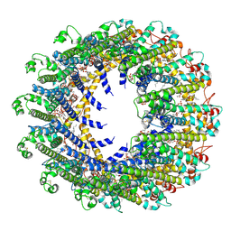
7DPT
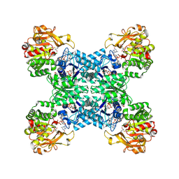 | | Structural basis for ligand binding modes of CTP synthase | | 分子名称: | 6-DIAZENYL-5-OXO-L-NORLEUCINE, ADENOSINE-5'-DIPHOSPHATE, CTP synthase, ... | | 著者 | Liu, J.L, Zhou, X, Guo, C.J, Chang, C.C. | | 登録日 | 2020-12-21 | | 公開日 | 2021-09-15 | | 実験手法 | ELECTRON MICROSCOPY (2.48 Å) | | 主引用文献 | Structural basis for ligand binding modes of CTP synthase.
Proc.Natl.Acad.Sci.USA, 118, 2021
|
|
7DPW
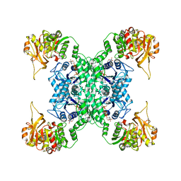 | | Structural basis for ligand binding modes of CTP synthase | | 分子名称: | CTP synthase, CYTIDINE-5'-TRIPHOSPHATE, MAGNESIUM ION | | 著者 | Liu, J.L, Zhou, X, Guo, C.J, Chang, C.C. | | 登録日 | 2020-12-21 | | 公開日 | 2021-09-15 | | 最終更新日 | 2024-06-05 | | 実験手法 | ELECTRON MICROSCOPY (2.65 Å) | | 主引用文献 | Structural basis for ligand binding modes of CTP synthase.
Proc.Natl.Acad.Sci.USA, 118, 2021
|
|
7D60
 | | Cryo-EM Structure of human CALHM5 in the presence of rubidium red | | 分子名称: | 1,2-DIOCTANOYL-SN-GLYCERO-3-PHOSPHATE, Calcium homeostasis modulator protein 5 | | 著者 | Liu, J, Guan, F.H, Wu, J, Wan, F.T, Lei, M, Ye, S. | | 登録日 | 2020-09-28 | | 公開日 | 2020-12-23 | | 実験手法 | ELECTRON MICROSCOPY (2.61 Å) | | 主引用文献 | Cryo-EM structures of human calcium homeostasis modulator 5.
Cell Discov, 6, 2020
|
|
8IB0
 | |
7E35
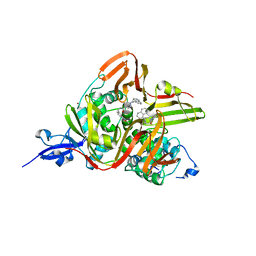 | | Crystal structure of the SARS-CoV-2 papain-like protease (PLPro) C112S mutant bound to compound S43 | | 分子名称: | N-[(3-acetamidophenyl)methyl]-1-[(1R)-1-naphthalen-1-ylethyl]piperidine-4-carboxamide, Non-structural protein 3, ZINC ION | | 著者 | Liu, J, Wang, Y, Xu, X, Pan, L. | | 登録日 | 2021-02-08 | | 公開日 | 2021-03-17 | | 最終更新日 | 2023-11-29 | | 実験手法 | X-RAY DIFFRACTION (2.4 Å) | | 主引用文献 | Development of potent and selective inhibitors targeting the papain-like protease of SARS-CoV-2.
Cell Chem Biol, 28, 2021
|
|
6K93
 | |
6K94
 | |
7WJ4
 | |
7WIZ
 | |
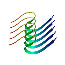
7WXI
 | | GPR domain of Drosophila P5CS filament with glutamate and ATPgammaS | | 分子名称: | Delta-1-pyrroline-5-carboxylate synthase, GAMMA-GLUTAMYL PHOSPHATE | | 著者 | Liu, J.L, Zhong, J, Guo, C.J, Zhou, X. | | 登録日 | 2022-02-14 | | 公開日 | 2022-03-30 | | 最終更新日 | 2024-06-26 | | 実験手法 | ELECTRON MICROSCOPY (4.2 Å) | | 主引用文献 | Structural basis of dynamic P5CS filaments.
Elife, 11, 2022
|
|
7WX4
 | |


